Journal of Animal Science and Technology
Korean Society of Animal Sciences and Technology
RESEARCH ARTICLE

Metabolic profiling of serum and urine in lactating dairy cows affected by subclinical ketosis using proton nuclear magnetic resonance spectroscopy

Jun Sik Eom1,#https://orcid.org/0000-0001-5360-0147, Shin Ja Lee1,2,#https://orcid.org/0000-0002-4224-1211, Hyun Sang Kim1https://orcid.org/0000-0002-3422-1990, Youyoung Choi3https://orcid.org/0000-0002-3417-8099, Seong Uk Jo3https://orcid.org/0000-0001-7901-0428, Sang Suk Lee4https://orcid.org/0000-0003-1540-7041, Eun Tae Kim5https://orcid.org/0000-0001-7486-5638, Sung Sill Lee1,2,3,*https://orcid.org/0000-0002-4621-4333
1Institute of Agriculture and Life Science, Gyeongsang National University, Jinju 52828, Korea
2University Centered Labs, Gyeongsang National University, Jinju 52828, Korea
3Division of Applied Life Science (BK21), Gyeongsang National Universitiy, Jinju 52828, Korea
4Ruminant Nutrition and Anaerobe Laboratory, Department of Animal Science and Technology, Sunchon National University, Suncheon 57922, Korea
5Dairy Science Division, National Institute of Animal Science, Rural Development Administration, Cheonan 31000, Korea
*Corresponding author: Sung Sill Lee, Division of Applied Life Science (BK21) & Institute of Agriculture and Life Science, Gyeongsang National University, Jinju 52828, Korea. Tel: +82-55-772-1883, E-mail: lss@gnu.ac.kr

These authors contributed equally to this work.

© Copyright 2022 Korean Society of Animal Science and Technology. This is an Open-Access article distributed under the terms of the Creative Commons Attribution Non-Commercial License (http://creativecommons.org/licenses/by-nc/4.0/) which permits unrestricted non-commercial use, distribution, and reproduction in any medium, provided the original work is properly cited.

Received: Dec 03, 2021; Revised: Feb 16, 2022; Accepted: Feb 21, 2022

Published Online: Mar 31, 2022

Abstract

Ketosis is associated with high milk yield during lactating or insufficient feed intake in lactating dairy cows. However, few studies have been conducted on the metabolomics of ketosis in Korean lactating dairy cows. The present study aimed to investigate the serum and urine metabolites profiling of lactating dairy cows through proton nuclear magnetic resonance (1H-NMR) spectroscopy and comparing those between healthy (CON) and subclinical ketosis (SCK) groups. Six lactating dairy cows were categorized into CON and SCK groups. All experimental Holstein cows were fed total mixed ration. Serum and urine samples were collected from the jugular vein of the neck and by hand sweeping the perineum, respectively. The metabolites in the serum and urine were determined using 1H-NMR spectroscopy. Identification and quantification of metabolites was performed by Chenomx NMR Suite 8.4 software. Metabolites statistical analysis was performed by Metaboanalyst version 5.0 program. In the serum, the acetoacetate level was significantly (p < 0.05) higher in the SCK group than in the CON group, and whereas acetate, galactose and pyruvate levels tended to be higher. CON group had significantly (p < 0.05) higher levels of 5-aminolevulinate and betaine. Indole-3-acetate, theophylline, p-cresol, 3-hydroxymandelate, gentisate, N-acetylglucosamine, N-nitrosodimethylamine, xanthine and pyridoxine levels were significantly (p < 0.05) higher in the urine of the SCK group than that in the CON group, which had higher levels of homogentisate, ribose, gluconate, ethylene glycol, maltose, 3-methyl-2-oxovalerate and glycocholate. Some significantly (p < 0.05) different metabolites in the serum and urine were associated with ketosis diseases, inflammation, energy balance and body weight. This study will be contributed useful a future ketosis metabolomics studies in Korea.

Keywords: Metabolites; Subclinical ketosis; Lactating dairy cow; Serum; Urine; Proton nuclear magnetic resonance spectroscopy

INTRODUCTION

Ketosis is common metabolic disease of dairy animals, and is associated with high milk yield during lactation or inadequate feed intake for energy that results in negative energy balance (NEB) [1]. Ketosis in lactating dairy cow decreases milk production and has negative effects on reproductive capacity [2]. In addition, such cows have a higher risk of developing periparturient disease such as lameness, mastitis, metritis, and retained placenta [36]. Ketosis is associated with an increased concentration of ketone bodies (acetoacetate, acetone and beta-hydroxybutyrate [BHB]) in the biofluid (milk, plasma, serum and urine) [7]. The underlying cause of the high concentration of ketone body metabolites are low blood glucose levels associated with hypoinsulinemia, which results in the mobilization of fatty acid (FA)s generated from adipose tissue [7]. Ketosis is classified into subclinical ketosis (SCK), clinical ketosis (CK), and type I and II ketosis. In SCK and CK, BHB concentration ranges from 1.2 to 1.4 mM/L and 2.6 to 3.0 mM/L, respectively [8,9]. Type I ketosis occurs between 3 and 6 weeks postpartum when the energy requirement for milk production is the highest [10]. Type II ketosis can lead to complications such as fatty liver via an increase in blood glucose and insulin levels because of excessive feed intake during the dry period [7,10]. Ketosis leads to considerable economic loss in the dairy industry [11]; therefore, research on the prevention and diagnosis of ketosis is required.

Metabolomics studies pertaining to ruminant biofluids including milk, plasma, rumen fluid, serum and urine are performed using many metabolite analyzers (nuclear magnetic resonance [NMR] spectroscopy, gas chromatography-mass spectrometry [GC-MS], liquid chromatography-mass spectrometry [LC-MS], etc.). Moreover, metabolomics studies pertaining to metabolic diseases (acidosis, ketosis etc.) have been conducted. Ketosis research using metabolites in the serum and plasma and the comparative study of metabolites in the plasma of CK, SCK and healthy lactating dairy cows have identified several metabolic pathways associated with CK and SCK by GC-MS, NMR and LC-MS [1113]. In another study, plasma metabolite profiling was performed for type I and II ketosis by proton NMR (1H-NMR) spectroscopy [14]. Another study compared the urine metabolites in healthy and SCK cows by NMR, direct injection GC-MS and LC-MS/MS [15]. However, there have been fewer urine-based research on ketosis in lactating dairy cows compared with those on other biofluids such as serum, plasma and milk. Metabolic profiling analysis of urine samples has identified biomarkers in various studies on humans [16,17]. Zhang et al. [15] reported a lacked of urinary metabolomics profiling for the identification of predictive biomarkers of ketosis in dairy cows. Therefore, it is necessary to study the urine metabolites of ketosis-induced dairy cows. Such studies will be useful in the future to search for biomarker candidates for ketosis diagnosis using blood (serum and plasma) and urine.

Recently, a metabolomics study on Hanwoo cattle and Holstein cows biofluids was performed by 1H-NMR spectroscopy in Korea [1820]. However, there have been few metabolomics studies on lactating dairy cow biofluids associated with ketosis Korea compared with those in other countries. In addition, in the Korea, the Holstein species are representing lactating dairy cow, and most important ruminant breeds in the Korean dairy industry [21]. Therefore, it is essential to conduct metabolomics studies that can help us to identify metabolic biomarkers biofluids that can diagnosis ketosis in the future.

We hypothesized that the serum and urine metabolites profiling of lactating dairy cows would be different between healthy (CON) and SCK groups. To test this hypothesis, we aimed to investigate the serum and urine metabolites profiling of lactating dairy cows using 1H-NMR spectroscopy and compare between two groups. This metabolic study will be helpful for developing strategies to reduce lactating dairy cow ketosis in Korea.

MATERIALS AND METHODS

Animals and sampling

CON and SCK Holstein lactating dairy cow general information (month-old, body weight, parity, milk yield, BHB concentration and number of experimental animals), total experimental day (14 days), diet adaptation period (4 days) blood BHB concentration monitoring period (9 days), blood and urine samples collected day (last day of the experiment) were same of previously Eom et al. study [22].

All experimental cows were fed total mixed ration (TMR). The consumed CON and SCK groups feed intake amount, same of previously Eom et al. study [22]. The results of chemical composition of the TMR is shown Table 1. Contents of acid detergent fiber, calcium, crude protein, dry matter, neutral detergent fiber and phosphorus in TMR was determined as described by Association of Official Analytical Communities [23,24] and Van Soest et al. [25] methods.

Table 1. Ingredients and nutrients of the experimental diets
Items Value (% of DM basis)
Ingredients (% of DM)
 Concentrate 15.3
 Soybean meal 2.40
 Corn silage 47.2
 Alfalfa hay 7.10
 Tall fescue 9.40
 Timothy 5.90
 Energy booster1) 7.10
 Cash gold1) 4.50
 Lyzin-Plus2) 0.20
 Limestone3) 0.20
 Zin care1) 0.10
 Supex-F1) 0.50
 Trace minerals4) 0.05
 Vitamins premix5) 0.05
Chemical composition (% of DM basis)
 DM (%) 53.2
 Crude protein 10.0
 Neutral detergent fiber 28.2
 Acid detergent fiber 16.9
 Calcium 0.40
 Phosphorus 0.15

Zin Care, Contained 16 GDU/g protease bromelain, 2.0 × 10⁸ cfu/g. Supex-F, Contained 99% protected fat from of palm oil (Cofavet, Cheonan, Korea).

Lyzin-Plus, Contained 6.0% Zn, 0.9% Cu, 1.4% Mn, 5.0% chelated glycine (A.N.Tech, Cheonan, Korea).

Sungshin minefield, Jeongseon, Korea.

Trace minerals, Contained 0.40% Mg; 0.20% K; 4.00% S; 0.08% Na; 0.03% Cl; 400 mg of Fe/kg; 60,042 mg of Zn/kg; 16,125 mg of Cu/kg; 42,375 mg of Mn/kg.

Vitamins premix, Provided approximately 5,000 KIU of vitamin A/kg; 1,000 KIU of vitamin D/kg; 33,500 mg of vitamin E/kg; 2,400 mg of vitamin C/kg.

DM, dry matter.

Download Excel Table

Blood samples collected and storage methods until 1H-NMR spectroscopy analysis was determined as described by Kim et al. [19] and Eom et al. [20] methods. Urine samples collected and storage methods until 1H-NMR spectroscopy analysis was determined as described by Kim et al. [19] and Eom et al. [20] methods.

Prepared proton nuclear magnetic resonance spectroscopy analyses

The 1H-NMR spectroscopy analysis of serum and urine samples was performed by following methods modified from Sun et al. [26], Jung et al. [27], Bertram et al. [28] and Jeong et al. [29] methods.

The spectra of two samples were obtained on a SPE-800 MHz NMR-MS Spectrometer (Bruker BioSpin AG, Fällanden, Switzerland) at 298 K using a 5 mm triple-resonance inverse cryoprobe with Z-gradients (Bruker BioSpin, Billerica, MA, USA) and method condition described by Kim et al. method [30].

Metabolites measurement, quantification, and statistical analysis

Serum and urine metabolites identification and quantification methods and data collected was performed by following methods modified from Kim et al. [19] and Eom et al. [20] methods. Metabolites data statistical analyses were using the Metaboanalyst version 5.0 program (https://www.metaboanalyst.ca), an open source R-based program for metabolomics. For the serum and urine metabolites analysis, when 50% of samples were under the identification limit or had at least 50% of missing values, they were eliminated from the analysis. The missing values were replaced by a value one-half of the minimum positive value from the original data. In addition, statistical analyses methods were determined as described by Kim et al. [19] and Eom et al. [20] methods. Univariate Student’s t-test were used to quantify difference between metabolite profiles of the serum and urine samples. In addition, principal component analysis (PCA), partial least square-discriminant analysis (PLS-DA), variable importance in projection (VIP) scores and metabolic pathway results were determined as described by Kim et al. [19] and Eom et al. [20] methods.

RESULTS

Multivariate data analysis

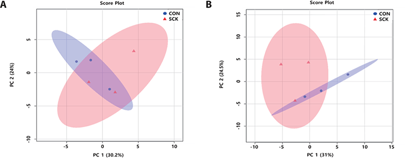
To analyze the variations in the serum and urine metabolites profiling of CON and SCK groups, we were performed PCA and PLS-DA. In the serum PCA score plot (Fig. 1A), two groups were not separated (PC 1 : 30.2%; PC 2 : 24%). In the urine PCA score plots (Fig. 1B) two groups were not separated (PC 1 : 31%; PC 2 : 24.5%).
jast-64-2-247-g1
Fig. 1. Principal components analysis score plot based on serum (A) and urine (B) metabolites data in healthy (CON) and subclinical ketosis (SCK) group by proton nuclear magnetic resonance spectroscopy analysis. On the score plot, each point represents an individual sample, with the blue dot representing the CON group (n=3), and the red triangle representing the SCK group (n=3). The abscissa and represent the variance associated with PC 1 and 2, respectively. PC, principal component.
Download Original Figure

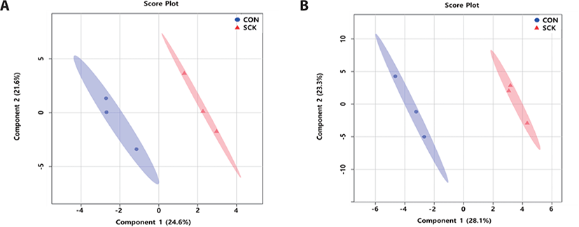
The serum PLS-DA score plots (Fig. 2A), for the two groups were clearly separated (component 1 : 24.6%; component 2 : 21.6%). The urine PLS-DA score plots (Fig. 2B) for two groups were clearly separated (component 1 : 28.1%; component 2 : 23.3%). These results show different in the concentration of serum and urine metabolites between CON and SCK groups.
jast-64-2-247-g2
Fig. 2. Partial least square-discriminant analysis score plot of serum (A) and urine (B) with healthy (CON) and subclinical ketosis (SCK) group by proton nuclear magnetic resonance spectroscopy analysis. The shaded ellipses represent the 95% confidence interval estimated from the score. On the score plot, each represents an individual sample, with blue dot representing the CON group (n=3), and red triangle representing the SCK group (n=3) The abscissa and ordinate represent the variance associated with component 1 and 2, respectively.
Download Original Figure
Detected and quantification of serum and urine metabolites

Supplementary Tables S1–S7 and, Supplementary Figs. 1 and 2 summarize the detected and quantified metabolites in the two groups. In the CON group, 98 metabolites were detected and divided into 12 chemical classes in the serum. In addition, a total of 52 metabolites were quantified. In the SCK group, 83 metabolites were detected and divided into 12 chemical classes in the serum. In addition, a total of 55 metabolites were quantified.

In the CON group, 144 metabolites were detected and divided into 13 chemical classes in the urine. In addition, a total of 93 metabolites were quantified. In the SCK group, 168 metabolites were detected and divided into 14 chemical classes in the urine. In addition, a total of 93 metabolites were quantified.

Differences in serum and urine metabolites between healthy and subclinical ketosis groups

Table 2 shows the significant trends (p < 0.05) and tendencies (0.05 ≤ p < 0.1) of different metabolites in the serum and urine of the two groups. Acetoacetate and succinate levels were significantly higher, whereas acetate, galactose and pyruvate levels tended to be higher but non-significantly in the serum of the SCK group than in the CON group. In contrast, 5-aminolevulinate (5-ALA) and betaine levels were significantly higher, whereas lactulose and 3-methylxanthine (3-MX) levels tended to be higher in the CON group than in the SCK group.

Table 2. Differential enrichment of metabolites content of serum and urine between healthy and subclinical ketosis group
Metabolites Classification CON/SCK p-value VIP score1) FC2)
Serum
 5-Aminolevulinate Carboxylic acid CON 6.21 × 10⁻³ 1.67 0.42
 Betaine Other CON 1.46 × 10⁻² 1.60 0.40
 Acetoacetate Carbohydrate SCK 2.44 × 10⁻² 1.85 −0.51
 Succinate Carbohydrate SCK 3.45 × 10⁻² 1.63 −0.42
 Lactulose Carbohydrate CON 5.04 × 10⁻² 0.94 0.16
 Acetate Carbohydrate SCK 6.98 × 10⁻² 0.79 −0.11
 3-Methylxanthine Other CON 7.63 × 10⁻² 1.82 0.47
 Galactose Carbohydrate SCK 8.24 × 10⁻² 1.58 −0.55
 Pyruvate Carbohydrate SCK 7.86 × 10⁻² 1.80 −0.49
Urine
 Indole-3-acetate Other SCK 9.34 × 10⁻⁴ 1.36 −0.39
 Homogentisate Benzoic acid CON 2.23 × 10⁻³ 1.81 0.69
 Ribose Carbohydrate CON 2.50 × 10⁻³ 1.88 0.76
 Gluconate Organic acid CON 5.97 × 10⁻³ 2.06 0.97
 Theophylline Other SCK 5.99 × 10⁻³ 2.01 −0.96
 p-Cresol Benzoic acid SCK 1.11 × 10⁻² 1.13 −0.29
 Ethylene glycol Lipid CON 1.56 × 10⁻² 2.55 1.67
 Maltose Carbohydrate CON 2.37 × 10⁻² 2.04 1.11
 3-Hydroxymandelate Benzoic acid SCK 2.72 × 10⁻² 1.73 −0.82
 Gentisate Benzoic acid SCK 2.96 × 10⁻² 1.94 −0.80
N-Acetylglucosamine Carbohydrate SCK 3.31 × 10⁻² 1.63 −0.72
 3-Methyl-2-oxovalerate Carboxylic acid CON 3.65 × 10⁻² 1.33 0.39
N-Nitrosodimethylamine Organic acid SCK 4.19 × 10⁻² 1.84 −0.74
 Glycocholate Lipid CON 4.22 × 10⁻² 1.82 0.76
 Xanthine Nucleoside, nucleotide SCK 4.25 × 10⁻² 0.90 −0.20
 Pyridoxine Other SCK 4.81 × 10⁻² 1.73 −0.89
 Nicotinurate Organic acid SCK 5.17 × 10⁻² 1.43 −0.47
 Acetoin Other SCK 5.35 × 10⁻² 1.58 −0.77
 Alanine Amino acid CON 6.71 × 10⁻² 0.70 0.13
 Trimethylamine N-oxide Aliphatic acylic compound SCK 6.97 × 10⁻² 1.11 −0.35
 3-Methylxanthine Other SCK 7.00 × 10⁻² 1.71 −0.96
 Indole-3-lactate Other SCK 8.18 × 10⁻² 1.34 −0.43
 Carnosine Amine SCK 8.20 × 10⁻² 1.21 −0.39
 3-Hydroxybutyrate Lipid SCK 9.03 × 10⁻² 1.54 −0.85

Variable importance in the projection obtained from partial least square-discriminant analysis model.

Fold change; Calculated as binary logarithm average concentration response ratio between CON and SCK group, where the positive value means that average concentration response of the metabolites in the former is larger than that in the latter and vice versa.

CON, healthy group; SCK, subclinical ketosis group; CON/SCK, Comparison between CON and SCK group.

Download Excel Table

Indole-3-acetate, theophylline, p-cresol, 3-hydroxymandelate, gentisate, N-acetylglucosamine, N-nitrosodimethylamine, xanthine and pyridoxine levels in the urine of the SCK group were significantly higher, and nicotinurate, acetoin, trimethylamine N-oxide, 3-MX, indole-3-lactate, carnosine and BHB levels tended to be higher compared with those in the CON group. In contrast, homogentisate, ribose, gluconate, ethylene glycol, maltose, 3-methyl-2-oxovalerate and glycocholate levels were significantly higher, and alanine level tended to be higher in the CON group than in the SCK group.

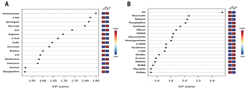
As shown in Figs. 3A and 3B, the evaluation of over to 1.5 VIP score of PLS-DA model showed 15 and 18 metabolites between the two groups of serum and urine, respectively. Acetoacetate, pyruvate and O-acetylcholine were higher VIP scores in the serum of the SCK group compared with those in the CON group. In contrast, 3-MX, syrinagate and arginine were higher VIP scores in the serum of the CON group compared with those in the SCK group. Theophylline, gentisate and N-nitrosodimethylamine were higher VIP scores in the urine of the SCK group compared with those in the CON group. In contrast, ethylene glycol, gluconate and maltose were higher VIP scores in the urine of the CON group compared with those in the SCK group.
jast-64-2-247-g3
Fig. 3. Variable importance in projection (VIP) scores of serum (A) and urine (B) metabolites in healthy (CON) and subclinical ketosis (SCK) group by proton nuclear magnetic resonance spectroscopy analysis. The selected metabolites were those with VIP score > 1.5. Heat map with red or blue boxes on the right indicates high and low abundance ratio, respectively, of the corresponding serum and urine metabolites in CON and SCK group. The VIP score was based on the partial least square-discriminant analysis model. Serum metabolites VIP score value: Acetoacetate, 1.8494; 3-MX, 1.8235; syringate, 1.8163; pyruvate, 1.798; ACh, 1.7379; arginine, 1.6952; 5-ALA, 1.6719; SUAC, 1.6445; succinate, 1.6309; betaine, 1.601; CrP, 1.5888; methionine, 1.5821; galactose, 1.5764; sucrose, 1.5189; glycylproline, 1.5164. Urine metabolites VIP score value: EG, 2.5494; gluconate, 2.0611; maltose, 2.0386; theophylline, 2.0136; gentisate, 1.937; ribose, 1.8785; NDMA, 1.8365; glycoholate, 1.8216; homogentisate, 1.8051; 3-HMA, 1.7327; pyridoxine, 1.7268; 3-MX, 1.7118; GlcNAc, 1.6289; acetoin, 1.5846; DMSO2, 1.5721; BHBA, 1.5414; glycolate, 1.5119; gallate, 1.5102. Metabolites abbreviation: 3-MX, 3-methylxanthine; ACh, O-acetylcholine; 5-ALA, 5-aminolevulinate; SUAC, succinylacetone; CrP, creatine phosphate; EG, ethylene glycol; NDMA, N-nitrosodimethylamine; 3-HMA, 3-hydroxymandelate; GlcNAc, N-acetylglucosamine; DMSO2, dimethyl sulfone; BHBA, 3-hydroxybutyrate.
Download Original Figure
Metabolic pathway analysis

In the serum profiling of including porphyrin and chlorophyll metabolism; glycine, serine and threonine metabolism; citrate cycle (tricarboxylic acid [TCA] cycle); and alanine, aspartate, and glutamate metabolism, four metabolic pathways significantly (p < 0.05) differed between the two groups. The following four metabolic pathways tendency (0.05 ≤ p < 0.1) differed in the serum between the two groups; such as tyrosine metabolism; butanoate metabolism; synthesis and degradation of ketone bodies; and valine, leucine and isoleucine degradation (Table 3 and Fig. 4A).
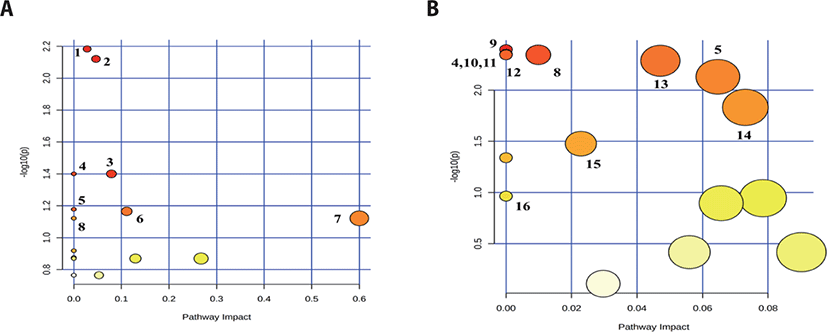
Table 3. Pathway analysis of significantly different serum metabolites compared with healthy and subclinical ketosis group
Metabolic pathway Total Cmpd Hits1) p-value −Log (p-value) FDR2) Impact3)
Porphyrin and chlorophyll metabolism 30 1 6.55 × 10⁻³ 2.18 6.06 × 10⁻² 0.03
Glycine, serine and threonine metabolism 34 3 7.57 × 10⁻³ 2.12 6.06 × 10⁻² 0.05
Citrate cycle (tricarboxylic acid [TCA] cycle) 20 2 3.98 × 10⁻² 1.40 0.15 0.08
Alanine, aspartate and glutamate metabolism 28 2 3.98 × 10⁻² 1.40 0.15 0.00
Tyrosine metabolism 42 2 6.64 × 10⁻² 1.18 0.15 0.00
Butanoate metabolism 15 2 6.84 × 10⁻² 1.17 0.15 0.11
Synthesis and degradation of ketone bodies 5 1 7.56 × 10⁻² 1.12 0.15 0.60
Valine, leucine and isoleucine degradation 40 1 7.56 × 10⁻² 1.12 0.15 0.00

The actually matched number from the user uploaded data.

The p-value adjusted using False Discovery Rate.

The pathway impact value calculated from pathway topology analysis.

Total Cmpd, The total number of compounds in the pathway.

Download Excel Table
jast-64-2-247-g4
Fig. 4. Metabolic pathway mapping significantly different serum (A) and urine (B) metabolites compared in healthy and subclinical ketosis group. The pathway impact analysis was performed using Metaboanalyst 5.0 software. The x-axis represents the pathway impact, and y-axis represents the pathway enrichment. The results are presented graphically as a bubble plot. The darker color and larger size represent higher p-value from enrichment analysis and greater impact from pathway topology analysis, respectively. Metabolic pathway name: 1, Porphrin and chlorophyll metabolism; 2, Glycine, serine and threonine metabolism; 3, Citrate cycle (tricarboxylic acid [TCA] cycle); 4, Alanine, aspartate and glutamate metabolism; 5, Tyrosine metabolism; 6, Butanoate metabolism; 7, Synthesis and degradation of ketone bodies; 8, Valine, leucine, and isoleucine degradation; 9, Ubiquinone and other terpenoid-quinone biosynthesis; 10, Selenocompound metabolism; 11, Aminoacyl-tRNA biosynthesis; 12, Valine, leucine and isoleucine biosynthesis; 13, Pentose phosphate pathway; 14, Starch and sucrose metabolism; 15, Primary bile acid biosynthesis; 16, Tryptophan metabolism.
Download Original Figure

In the urine, the following 11 metabolic pathways significantly (p < 0.05) differed between the two groups; such as ubiquinone and other terpenoid-quinone biosynthesis; alanine, aspartate and glutamate metabolism; selenocompound metabolism; aminoacyl-tRNA biosynthesis; valine, leucine and isoleucine degradation; valine, leucine and isoleucine biosynthesis; pentose phosphate pathway; tyrosine metabolism; starch and sucrose metabolism; primary bile and biosynthesis; and tryptophan metabolism (Table 4 and Fig. 4B).

Table 4. Pathway analysis of significantly different urine metabolites compared with healthy and subclinical ketosis group
Metabolic pathway Total Cmpd Hits1) p-value −Log (p-value) FDR2) Impact3)
Ubiquinone and other terpenoid-quinone biosynthesis 9 1 4.02 × 10⁻³ 2.40 1.33 × 10⁻² 0.00
Alanine, aspartate and glutamate metabolism 28 1 4.47 × 10⁻³ 2.35 1.33 × 10⁻² 0.00
Selenocompound metabolism 20 1 4.47 × 10⁻³ 2.35 1.33 × 10⁻² 0.00
Aminoacyl-tRNA biosynthesis 48 1 4.47 × 10⁻³ 2.35 1.33 × 10⁻² 0.00
Valine, leucine and isoleucine degradation 40 1 4.52 × 10⁻³ 2.34 1.33 × 10⁻² 0.01
Valine, leucine and isoleucine biosynthesis 8 1 4.52 × 10⁻³ 2.34 1.33 × 10⁻² 0.00
Pentose phosphate pathway 22 2 5.17 × 10⁻³ 2.29 1.33 × 10⁻² 0.05
Tyrosine metabolism 42 2 7.41 × 10⁻³ 2.13 1.67 × 10⁻² 0.06
Starch and sucrose metabolism 18 1 1.47 × 10⁻² 1.83 2.95 × 10⁻² 0.07
Primary bile acid biosynthesis 46 1 3.33 × 10⁻² 1.48 6.00 × 10⁻² 0.02
Tryptophan metabolism 41 1 4.57 × 10⁻² 1.34 7.48 × 10⁻² 0.00

The actually matched number from the user uploaded data.

The p-value adjusted using False Discovery Rate.

The pathway impact value calculated from pathway topology analysis.

Total Cmpd, The total number of compounds in the pathway.

Download Excel Table

DISCUSSION

In lactating dairy cows with NEB, the increased glycine concentration in plasma is related to the breakdown of muscle protein [31] or to the de novo synthesis of glycine from threonine and serine [32]. Shibano et al. [33] reported that glycine in the serum could be used as a marker for EB and metabolic position in lactating dairy cows. In addition, the ratio of glycine to alanine concentration in the serum was used as a biomarker for malnutrition in lactating dairy cows during early lactation [33]. In this study, glycine concentration was higher in the SCK group. Alanine concentration was higher in the CON group; however, not significance different (p > 0.05). High concentration of glycine, kynurenine, and pantothenate and low concentration of arginine as a novel biomarker for NEB diagnosis [34]. In addition, the low concentration of arginine in lactating dairy cows with NEB induces an increase in nitrogen oxide concentration with an attendant increase in blood flow, which is useful for higher nutrients supply for milk production in the mammary gland [35]. In this study, pantothenate concentration was higher in the SCK group; however, not significance different (p > 0.05). Arginine was quantified only CON group and the VIP score was high in the CON group. The substrates for de novo synthesis of FA are BHB and acetate and are used by the mammary epithelial cells to synthesize short and medium chain FAs and sixteen-carbon FAs [36]. During the NEB period, the de novo synthesis of FA decreases and the body commence to use its own storage of energy [37]. In addition, type I and II ketosis have higher concentrations of acetate and BHB [14]. In this study, BHB concentration was higher in the SCK group; however, not significance different (p- > 0.05), and the acetate concentration in the SCK group was tended to be higher (0.05 ≤ p < 0.1) compared with the CON group. Low concentrations of blood glucose are related to hypoinsulinemia, which subsequently activates FA mobilization from tissues, thereby increasing the concentration of ketone body metabolites [7]. Carocho et al. [38] reported that the caloric of 100 g glucose and sucrose led to different peaks in blood glucose concentration. Sucrose is a disaccharide, comprising one molecule of glucose and one molecule of fructose [39]. Therefore, sucrose is connected to the concentration of glucose in the blood. In this study, glucose concentration was higher in the CON group; however, the difference was not significant (p > 0.05). In addition, sucrose was quantified in the CON group but not in the SCK group, and the VIP score was also higher in the CON group. Blood BHB, acetoacetate, and acetone are associated with the incomplete beta-oxidation of mobilized excess of fats that, result in ketosis [40,41]. Among them, BHB and acetone concentrations in the plasma are useful in the diagnosis of SCK in lactating dairy cows during early lactation [42,43]. In this study, BHB and acetone concentrations were higher in the SCK group; however, the difference was not significant (p > 0.05). Acetoacetate concentration was significantly (p- < 0.05; VIP score : 1.85) higher in the SCK group.

5-ALA is a dietary supplement for livestock that can affect the synthesis of heme and positively influence the iron status of hemoglobin in animals [44]. In addition, 5-ALA supplementation improved milk protein, fat and casein in dairy cows [45,46]. 5-ALA in blood is a product of condensing succinyl-CoA and glycine through the catalytic activity of 5-ALA synthase [47]. Hendawy et al. [48] reviewed biological activities of 5-ALA and reported, for example, antioxidant, anti-inflammatory, and immunomodulator activities. Nuclear factor kappa-light-chain-enhancer of activated B cells (NF-κβ) induces a variety of genes that encode proteins involved in inflammation including tumor necrosis factor (TNF), interleukin-1 (IL-1), and interleukin-23 [49]. Betaine supplementation improved milk yield, fat and FA synthesis [50]. Betaine in serum has shown several anti-inflammatory effects including the inhibition of NF-κβ [49]. Ametaj et al. [51] reported a potential role of immune factors in triggering systemic inflammation during the transition period in the pathobiology of metabolic disorders (e.g, concurrent disease with type II ketosis). In this study, 5-ALA (p < 0.01; VIP score : 1.67) and betaine (p < 0.05; VIP score : 1.60) concentration were significantly lower in the SCK group. Therefore, 5-ALA and betaine levels in the serum of lactating dairy cows are potential biomarkers for the diagnosis of ketosis. However, further research is needed on the relationship between the two metabolite (5-ALA and betaine) and ketosis diagnosis.

SCK involves an increase in the levels of ketone body metabolites in the urine, and the distinct signs of CK disease are absent [7]. Recently, the acetoacetate level in urine was used as a biomarker for ketosis diagnosis in the dairy industry [15]. This method is a quantitative test limited by its short sensitivity, and is used only for examine purposes [52]. In this study, acetoacetate and acetone concentrations were higher in the SCK group; however, the difference was not significantly (p > 0.05). The BHB concentration tended to be higher (0.05 ≤ p < 0.1), as did the VIP score (1.54) was also higher in the SCK group. Kawasaki et al. [53] reported that urine fructose concentration decreases during ketosis in patients. Acute administration of fructose promotes other adverse metabolic diseases, including hyperuricemia and lactic acidosis [54]. Therefore, the fructose level in the urine of lactating dairy cows might be considered a potential biomarker for the diagnosis of ketosis. In this study, fructose concentration was higher in the CON group; however, the difference was not significantly (p > 0.05). Amino acids are substantial precursors for associated with gluconeogenesis and ketogenesis [55,56], and crucial moderator or intervening in diverse metabolic pathways, including cell signaling, immunity, growth, maintenance and oxidative stress [5759]. Therefore, amino acid metabolism is essential for sustained condition and for preventing diseases (metabolic and contagious) [15]. Pantothenate has importance metabolites in production of carbohydrate and FA metabolism associated with energy [60]. Zhang et al. [15] reported that amino acid metabolites (arginine, aspartic acid, glutamate, glycine, alanine, cysteine, isoleucine, lysine, phenylalanine, and tyrosine) and carnosine, N-acetylglutamate, 1-methylhistidine, 3-methylhistidine and pantothenate were higher concentration in the urine of normal cow. In this study, glycine, 3-methylhistidine and pantothenate concentrations were higher in the CON group; however, the difference was not significantly (p > 0.05). Alanine concentration tended to be higher (0.05 ≤ p < 0.1) in the CON group. In addition, aspartate and glutamate metabolites was not significance different (p > 0.05) between the two groups.

Homogentisate in urine is produced through the catabolism of phenylalanine by homogentisate 1,2 dioxygenase (HGD) [61], and HGD affects body weight and sirloin cross-sectional area in cattle [62]. Holtenius and Holtenius [10] reported that CK in lactating dairy cows reduced milk yield and body weight. In this study, homogentisate concentration was significantly (p < 0.01; VIP score : 1.81) higher in the CON group. Theophylline is a xanthine-based metabolite and, an intermediate product in the metabolic process of caffeine and 3-MX. This metabolite can be excreted through the kidneys and has a diuretic effect [63,64] and can cause a variety of side effects in cows, such as acid-base and electrolyte imbalances [65]. In this study, theophylline concentration was significantly (p < 0.01; VIP score : 2.01) higher and 3-MX concentration was tended to be higher (0.05 ≤ p < 0.1; VIP score : 1.71) in the SCK group. The p-cresol concentration in urine may reflect the intake of dietary phenylalanine and tyrosine in non-ruminants and thus may be a proxy of the overall N intake [66]. However, approximately half of the tyrosine content of rumen-administered casein was excreted as p-cresol [67]. Therefore, an excessively high concentration in the urine may have a negative effect on nitrogen metabolism in ruminants. In this study, p-cresol concentration was significantly (p < 0.05) higher in the SCK group. Since the research on urine metabolites related to ketosis in lactating dairy cow is insufficient the relationship between metabolites and ketosis shown in this study will be helpful for minimizing the incidence of the disease. In addition, homogentisate, theophylline, 3-MX and p-cresol levels in the urine of lactating dairy cows are potential biomarkers for the diagnosis of ketosis. However, further research is needed on the relationship between the four metabolite (homogentisate, theophylline, 3-MX and p-cresol) and ketosis diagnosis.

The metabolites profiling of CON and SCK group lactating dairy cows were investigated by 1H-NMR spectroscopy. In the serum, associated with inflammation (5-ALA and betaine) and positive energy balance (arginine) metabolites was high concentration in the CON group, whereas ketone bodies including acetoacetate and acetate were high concentration in the SCK group. In the urine, associated with gluconeogenesis (amino acids; alanine) and body weight (homogentisate) were high concentration in the CON group, whereas ketone bodies including BHB was high concentration in SCK group. In Korea, studies on metabolic profiling by 1H-NMR spectroscopy are inadequate. Therefore, this study will contribute to future ketosis metabolomics studies in Korea by serving as a reference guide.

SUPPLEMENTARY MATERIALS

Supplementary Tables

jast-64-2-247-suppl1.pdf

Competing interests

No potential conflict of interest relevant to this article was reported.

Funding sources

This work was carried out with the support of “Cooperative Research Program for Agriculture Science and Technology Development (Project No. PJ01503902)” Rural Development Administration, Korea.

Acknowledgements

Not applicable.

Availability of data and material

Upon reasonable request, the datasets of this study can be available from the corresponding author.

Authors’ contributions

Conceptualization: Eom JS, Lee SJ, Lee Sung Sill.

Data curation: Eom JS, Kim HS, Choi Y.

Formal analysis: Eom JS, Kim HS, Choi Y, Jo SU.

Methodology: Lee SJ, Lee Sang Suk, Lee Sung Sill.

Software: Eom JS, Kim HS.

Validation: Eom JS, Kim HS, Jo SU, Kim ET.

Investigation: Eom JS, Kim HS, Choi Y, Jo SU.

Writing - original draft: Eom JS.

Writing - review & editing: Eom JS, Lee SJ, Kim HS, Choi Y, Jo SU, Lee Sang Suk, Kim ET, Lee Sung Sill.

Ethics approval and consent to participate

Animal care and experimental procedures were conducted according to the guideline of the National Institute of Animal Science (NIAS; Rural Development Administration, Cheonan 31000, Korea), Animal Care and Use Committee (Approval number: NIAS-2017-249).

REFERENCES

1.

Grummer RR. Impact of changes in organic nutrient metabolism on feeding the transition dairy cow. J Anim Sci. 1995; 73:2820-33

2.

Baird GD. Primary ketosis in the high-producing dairy cow: clinical and subclinical disorders, treatment, prevention, and outlook. J Dairy Sci. 1982; 65:1-10

3.

Duffield TF, Lissemore KD, McBride BW, Leslie KE. Impact of hyperketonemia in early lactation dairy cows on health and production. J Dairy Sci. 2009; 92:571-80

4.

McArt JAA, Nydam DV, Ospina PA, Oetzel GR. A field trial on the effect of propylene glycol on milk yield and resolution of ketosis in fresh cows diagnosed with subclinical ketosis. J Dairy Sci. 2011; 94:6011-20

5.

Ospina PA, Nydam DV, Stokol T, Overton TR. Evaluation of nonesterified fatty acids and β-hydroxybutyrate in transition dairy cattle in the northeastern United States: critical thresholds for prediction of clinical diseases. J Dairy Sci. 2010; 93:546-54

6.

Raboisson D, Mounié M, Maigné E. Diseases, reproductive performance, and changes in milk production associated with subclinical ketosis in dairy cows: a meta-analysis and review. J Dairy Sci. 2014; 97:7547-63

7.

Zhang G, Ametaj BN. Ketosis an old story under a new approach. Dairy. 2020; 1:42-60

8.

Suthar VS, Canelas-Raposo J, Deniz A, Heuwieser W. Prevalence of subclinical ketosis and relationships with postpartum diseases in European dairy cows. J Dairy Sci. 2013; 96:2925-38

9.

Duffield T. Subclinical ketosis in lactating dairy cattle. Vet Clin North Am Food Anim Pract. 2000; 16:231-53

10.

Holtenius P, Holtenius K. New aspects of ketone bodies in energy metabolism of dairy cows: a review. Zentralbl Veterinarmed A. 1996; 43:579-87

11.

Sun LW, Zhang HY, Wu L, Shu S, Xia C, Xu C, et al. 1H-Nuclear magnetic resonance-based plasma metabolic profiling of dairy cows with clinical and subclinical ketosis. J Dairy Sci. 2014; 97:1552-62

12.

Zhang H, Wu L, Xu C, Xia C, Sun L, Shu S. Plasma metabolomic profiling of dairy cows affected with ketosis using gas chromatography/mass spectrometry. BMC Vet Res. 2013; 9:186

13.

Li Y, Xu C, Xia C, Zhang H, Sun L, Gao Y. Plasma metabolic profiling of dairy cows affected with clinical ketosis using LC/MS technology. Vet Q. 2014; 34:152-8

14.

Xu C, Li Y, Xia C, Zhang HY, Sun L, Xu CC. 1H NMR-based plasma metabolic profiling of dairy cows with type I and type II ketosis. Pharm Anal Acta. 2015; 6:1000328

15.

Zhang G, Mandal R, Wishart DS, Ametaj BN. A multi-platform metabolomics approach identifies urinary metabolite signatures that differentiate ketotic from healthy dairy cows. Front Vet Sci. 2021; 8:595983

16.

Emwas AH, Luchinat C, Turano P, Tenori L, Roy R, Salek RM, et al. Standardizing the experimental conditions for using urine in NMR-based metabolomic studies with a particular focus on diagnostic studies: a review. Metabolomics. 2015; 11:872-94

17.

Emwas AH, Roy R, McKay RT, Ryan D, Brennan L, Tenori L, et al. Recommendations and standardization of biomarker quantification using NMR-based metabolomics with particular focus on urinary analysis. J Proteome Res. 2016; 15:360-73

18.

Eom JS, Lee SJ, Kim HS, Choi YY, Kim SH, Lee YG, et al. Metabolomics comparison of Hanwoo (Bos taurus coreanae) biofluids using proton nuclear magnetic resonance spectroscopy. Metabolites. 2020; 10:333

19.

Kim HS, Kim ET, Eom JS, Choi YY, Lee SJ, Lee SS, et al. Exploration of metabolite profiles in the biofluids of dairy cows by proton nuclear magnetic resonance analysis. PLOS ONE. 2021; 16e0246290

20.

Eom JS, Kim ET, Kim HS, Choi YY, Lee SJ, Lee SS, et al. Metabolomics comparison of rumen fluid and milk in dairy cattle using proton nuclear magnetic resonance spectroscopy. Anim Biosci. 2021; 34:213-22

21.

KOSIS [Korean statistical information service]. Population of cows by breed [Internet]. 2019[cited 2022 Jan 8]https://kosis.kr/statHtml/statHtml.do?orgld=101&tblld=DT_1NGB503&conn_path=12

22.

Eom JS, Kim HS, Lee SJ, Choi YY, Jo SU, Kim J, et al. Metabolic profiling of rumen fluid and milk in lactating dairy cattle influenced by subclinical ketosis using proton nuclear magnetic resonance spectroscopy. Animals. 2021; 11:2526

23.

AOAC [Association of Official Analytical Chemists] International. Official methods of analysis of the AOAC International. 17th ed Washington DC: AOAC International. 2003

24.

AOAC [Association of Official Analytical Chemists] International. Official methods of analysis of the AOAC International. 18th ed Washington DC: AOAC International. 2005

25.

Van Soest PJ, Robertson JB, Lewis BA. Methods for dietary fiber, neutral detergent fiber, and nonstarch polysaccharides in relation to animal nutrition. J Dairy Sci. 1991; 74:3583-97

26.

Sun Y, Xu C, Li C, Xia C, Xu C, Wu L, et al. Characterization of the serum metabolic profile of dairy cows with milk fever using 1H-NMR spectroscopy. Vet Q. 2014; 34:159-63

27.

Jung YS, Hyeon JS, Hwang GS. Software-assisted serum metabolite quantification using NMR. Anal Chim Acta. 2016; 934:194-202

28.

Bertram HC, Yde CC, Zhang X, Kristensen NB. Effect of dietary nitrogen content on the urine metabolite profile of dairy cows assessed by nuclear magnetic resonance (NMR)-based metabolomics. J Agric Food Chem. 2011; 59:12499-505

29.

Jeong JY, Hwang GS, Park JC, Kim DH, Ha MN. 1H NMR-based urinary metabolic profiling of gender and diurnal variation in healthy Korean subjects. Environ Health Toxicol. 2010; 25:295-306

30.

Kim MS, Kim IY, Sung HR, Nam M, Kim YJ, Kyung DS, et al. Metabolic dysfunction following weight regain compared to initial weight gain in a high-fat diet-induced obese mouse model. J Nutr Biochem. 2019; 69:44-52

31.

Doepel L, Lapierre H, Kennelly JJ. Peripartum performance and metabolism of dairy cows in response to prepartum energy and protein intake. J Dairy Sci. 2002; 85:2315-34

32.

Amelio I, Cutruzzolá F, Antonov A, Agostini M, Melino G. Serine and glycine metabolism in cancer. Trends Biochem Sci. 2014; 39:191-8

33.

Shibano K, Kawamura S, Hakamada R, Kawamura Y. The relationship between changes in serum glycine and alanine concentrations in non-essential amino acid and milk production in the transition period in dairy cows. J Vet Med Sci. 2005; 67:191-3

34.

Xu W, Vervoort J, Saccenti E, Kemp B, van Hoeij RJ, van Knegsel ATM. Relationship between energy balance and metabolic profiles in plasma and milk of dairy cows in early lactation. J Dairy Sci. 2020; 103:4795-805

35.

Kim SW, Wu G. Regulatory role for amino acids in mammary gland growth and milk synthesis. Amino Acids. 2009; 37:89-95

36.

Lock AL, Bauman DE. Modifying milk fat composition of dairy cows to enhance fatty acids beneficial to human health. Lipids. 2004; 39:1197-206

37.

van Knegsel ATM, van den Brand H, Dijkstra J, Tamminga S, Kemp B. Effect of dietary energy source on energy balance, production, metabolic disorders and reproduction in lactating dairy cattle. Reprod Nutr Dev. 2005; 45:665-88

38.

Carocho M, Morales P, Ferreira ICFR. Sweeteners as food additives in the XXI century: a review of what is known, and what is to come. Food Chem Toxicol. 2017; 107:302-17

39.

Grüneis V, Schweiger K, Galassi C, Karl CM, Treml J, Ley JP, et al. Sweetness perception is not involved in the regulation of blood glucose after oral application of sucrose and glucose solutions in healthy male subjects. Mol Nutr Food Res. 2021; 65:2000472

40.

Cooke RF, Silva Del Río N, Caraviello DZ, Bertics SJ, Ramos MH, Grummer RR. Supplemental choline for prevention and alleviation of fatty liver in dairy cattle. J Dairy Sci. 2007; 90:2413-8

41.

Zhou Z, Loor JJ, Piccioli-Cappelli F, Librandi F, Lobley GE, Trevisi E. Circulating amino acids in blood plasma during the peripartal period in dairy cows with different liver functionality index. J Dairy Sci. 2016; 99:2257-67

42.

Leblanc S. Monitoring metabolic health of dairy cattle in the transition period. J Reprod Dev. 2010; 56:S29-35

43.

Sato H. Increased blood concentration of isopropanol in ketotic dairy cows and isopropanol production from acetone in the rumen. Anim Sci J. 2009; 80:381-6

44.

Chen YJ, Kim IH, Cho JH, Min BJ, Yoo JS, Wang Q. Effect of δ-aminolevulinic acid on growth performance, nutrient digestibility, blood parameters and the immune response of weanling pigs challenged with Escherichia coli lipopolysaccharide. Livest Sci. 2008; 114:108-16

45.

Wang JP, Kim HJ, Chen YJ, Yoo JS, Cho JH, Kang DK, et al. Effects of delta-aminolevulinic acid and vitamin C supplementation on feed intake, backfat, and iron status in sows. J Anim Sci. 2009; 87:3589-95

46.

Hendawy AO, Shirai M, Takeya H, Sugimura S, Miyanari S, Taniguchi S, et al. Effects of 5-aminolevulinic acid supplementation on milk production, iron status, and immune response of dairy cows. J Dairy Sci. 2019; 102:11009-15

47.

Fujino M, Nishio Y, Ito H, Tanaka T, Li XK. 5-Aminolevulinic acid regulates the inflammatory response and alloimmune reaction. Int Immunopharmacol. 2016; 37:71-8

48.

Hendawy AO, Khattab MS, Sugimura S, Sato K. Effects of 5-aminolevulinic acid as a supplement on animal performance, iron status, and immune response in farm animals: a review. Animals. 2020; 10:1352

49.

Go EK, Jung KJ, Kim JY, Yu BP, Chung HY. Betaine suppresses proinflammatory signaling during aging: the involvement of nuclear factor-κB via nuclear factor-inducing kinase/IκB kinase and mitogen-activated protein kinases. J Gerontal Biol Sci. 2005; 60A:1252-64

50.

Peterson SE, Rezamand P, Williams JE, Price W, Chahine M, McGuire MA. Effects of dietary betaine on milk yield and milk composition of mid-lactation Holstein dairy cows. J Dairy Sci. 2012; 95:6557-62

51.

Ametaj BN, Bradford BJ, Bobe G, Nafikov RA, Lu Y, Young JW, et al. Strong relationships between mediators of the acute phase response and fatty liver in dairy cows. Can J Anim Sci. 2005; 85:165-75

52.

Oetzel GR. Monitoring and testing dairy herds for metabolic disease. Vet Clin North Am Food Anim Pract. 2004; 20:651-74

53.

Kawasaki T, Igarashi K, Ogata N, Oka Y, Ichiyanagi K, Yamanouchi T. Markedly increased serum and urinary fructose concentrations in diabetic patients with ketoacidosis or ketosis. Acta Diabetol. 2012; 49:119-23

54.

Woods HF, Alberti KGMM. Dangers of intravenous fructose. Lancet. 1972; 300:1354-7

55.

D’Mello JPF. Amino acids in animal nutrition. 2nd ed Waillingford, Oxon: CABI. 2003

56.

Di Pasquale MG. Amino acids and proteins for the athlete: the anabolic edge. 2nd ed Boca Raton, FL: CRC Press. 2007

57.

Galli F. Amino acid and protein modification by oxygen and nitrogen species. Amino Acids. 2012; 42:1-4

58.

Li P, Yin YL, Li D, Kim SW, Wu G. Amino acids and immune function. Br J Nutr. 2007; 98:237-52

59.

Moriwaki H, Miwa Y, Tajika M, Kato M, Fukushima H, Shiraki M. Branched-chain amino acids as a protein- and energy-source in liver cirrhosis. Biochem Biophys Res Commun. 2004; 313:405-9

60.

Ragaller V, Lebzien P, Südekum KH, Hüther L, Flachowsky G. Pantothenic acid in ruminant nutrition: a review. J Anim Physiol Anim Nutr. 2011; 95:6-16

61.

Rodríguez JM, Timm DE, Titus GP, Beltrán-Valero de Bernabé D, Criado O, Mueller HA, et al. Structural and functional analysis of mutations in alkaptonuria. Hum Mol Genet. 2000; 9:2341-50

62.

Zhou G, Dudgeon C, Li M, Cao Y, Zhang L, Jin H. Molecular cloning of the HGD gene and association of SNPs with meat quality traits in Chinese red cattle. Mol Biol Rep. 2010; 37:603-11

63.

Kanazawa H, Atsumi R, Matsushima Y, Kizu J. Determination of theophylline and its metabolites in biological samples by liquid chromatography–mass spectrometry. J Chromatogr A. 2000; 870:87-96

64.

American Society of Health-System Pharmacists. AHFS drug information 2016. Bethesda, MD: American Society of Health-System Pharmacists. 2016

65.

Chalmeh A, Mazrouei Sebdani M. Furosemide induced electrocardiographic alterations in high producing Holstein dairy cows. Iran J Rumin Health Res. 2016; 1:46-58

66.

De Pascali SA, Gambacorta L, Oswald IP, Del Coco L, Solfrizzo M, Fanizzi FP. 1H NMR and MVA metabolomic profiles of urines from piglets fed with boluses contaminated with a mixture of five mycotoxins. Biochem Biophys Rep. 2017; 11:9-18

67.

Martin AK. The origin of urinary aromatic compounds excreted by ruminants: 3. the metabolism of phenolic compounds to simple phenols. Br J Nutr. 1982; 48:497-507