Journal of Animal Science and Technology
Korean Society of Animal Sciences and Technology
RESEARCH ARTICLE

Prediction of male fertility using Ras-related proteins

Jeong-Won Bae1https://orcid.org/0000-0002-7030-7767, Ju-Mi Hwang1https://orcid.org/0000-0003-1776-9759, Woo-Sung Kwon1,*https://orcid.org/0000-0002-0848-7189
1Department of Animal Science and Biotechnology, Kyungpook National University, Sangju 37224, Korea
*Corresponding author: Woo-Sung Kwon, Department of Animal Science and Biotechnology, Kyungpook National University, Sangju 37224, Korea. Tel: +82-54-530-1942, E-mail: wskwon@knu.ac.kr

© Copyright 2022 Korean Society of Animal Science and Technology. This is an Open-Access article distributed under the terms of the Creative Commons Attribution Non-Commercial License (http://creativecommons.org/licenses/by-nc/4.0/) which permits unrestricted non-commercial use, distribution, and reproduction in any medium, provided the original work is properly cited.

Received: Aug 09, 2022; Revised: Sep 22, 2022; Accepted: Oct 04, 2022

Published Online: Nov 30, 2022

Abstract

Identifying effective biomarkers for the diagnosis of male fertility is crucial for improving animal production and treating male infertility in humans. Ras-related proteins (Rab) are associated with morphological and motion kinematic functions in spermatozoa. Moreover, Rab2A, a Rab protein, is a possible male fertility-related biomarker. The present study was designed to identify additional fertility-related biomarkers among the various Rab proteins. First, the expression of Rab proteins (Rab3A, 4, 5, 8A, 9, 14, 25, 27A, and 34A) from 31 duroc boar spermatozoa was measured before and after capacitation; correlation between Rab protein expression and litter size was evaluated by statistical analysis. The results showed that the expression of Rab3A, 4, 5, 8A, 9, and 25 before capacitation and Rab3A, 4, 5, 8A, 9, and 14 after capacitation were negatively correlated with litter size. Moreover, depending on the cut-off values calculated by receiver operating curves, an increase in litter size was observed when evaluating the ability of the Rab proteins to forecast litter size. Therefore, we suggest that Rab proteins may be potential fertility-related biomarkers that could help select superior sires in the livestock industry.

Keywords: Ras-related proteins; Biomarkers; Prediction; Male fertility; Pig

INTRODUCTION

To date, various efforts have been made to identify biomarkers linked to fertility mechanisms as well as to predict and diagnose male infertility at the molecular level [1-3]. However, the efficiency of the biomarkers varies, and they are not validated enough to be applied in the animal industry and to humans. Most biomarkers have not been fully studied, to enable directly determining cell functions and identifying the underlying mechanisms. Recently, Ras-related proteins (Rab), which are members of the Ras superfamily of monomeric G proteins, were identified as validated biomarkers that could predict male fertility. In 2015, Kwon et al. reported the use of Rab2A for evaluation of boar fertility [1, 2]. Rab proteins are known to be involved in acrosomal biogenesis and exocytosis. Rab proteins play a role in preserving and establishing the Golgi structure, and the Golgi apparatus is derived from the acrosome during spermatogenesis [4-6]. In addition, Rab proteins are not only linked to acrosome formation and reaction but also to sperm motility capacitation conditions [5]. Thus, the importance of the Rab proteins for male reproduction has been emphasized by several studies to identify the function of Rab proteins in spermatozoa of humans and animals [5-8]. Moreover, recently, the mechanism of Rab protein signaling has been identified in spermatozoa [6].

Previous studies have shown that Rab proteins are associated with morphological and motion kinematic functions in spermatozoa [5,6]. Further, Rab2A is a validated biomarker that aids in the prognosis and diagnosis of boar fertility [1,2]. Therefore, this study was designed to prove the application of other Rab proteins in predicting boar fertility. In the present study, the expression of Rab proteins (Rab3A, 4, 5, 8A, 9, 14, 25, 27A, and 34A) in 31 boar spermatozoa was evaluated before and after capacitation. Statistical analysis was performed to assess the correlation between Rab protein expression and in vivo fertility. In addition, the sensitivity, specificity, negative predictive value, positive predictive value, and overall accuracy of the Rab proteins were analyzed based on receiver operating curves (ROCs). Finally, artificial insemination was performed after predicting litter size based on the expression of Rab proteins, and the average litter size was evaluated.

MATERIALS AND METHODS

All processes were performed in accordance with the guidelines and were approved by the Institutional Animal Care and Use Committee of Kyungpook National University.

Artificial insemination

To produce F1 pigs, artificial insemination was performed at a commercial pig farm (Gyeongsan Swine Gene, Gyeongsan, Korea). Breeding environments (light, ventilation, and temperature) were controlled to exclude seasonal effects. Duroc semen were gathered by the gloved hand technique once per week [9], then diluted with Beltsville Thawing Solution for AI (20 × 109 sperm cells/100 mL). In vivo fertility data of 123 Duroc sows (mean age: 29.5 ± 0.53; range: 20–39 months; farrowing rate = 91.37 ± 1.35) were provided by Gyeongsan Swine Gene. Since the litter size of first and older parities sows is generally lower than that of other parities [10], 2–5 parity Duroc sows were randomly selected and inseminated with semen collected from a boar. Trained technicians artificially inseminated sperm cells (20 × 109 sperm cells) twice per estrus in the cervix.

Preparation and treatment of spermatozoa

Semen samples were collected three times per Duroc boar in different periods from randomly selected 31 Duroc boars (average litter size = 13.76 ± 0.38). The semen samples were centrifuged at 500×g for 20 min with a Percoll (70% and 35% [v/v], Sigma, St Louis, MO, USA) [5,11,12]. Samples were incubated in medium 199 (containing sodium bicarbonate [2.2 g/L], D-glucose [3.05 mM], calcium lactate [2.92 mM], sodium pyruvate [0.91 mM], fetal bovine serum [10%], and heparin [10 µg/mL]; Sigma) for 60 min at 37°C under 5% CO2 in air, to induce capacitation [5].

Enzyme-linked immunosorbent assay

The expression of Rab proteins in Duroc spermatozoa was determined by enzyme-linked immunosorbent assay (ELISA) [5, 11, 13]. Total proteins were extracted with rehydration buffer (urea [7 M], thiourea [2 M], 3-[(3-cholamidopropyl) dimethylammonio]-1-propane sulfonate [4%], octyl b-D-glucopyranoside [1%], PMSF [24 mM], dithiothreitol [1%], Triton X-100 [0.05 %], and bromophenol blue [0.002 %]; Sigma) at 4°C for 1 h as previously described [2,5,14]. The final concentration of total proteins was calculated using the Bradford protein-binding protocol [15]. Extracted protein (50 µg/well) was loaded into plates and incubated overnight at 4°C. The plates were washed with 0.05% Tween-20 (PBST) and blocked with a blocking solution (1% bovine serum albumin [BSA] in PBST) for 90 min at 37°C. The plates were incubated with anti-Rab3A, 4, 5, 8A, 9, 11, 14, 25, 27A, and 34 antibodies (1 : 5,000; Abcam, Cambridge, UK) for 90 min at 37°C. Then, the plates were incubated with goat anti-rabbit IgG H&L (HRP) antibody (1 : 5,000; Abcam) for 90 min at 37°C. Finally, tetramethylbenzidine solution (Sigma) was used to activate peroxidase for 15 min at room temperature (RT). Then, the activation was terminated with sulfuric acid (1 N). Rab protein signals (450 nm) were detected by a microplate reader (Gemini Em; Molecular Devices Corporation, Sunnyvale, CA, USA). We then excluded the background signal (0.038) from the detected all data.

Quality assessment

Sensitivity, specificity, negative predictive value, and positive predictive value, have been used in screening tests to evaluate quality assessment [1,2,11,13]. Sensitivity was the percentage that was correctly identified as having litter size when predicted litter size ≥ 14 based on the expression level of Rab proteins. The specificity was the percentage that was correctly identified as having litter size when predicted litter size < 14 based on the expression level of Rab proteins. The negative predictive value was the percentage when more than the specific expression level of Rab proteins, litter size was actually predicted < 14 among results predicted litter size ≥ 14 or < 14. The positive predictive value was the percentage when equal to or less than the specific expression level of Rab proteins, litter size was actually predicted ≥ 14 among results predicted litter size ≥ 14 or < 14.

Statistical analysis

Statistical analyses were performed using SPSS (version 25.0, Chicago, IL, USA). Correlation between expression of Rab proteins and litter size were analyzed using Pearson correlation coefficients. The expression of Rab proteins as a function of litter size ≥ 14 or < 14 was evaluated using ROCs. The cut-off value calculated by ROCs was relation to maximized sensitivity and specificity [1,2,11,12]. Finally, Student’s two-tailed t-test was applied to compare litter size predicted by cut-off value. Differences were considered significant at p < 0.05. All data are expressed as the mean ± SEM.

RESULTS AND DISCUSSION

Rab proteins that are concerned in Golgi trafficking play important roles in preserving and establishing the Golgi structure in eukaryotic cells. [4-6, 16]. In particular, Rab proteins induce acrosome biogenesis from the Golgi apparatus during spermatogenesis [17-19], and they are involved in acrosome exocytosis after capacitation [7,20,21]. Recently, it was confirmed that Rab proteins are present in the sperm tail and head [6]. In addition, it has been reported that Rab protein mechanism in spermatozoa is independent of that in general eukaryotic cells [6]. Moreover, Rab proteins are directly correlated with motion parameters and capacitation status before and after capacitation [5]. Rab proteins play a critical role in male fertility by being involved in capacitation status and sperm motility. However, further validation is needed to evaluate the association between fertility and Rab proteins. Therefore, the present study aimed to discover biomarkers for the prediction and diagnosis of male fertility by an analysis of the correlation between the expression of Rab proteins and litter size following capacitation.

Correlation between expression of Rab proteins and litter size before capacitation

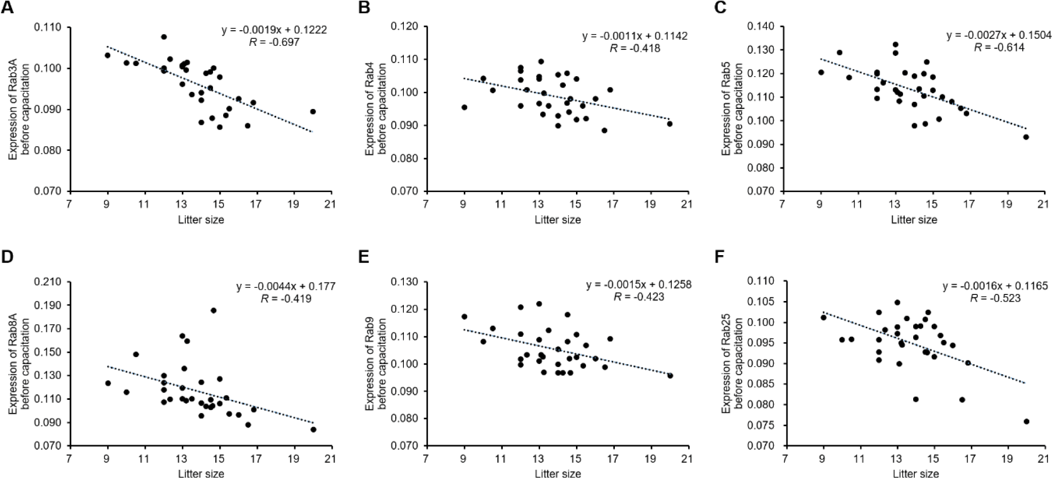
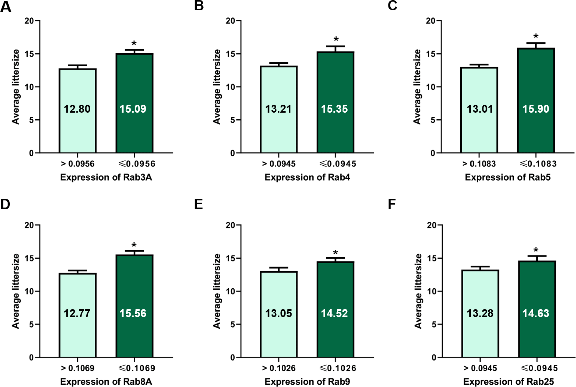
The expression of Rab3A (r = −0.697, p < 0.01), Rab4 (r = −0.418, p < 0.05), Rab5 (r = −0.614, p < 0.01), Rab8A (r = −0.419, p < 0.05), Rab9 (r = −0.423, p < 0.05), and Rab25 (r = −0.523, p < 0.01) was negatively correlated with litter size (Table 1 and Fig. 1). According to the ROCs, the cut-off values of Rab proteins for the 14 litter sizes were as follows: Rab3A = 0.0956, Rab4 = 0.0945, Rab5 = 0.1083; Rab8A = 0.1069, Rab9 = 0.1026; and Rab25, 0.0945 (Table 2 and Figs. 2 and 3). The average litter size with Rab3A > 0.0956 was 12.80 and ≤ 0.0956 was 15.09 (p < 0.05, Fig. 3A). The average litter size with Rab4 > 0.0945 was 13.21 and ≤ 0.0945 was 15.35 (p < 0.05, Fig. 3B). The average litter size with Rab5 > 0.1083 was 13.01 and ≤ 0.1083 was 15.90 (p < 0.05, Fig. 3C). The average litter size with Rab8A > 0.1069 was 12.77 and ≤ 0.1069 was 15.56 (p < 0.05, Fig. 3D). The average litter size with Rab9 > 0.1026 was 13.05 was ≤ 0.1026 was 14.52 (p < 0.05, Fig. 3E). The average litter size with Rab25 > 0.0945 was 13.28 was ≤ 0.0945 was 14.63 (p < 0.05, Fig. 3F). In addition, the overall accuracies of Rab3A, 4, 5, 8A, 9, and 25 before capacitation were 77.42, 67.74, 74.19, 83.87, 67.74, and 58.06 %, respectively (Table 2). The acrosome is derived from the Golgi apparatus and a lysosome-like structure is established during spermatogenesis [4, 16]. Because Rab proteins are involved in the establishment of the acrosome by regulating Golgi trafficking, they are key proteins in acrosome formation [4-6, 16]. Almost all Rab proteins are expressed in the sperm head and tail [6, 7, 22]. Further, Rab4 is correlated with the integrity of the sperm head structure, and Rab5 is correlated with various motion parameters in spermatozoa [5]. Our results showed that several Rab proteins (Rab3A, 4, 5, 8A, 9, and 25) are correlated with litter size before capacitation. Rab proteins play key roles in male fertility as they are involved in establishing sperm cell structure and function, including the structural integrity and motility of sperms. Moreover, five Rab proteins (Rab3A, 4, 5, 8A, 9, and 25) were validated based on their expression to predict fertility in the present study. It was confirmed that the litter size increased with the specific expression of Rab protein. Therefore, Rab3A, 4, 5, 8A, 9, and 25 may be used as new biomarkers for the prediction and diagnosis of male fertility. Particularly, Rab5 resulted in the highest increase in litter size and the third-highest overall accuracy. In addition, Rab8A resulted in the highest overall accuracy and the second-highest increase in litter size. Therefore, Rab5 and Rab8A are anticipated to be useful for developing efficient fertility-related biomarkers.

Table 1. Correlation between littersize and expression of Rab proteins before capacitation
Litter size Rab3A Rab4 Rab5 Rab8A Rab9 Rab11 Rab14 Rab25 Rab27A Rab34A
Litter size 1 −0.697** −0.418* −0.614** −0.419* −0.423* −0.253 −0.202 −0.523** −0.173 0.045
Rab3A 0.660** 0.556** 0.450* 0.335 0.216 −0.030 0.510** 0.438* 0.217
Rab4 0.369* 0.221 0.316 0.000 −0.245 0.364* 0.387* 0.460**
Rab5 0.567** 0.340 0.316 0.101 0.668** 0.302 0.190
Rab8A −0.095 0.066 0.178 0.405* −0.083 −0.089
Rab9 0.465** −0.208 0.481** 0.493** 0.042
Rab11 0.255 0.465** 0.157 −0.054
Rab14 0.141 −0.262 −0.027
Rab25 0.455* 0.057
Rab27A 0.448*

p < 0.05,

p < 0.01.

Rab, Ras-related proteins.

Download Excel Table
Table 2. Correlation between expression of Rab proteins and litter size before capacitation
Litter size ≥ 14 Litter size < 14
Rab3A
 Expression of Rab3A ≤ 0.0956 11 2
 Expression of Rab3A > 0.0956 5 13
 Sensitivity 68.75
 Specificity 86.67
 Negative predictive value 72.22
 Positive predictive value 84.62
 Overall accuracy 77.42
Rab4
 Expression of Rab4 ≤ 0.0945 7 1
 Expression of Rab4 > 0.0945 9 14
 Sensitivity 43.75
 Specificity 93.33
 Negative predictive value 60.87
 Positive predictive value 87.50
 Overall accuracy 67.74
Rab5
 Expression of Rab5 ≤ 0.1083 8 0
 Expression of Rab5 > 0.1083 8 15
 Sensitivity 50.00
 Specificity 100.00
 Negative predictive value 65.22
 Positive predictive value 100.00
 Overall accuracy 74.19
Rab8A
 Expression of Rab8A ≤ 0.1069 11 0
 Expression of Rab8A > 0.1069 5 15
 Sensitivity 68.75
 Specificity 100.00
 Negative predictive value 75.00
 Positive predictive value 100.00
 Overall accuracy 83.87
Rab9
 Expression of Rab9 ≤ 0.1026 10 5
 Expression of Rab9 > 0.1026 5 11
 Sensitivity 66.67
 Specificity 68.75
 Negative predictive value 68.75
 Positive predictive value 66.67
 Overall accuracy 67.74
Rab25
 Expression of Rab25 ≤ 0.0945 10 5
 Expression of Rab25 > 0.0945 6 10
 Sensitivity 62.50
 Specificity 66.67
 Negative predictive value 62.50
 Positive predictive value 66.67
 Overall accuracy 64.52

Sensitivity = [A / (A + C)] × 100; Specificity = [D / (B + D)] × 100; Positive predictive value = [A / (A + B)] × 100; Negative predictive value = [C / (C + D)] × 100; and overall accuracy = [(A + D) / (A + B + C + D)] × 100.

Download Excel Table
jast-64-6-1024-g1
Fig. 1. Correlation between expression of Rab proteins and litter size before capacitation. (A) Correlation between Rab3A in spermatozoa and litter size before capacitation. (B) Correlation between Rab4 in spermatozoa and litter size before capacitation. (C) Correlation between Rab5 in spermatozoa and litter size before capacitation. (D) Correlation between Rab8A in spermatozoa and litter size before capacitation. (E) Correlation between Rab9 in spermatozoa and litter size before capacitation. (F) Correlation between Rab25 in spermatozoa and litter size before capacitation. n = 3.
Download Original Figure
jast-64-6-1024-g2
Fig. 2. Receiver operating characteristic (ROC) curves for expression of Rab proteins. (A) ROC curve for expression of Rab3A, 4, 5, 8A, 9, and 25 before capacitation. (B) ROC curve for expression of Rab3A, 4, 5, 8A, 9, and 14 after capacitation.
Download Original Figure
jast-64-6-1024-g3
Fig. 3. Average litter size based on expression of Rab proteins before capacitation. (A) Average litter size according to the expression of Rab3A (cut-off value = 0.0956). (B) Average litter size according to the expression of Rab4 (cut-off value = 0.0945). (C) Average litter size according to the expression of Rab5 (cut-off value = 0.1083). (D) Average litter size according to the expression of Rab8A (cut-off value = 0.1069). (E) Average litter size according to the expression of Rab9 (cut-off value = 0.1026). (F) Average litter size according to the expression of Rab25 (cut-off value = 0.0956). Data represent the mean ± SEM, *p < 0.05.
Download Original Figure
Correlation between expression of Rab proteins and litter size after capacitation

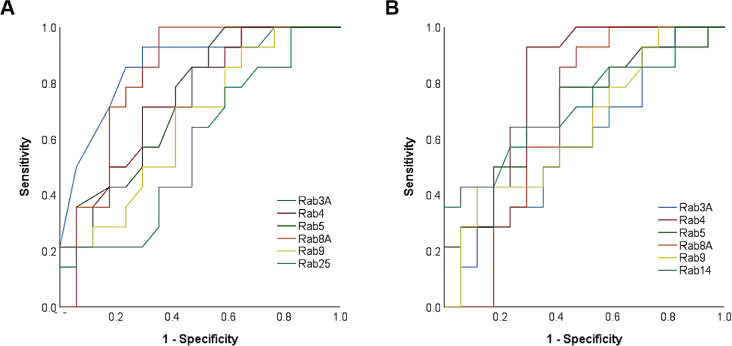
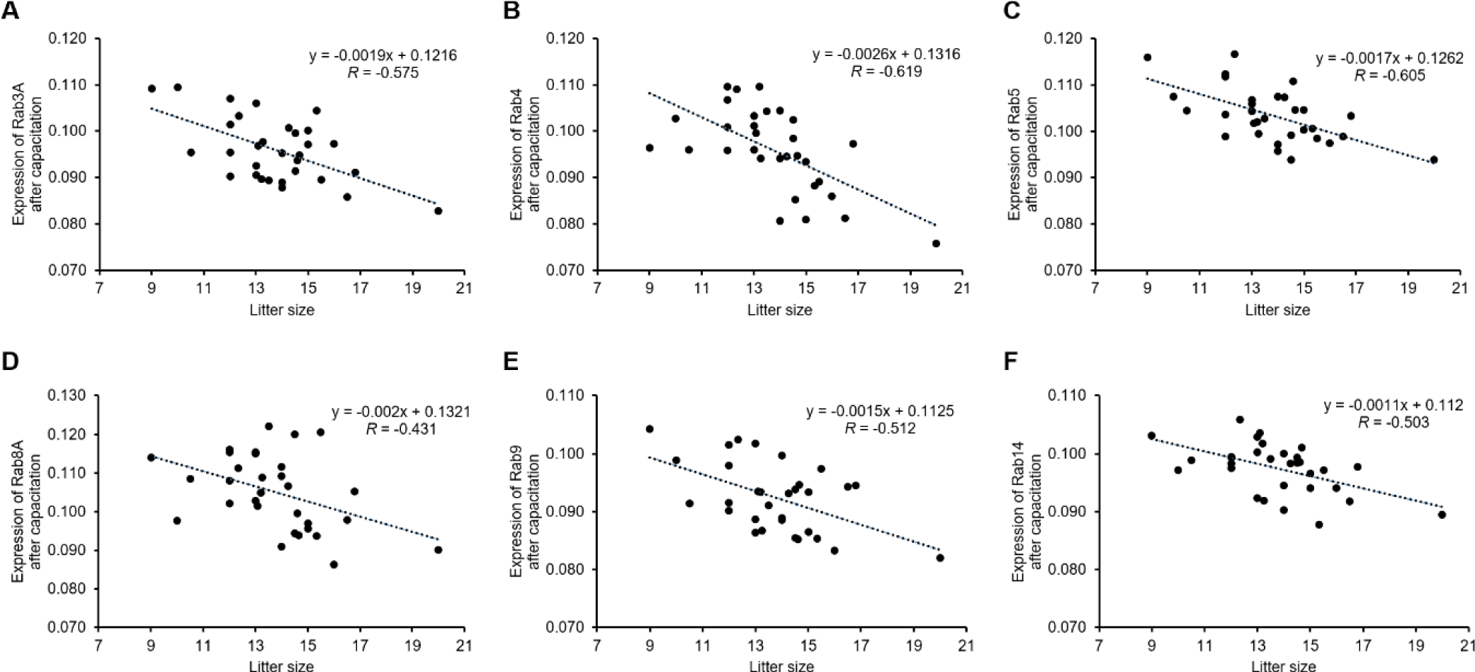
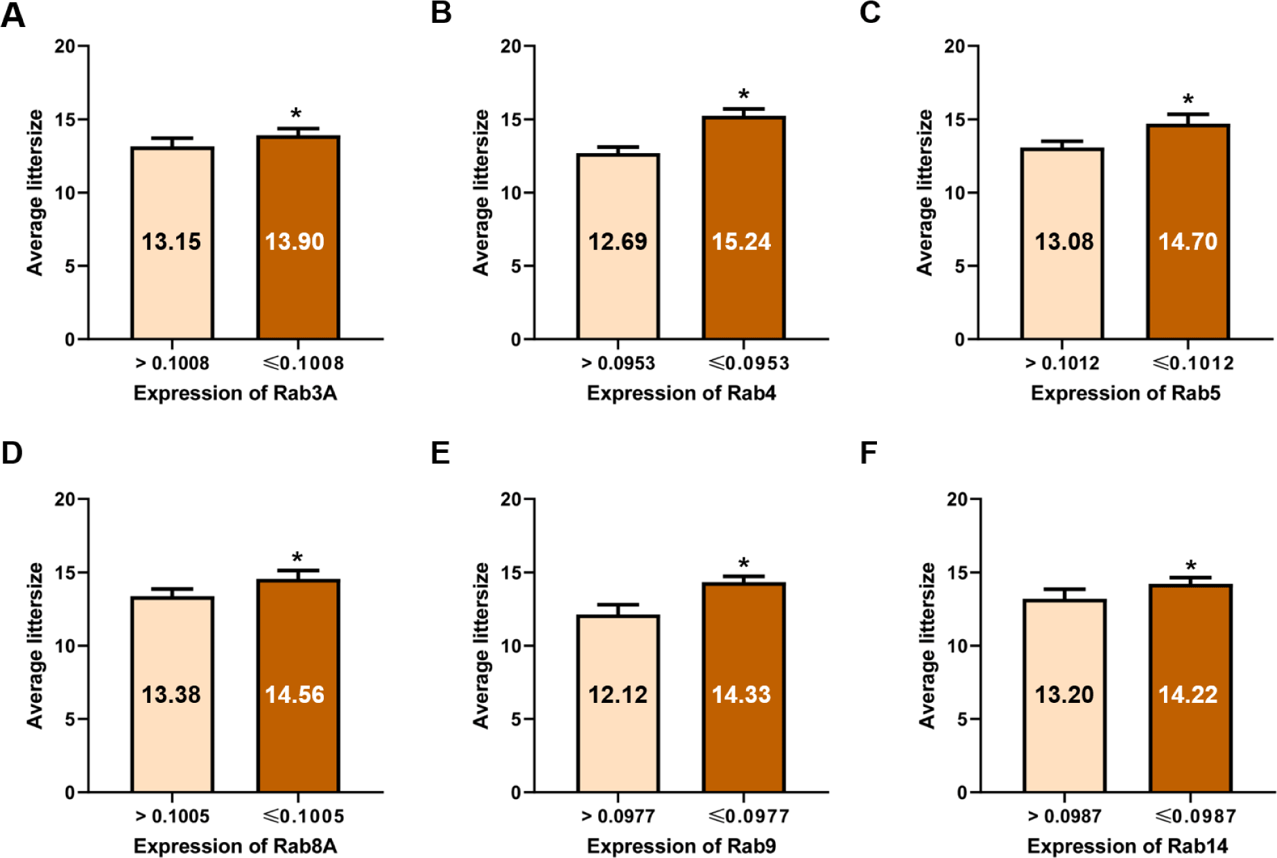
After inducing capacitation, most Rab proteins, except Rab25, were negatively correlated with litter size, similar to what was observed before capacitation. The expression of Rab3A (r = −0.575, p < 0.01), Rab4 (r = −0.619, p < 0.01), Rab5 (r = −0.605, p < 0.01), Rab8A (r = −0.431, p < 0.05), and Rab9 (r = −0.512, p < 0.01) was negatively correlated with litter size after capacitation (Table 3 and Fig. 4). Rab14 expression was negatively correlated after capacitation (r = −0.503, p < 0.01, Table 3 and Fig. 4). According to the ROCs, the cut-off values of Rab proteins for the 14 litter sizes were as follows: Rab3A = 0.1008, Rab4 = 0.0953, Rab5 = 0.1012, Rab8A = 0.1005, Rab9 = 0.0977, and Rab14, 0.0987 (Table 4 and Figs. 2 and 5). The average litter size with Rab3A > 0.1008 was 13.15 and ≤ 0.1008 was 13.90 (p < 0.05, Fig. 5A). The average litter size with Rab4 > 0.0953 was 12.69 and ≤ 0.0953 was 15.24 (p < 0.05, Fig. 5B). The average litter size with Rab5 > 0.1012 was 13.08 and ≤ 0.1012 was 14.70 (p < 0.05, Fig. 5C). The average litter size with Rab8A > 0.1005 was 13.38 and ≤ 0.1005 was 14.56 (p < 0.05, Fig. 5D). The average litter size with Rab9 > 0.0977 was 12.12 and ≤ 0.0977 was 14.33 (p < 0.05, Fig. 5E). The average litter size with Rab14 > 0.0987 was 13.20 and ≤ 0.0987 was 14.22 (p < 0.05, Fig. 5F). In addition, overall accuracies of Rab3A, 4, 5, 8A, 9, and 14 after capacitation were 59.38, 83.87, 70.97, 75.76, 64.52, and 70.97 %, respectively (Table 4). Similar to ejaculated spermatozoa, most Rab proteins also exist after capacitation [6]. In particular, Rab3A is correlated with capacitation status after capacitation [5]. Rab3A is well known for regulating acrosome exocytosis at end of capacitation [7, 22, 23]. In addition, Rab5 and 14 correlate with sperm motion parameters after capacitation [5]. Therefore, it may be considered that the Rab proteins are also playing an important role in male fertility by association with sperm motility and capacitation status. In the present study, the expression of Rab3A, 4, 5, 8A, 9, and 14 correlated with litter size after capacitation. Therefore, these five Rab proteins may be used to predict and diagnose male fertility. The most efficient Rab protein was Rab4, which resulted in the highest increase in litter size and overall accuracy after capacitation. Therefore, our results suggest that Rab4 may be a strong fertility-related biomarker after capacitation to evaluate male fertility.

Table 3. Correlation between littersize and expression of Rab proteins after capacitation
Litter size Rab3A Rab4 Rab5 Rab8A Rab9 Rab11 Rab14 Rab25 Rab27A Rab34A
Litter size 1 −0.575** −0.619** −0.605** −0.431* −0.512** −0.089 −0.503** 0.019 −0.014 −0.011
Rab3A 0.203 0.378* −0.105 0.280 −0.084 0.240 0.008 −0.019 0.019
Rab4 0.380* 0.501** 0.374* 0.197 0.602** −0.113 0.297 0.202
Rab5 0.268 0.482** −0.085 0.571** −0.037 −0.040 −0.083
Rab8A 0.389* 0.281 0.359* −0.207 0.375* 0.078
Rab9 0.308 0.295 0.007 0.272 −0.344
Rab11 −0.258 0.278 0.489** −0.086
Rab14 −0.088 0.020 −0.011
Rab25 0.191 −0.187
Rab27A 0.083

p < 0.05,

p < 0.01.

Rab, Ras-related proteins.

Download Excel Table
Table 4. Correlation between expression of Rab proteins and litter size after capacitation
Litter size ≥ 14 Litter size < 14
Rab3A
 Expression of Rab3A ≤ 0.1008 15 11
 Expression of Rab3A > 0.1008 2 4
 Sensitivity 88.24
 Specificity 26.67
 Negative predictive value 66.67
 Positive predictive value 57.69
 Overall accuracy 59.38
Rab4
 Expression of Rab4 ≤ 0.0953 12 1
 Expression of Rab4 > 0.0953 4 14
 Sensitivity 75.00
 Specificity 93.33
 Negative predictive value 77.78
 Positive predictive value 92.31
 Overall accuracy 83.87
Rab5
 Expression of Rab5 ≤ 0.1012 10 3
 Expression of Rab5 > 0.1012 6 12
 Sensitivity 62.50
 Specificity 80.00
 Negative predictive value 66.67
 Positive predictive value 76.92
 Overall accuracy 70.97
Rab8A
 Expression of Rab8A ≤ 0.1005 11 1
 Expression of Rab8A > 0.1005 7 14
 Sensitivity 61.11
 Specificity 93.33
 Negative predictive value 66.67
 Positive predictive value 91.67
 Overall accuracy 75.76
Rab9
 Expression of Rab9 ≤ 0.0977 15 8
 Expression of Rab9 > 0.0977 3 5
 Sensitivity 83.33
 Specificity 38.46
 Negative predictive value 62.50
 Positive predictive value 65.22
 Overall accuracy 64.52
Rab14
 Expression of Rab14 ≤ 0.0987 12 5
 Expression of Rab14 > 0.0987 4 10
 Sensitivity 75.00
 Specificity 66.67
 Negative predictive value 71.43
 Positive predictive value 70.59
 Overall accuracy 70.97

Sensitivity = [A / (A + C)] × 100; Specificity = [D / (B + D)] × 100; Positive predictive value = [A / (A + B)] × 100; Negative predictive value = [C / (C + D)] × 100; and overall accuracy = [(A + D) / (A + B + C + D)] × 100.

Download Excel Table
jast-64-6-1024-g4
Fig. 4. Correlation between expression of Rab proteins and litter size after capacitation. (A) Correlation between Rab3A in spermatozoa and litter size after capacitation. (B) Correlation between Rab4 in spermatozoa and litter size after capacitation. (C) Correlation between Rab5 in spermatozoa and litter size after capacitation. (D) Correlation between Rab8A in spermatozoa and litter size after capacitation. (E) Correlation between Rab9 in spermatozoa and litter size after capacitation. (F) Correlation between Rab14 in spermatozoa and litter size after capacitation. n = 3.
Download Original Figure
jast-64-6-1024-g5
Fig. 5. Average litter size based on expression of Rab proteins after capacitation. (A) Average litter size according to the expression of Rab3A (cut-off value = 0.1008). (B) Average litter size according to the expression of Rab4 (cut-off value = 0.0953). (C) Average litter size according to the expression of Rab5 (cut-off value = 0.1012). (D) Average litter size according to the expression of Rab8A (cut-off value = 0.1005). (E) Average litter size according to the expression of Rab9 (cut-off value = 0.0977). (F) Average litter size according to the expression of Rab25 (cut-off value = 0.0987). Data represent the mean ± SEM, *p < 0.05.
Download Original Figure

CONCLUSION

Taken together, our results showed that the expression of six Rab proteins before (Rab3A, 4, 5, 8A, 9, and 25) and after capacitation (Rab3A, 4, 5, 8A, 9, and 14) was correlated with litter size. In addition, an increase in litter size was confirmed while evaluating the ability of the individual Rab proteins to predict litter size based on the cut-off value calculated by ROCs. The Rab8A protein had the highest overall accuracy before capacitation and Rab4 had the highest overall accuracy after capacitation (both 83.87%). Our results suggest that Rab proteins are correlated with litter size and may be applied as fertility-related biomarkers. We anticipate that it may be possible to improve productivity in pigs, as well as other domestic animals, using Rab proteins as biomarkers to analyze male fertility and select superior sires. Moreover, it suggests that Rab proteins may be applied to humans for the prediction and diagnosis of male fertility, as well as for identifying the cause of idiopathic male infertility/subfertility in patients.

Competing interests

No potential conflict of interest relevant to this article was reported.

Funding sources

This study was conducted with the support of the Gyeongsangbuk-do agricultural and fishery R&D activation project.

Acknowledgements

Not applicable.

Availability of data and material

Upon reasonable request, the datasets of this study can be available from the corresponding author.

Authors’ contributions

Conceptualization: Bae JW, Kwon WS.

Data curation: Bae JW, Hwang JM, Kwon WS.

Formal analysis: Bae JW, Hwang JM, Kwon WS.

Methodology: Bae JW, Hwang JM, Kwon WS.

Software: Bae JW, Hwang JM, Kwon WS.

Validation: Bae JW, Kwon WS.

Investigation: Bae JW, Hwang JM, Kwon WS.

Writing - original draft: Bae JW, Hwang JM, Kwon WS.

Writing - review & editing: Bae JW, Hwang JM, Kwon WS.

Ethics approval and consent to participate

All processes were performed in accordance with the guidelines and approved by Institutional Animal Care and Use Committee of Kyungpook National University (KNU 2021-207).

REFERENCES

1.

Kwon WS, Rahman MS, Ryu DY, Park YJ, Pang MG. Increased male fertility using fertility-related biomarkers. Sci Rep. 2015; 5:15654

2.

Kwon WS, Rahman MS, Lee JS, Yoon SJ, Park YJ, Pang MG. Discovery of predictive biomarkers for litter size in boar spermatozoa. Mol Cell Proteomics. 2015; 14:1230-40

3.

Pang WK, Son JH, Ryu DY, Rahman MS, Park YJ, Pang MG. Heat shock protein family D member 1 in boar spermatozoa is strongly related to the litter size of inseminated sows. J Anim Sci Biotechnol. 2022; 13:42

4.

Liu S, Storrie B. Are Rab proteins the link between Golgi organization and membrane trafficking?. Cell Mol Life Sci. 2012; 69:4093-106

5.

Bae JW, Yi JK, Jeong EJ, Lee WJ, Hwang JM, Kim DH, et al. Ras-related proteins (Rab) play significant roles in sperm motility and capacitation status. Reprod Biol. 2022; 22:100617

6.

Bae JW, Kim SH, Kim DH, Ha JJ, Yi JK, Hwang S, et al. Ras-related proteins (Rab) are key proteins related to male fertility following a unique activation mechanism. Reprod Biol. 2019; 19:356-62

7.

Bustos MA, Lucchesi O, Ruete MC, Mayorga LS, Tomes CN. Rab27 and Rab3 sequentially regulate human sperm dense-core granule exocytosis. Proc Natl Acad Sci USA. 2012; 109:E2057-66

8.

Mruk DD, Lau ASN. RAB13 participates in ectoplasmic specialization dynamics in the rat testis. Biol Reprod. 2009; 80:590-601

9.

Almond G. The swine A.I. book: a field and laboratory technicians’ guide to artificial insemination in swine. Raleigh, NC: Morgan Morrow Press. 1998

10.

Tummaruk P, Lundeheim N, Einarsson S, Dalin AM. Effect of birth litter size, birth parity number, growth rate, backfat thickness and age at first mating of gilts on their reproductive performance as sows. Anim Reprod Sci. 2001; 66:225-37

11.

Kwon WS, Shin DH, Ryu DY, Khatun A, Rahman MS, Pang MG. Applications of capacitation status for litter size enhancement in various pig breeds. Asian-Australas J Anim Sci. 2018; 31:842-50

12.

Kwon WS, Rahman MS, Lee JS, You YA, Pang MG. Improving litter size by boar spermatozoa: application of combined H33258/CTC staining in field trial with artificial insemination. Andrology. 2015; 3:552-7

13.

Kwon WS, Rahman MS, Ryu DY, Khatun A, Pang MG. Comparison of markers predicting litter size in different pig breeds. Andrology. 2017; 5:568-77

14.

Kwon WS, Rahman MS, Lee JS, Kim J, Yoon SJ, Park YJ, et al. A comprehensive proteomic approach to identifying capacitation related proteins in boar spermatozoa. BMC Genomics. 2014; 15:897

15.

Bradford MM. A rapid and sensitive method for the quantitation of microgram quantities of protein utilizing the principle of protein-dye binding. Anal Biochem. 1976; 72:248-54

16.

Goud B, Liu S, Storrie B. Rab proteins as major determinants of the Golgi complex structure. Small GTPases. 2018; 9:66-75

17.

Nakamura Y, Asano A, Hosaka Y, Takeuchi T, Iwanaga T, Yamano Y. Expression and intracellular localization of TBC1D9, a Rab GTPase-accelerating protein, in mouse testes. Exp Anim. 2015; 64:415-24

18.

Ramalho-Santos J, Moreno RD, Wessel GM, Chan EKL, Schatten G. Membrane trafficking machinery components associated with the mammalian acrosome during spermiogenesis. Exp Cell Res. 2001; 267:45-60

19.

Smith CE, Hermo L, Fazel A, Lalli MF, Bergeron JJM. Ultrastructural distribution of NADPase within the Golgi apparatus and lysosomes of mammalian cells. Prog Histochem Cytochem. 1990; 21:V-VII 1-120

20.

Lopez CI, Belmonte SA, De Blas GA, Mayorga LS. Membrane-permeant Rab3A triggers acrosomal exocytosis in living human sperm. FASEB J. 2007; 21:4121-30

21.

Ruete MC, Lucchesi O, Bustos MA, Tomes CN. Epac, Rap and Rab3 act in concert to mobilize calcium from sperm’s acrosome during exocytosis. Cell Commun Signal. 2014; 12:43

22.

Yunes R, Tomes C, Michaut M, De Blas G, Rodriguez F, Regazzi R, et al. Rab3A and calmodulin regulate acrosomal exocytosis by mechanisms that do not require a direct interaction. FEBS Lett. 2002; 525:126-30

23.

Yunes R, Michaut M, Tomes C, Mayorga LS. Rab3A triggers the acrosome reaction in permeabilized human spermatozoa. Biol Reprod. 2000; 62:1084-9