Journal of Animal Science and Technology
Korean Society of Animal Sciences and Technology
RESEARCH ARTICLE

Inhibitory effects of Porphyra dentata extract on 3T3-L1 adipocyte differentiation

Su-Young Choi1,#https://orcid.org/0000-0002-6568-9246, Su Yeon Lee2,#https://orcid.org/0000-0003-4799-1393, Da hye Jang2https://orcid.org/0000-0002-6139-8310, Suk Jun Lee3https://orcid.org/0000-0003-0216-209X, Jeong-Yong Cho2,*https://orcid.org/0000-0002-2048-5661, Sung-Hak Kim1,*https://orcid.org/0000-0003-4882-8600
1Department of Animal Science, Chonnam National University, Gwangju 61186, Korea
2Department of Food Science and Technology, Chonnam National University, Gwangju 61186, Korea
3Department of Biomedical Laboratory Science, College of Health & Medical Sciences, Cheongju University, Chungbuk 28503, Korea
*Corresponding author: Jeong-Yong Cho, Department of Food Science and Technology, Chonnam National University, Gwangju 61186, Korea., Tel: +82-62-530-2143, E-mail: jyongcho17@jnu.ac.kr
*Corresponding author: Sung-Hak Kim, Department of Animal Science, Chonnam National University, Gwangju 61186, Korea., Tel: +82-62-530-2115, E-mail: sunghakkim@jnu.ac.kr

#These authors contributed equally to this work.

© Copyright 2020 Korean Society of Animal Science and Technology. This is an Open-Access article distributed under the terms of the Creative Commons Attribution Non-Commercial License (http://creativecommons.org/licenses/by-nc/4.0/) which permits unrestricted non-commercial use, distribution, and reproduction in any medium, provided the original work is properly cited.

Received: Sep 08, 2020; Revised: Sep 26, 2020; Accepted: Oct 02, 2020

Published Online: Nov 30, 2020

Abstract

This study was aimed to investigate the inhibitory effects of Porphyra dentata (P. dentata) extract on the adipogenesis of 3T3-L1 cells and evaluate its anti-obesity effect. The proliferation of 3T3-L1 cells and differentiation of adipocytes under treatment of P. dentata extract was examined by measuring the cell viability using alamarBlue assay and lipid droplets by Oil Red O staining. Results showed that P. dentata extract has no cytotoxicity effect and lipid droplets formation decreased in a concentration-dependent manner in 3T3-L1 cells. It has been confirmed that transcription factors affecting lipid accumulation and anti-adipogenic effects during cell differentiation are linked to P. dentata extract. We observed that P. dentata shows lowering the mRNA expression of peroxisome proliferator-activated receptor γ2 (PPARγ2), CCAAT/enhancer binding protein α (C/EBPα) that adipogenesis-associated key transcription factors and inhibiting adipogenesis in the early stages of differentiation. Treating the cells with P. dentata did not only suppressed PPARγ2 and C/EBPα but also significantly decreased the mRNA expression of adiponectin, Leptin, fatty acid synthase, adipocyte protein 2, and Acetyl-coA carboxylase 1. Overall, the P. dentata extract demonstrated inhibitory property in adipogenesis, which has a potential effect in anti-obesity in 3T3-L1 cells.

Keywords: 3T3-L1; Adipocyte differentiation; Adipogenesis; Porphyra dentata; Anti-obesity

INTRODUCTION

Obesity is one of the biggest health problems in the world today and the number of obese people are increasing in all over the world [1,2]. Obesity is defined as an increase in body weight [3] caused excessive accumulation of fat cells due to adipocyte differentiation [4]. Hence, it is also closely linked to metabolic diseases such as type 2 diabetes (T2D), liver disease, cardiovascular disease (CVD), cancers, hypertension and other disorders which increased these disease occurrence [5,6]. Accumulation of fat cells that cause obesity is by the differentiation of adipocytes called adipogenesis. Peroxisome proliferator-activated receptor γ (PPARγ) and CCAAT/enhancer-binding protein (C/EBP) transcription factor family are key players to regulate the differentiation of preadipocytes by inducing adipogenic related genes including adiponectin (ADIPOQ), leptin, fatty acid synthase (FAS), adpocyte protein 2 (aP2), and acetyl-coA carboxylase 1 (ACC) [710].

Laver, an edible seaweed species belonging to the genus Porphya, is commonly grown and consumed in Korea, China, and Japan. Laver is a rich source of vitamins, minerals, polysaccharides, phenolic compounds and mycosporine-like amino acids (MAAs) [11]. The polysaccharides components include laminarin and fucoidan while phenolic compounds present includes epigallocatechin gallate (EGCG) and catechin. Moreover, MAAs present in laver are mycosporine, shinorine, and porphyra-334. Several studies have shown that laver has antioxidant [12], anti-ultraviolet [13], anti-inflammatory [14], and antitumor [15] effects because of its bioactive compounds present. Marine algae, especially seaweeds are a promising source of anti-obesity agent [16] and anti-obesity effects are reported in various kinds of seaweed (brown, red and green) [17]. Also polysaccharides and phenol compounds are also reported to have anti-obesity effects [18,19].

Porphyra dentata (P. dentata) used in this study, is a kind of red algae and belongs to the Bangiaceae and Pyropia genus and an edible red seaweed in eastern Asian countries [20]. P. dentata contains polysaccharides and phenolic compound such as fucoidan, EGCG, and catechin [21]. It was reported that it has anti-inflammatory activity by suppressing nitric oxide production in LPS-stimulated macrophage [21]. As reported, although the components of P. dentata have a potential to regulate adipogenesis, anti-obesity effect on this extract have not been addressed.

The purpose of this study is to evaluate whether the P. dentata extract has inhibitory effects on adipogenesis from preadipocyte to mature adipocyte in 3T3-L1 cells.

MATERIALS AND METHODS

Chemicals and cell

Dulbecco’s Modified Eagle’s Medium (DMEM; high glucose) was purchased from HycloneTM (Logan, UT, USA). Fetal bovine serum (FBS) were purchased from Gibco-BRL (Gaithersburg, MD, USA). 3-Isobutyl-1-methylxanthine (IBMX), dexamethasone (DEX), pioglitazone, insulin, Oil Red O powder, and dimethyl sulfoxide (DMSO) were purchased from Sigma-Aldrich (St. Louis, MO, USA). Formaldehyde Solution for 4% formalin was purchased from Fujifilm Wako Pure Chemical (Osaka, Japan). 2-propanol-GR and ethanol were purchased Merck (Kenilworth, NJ, USA). The 3T3-L1 cells were purchased from American Type Culture Collection (Rockville, CT, USA).

Preparation of the Porphyra dentata extract

Dried P. dentata was obtained from Mokpo Marine Food-industry Research Center (Mokpo, Korea). The dried P. dentata (50 g) was extracted with 1.5 L of 50% aqueous methanol (MeOH) at room temperature for one day and then filtered. The residues were re-extracted with 0.75 L 50% methanol and then filtered. The combined filtrates were evaporated at 38°C under a vacuum. The 50% MeOH extracts of P. dentata were deposited at −20°C until use in experiment.

Cell culture and differentiation

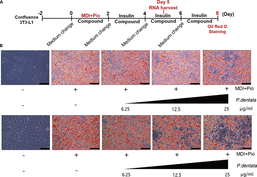
3T3-L1 preadipocytes (ATCC®, CL-173TM) were cultured in DMEM (high glucose) supplemented with 10% FBS, 1% penicillin and streptomycin (Welgene, Gyeongsan, Korea) at 37°C in 5% CO2. For experiment, cells were seeded in 6-well plates at a density of 1.0 × 10⁵ cells/well and grown to confluence. Forty-eight hours after confluence (day 0), adipogenesis was induced by adding differentiation medium (DMEM; high glucose containing 10% FBS, 0.5 mM 3-isobutyl-1-methylxanthine; IBMX, 1 μM Dexamethasone; DEX, 1 μM Pioglitazone, 10 μg/mL insulin, 1 μL/mL dimethyl sulfoxide; DMSO) for 48 h. Every two days, the medium was changed with DMEM; high glucose supplemented 10% FBS, 10 μg/mL insulin, 1 μL/mL DMSO until 8 days. The pre-adipocytes were maintained and changed medium for every 48 h with DMEM; high glucose, 10% FBS, and 1 μL/mL DMSO medium. To investigate the effects of P. dentata on adipocyte differentiation, cell culture was treated P. dentata 50% MeOH extract in different concentrations (6.25, 12.5, and 25 μg/mL) in the differentiation medium for every two days, from the beginning to the end of the experiment. After 5 days of treatment with P. dentata 50% MeOH extract. 3T3-L1 adipocyte cells were harvested for Real-time quantitative polymerase chain reaction (RT-qPCR) and after 8 days the 3T3-L1 adipocyte cells were fixed in 4% formalin for Oil Red O staining.

Cell viability assay

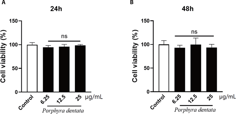
3T3-L1 cells were seeded in 96-well plates at a density of 7.5 × 10² cells/well containing 200 μL of 10% FBS-DMEM; high glucose. After cell seeding, P. dentata 50% MeOH extract was added by concentration dependent (6.25, 12.5, and 25 μg/mL). After 24 and 48hr after addition of the extract, alamarBlueTM Cell Viability Reagent (ThermoFisher Scientific, Waltham, MA, USA) was added and then fluorescence value was measured by SYNERGY multi-mode reader (BioTek, Seoul, Korea). Viability of cells was measured using alamarBlue assay according manufacturer instructions.

Oil Red O staining of lipid droplets

To measure the cell lipid droplets, the 3T3-L1 cells were stained with Oil Red O solution. 3T3-L1 cells were washed twice with PBS and adherent cells were fixed in 4% formalin for 10 min at room temperature. The 4% formalin was discarded and fresh 4% formalin was added and incubated for 1h at room temperature. After 1hr, is was washed with tertiary distilled water. The cells were added with 60% isopropanol and let it stand for 5 min at room temperature. After 5 min, 60% isopropanol was discarded and the cells were allowed dry completely at room temperature. After drying, 1 ml Oil Red O solution was added to each well and incubated at room temperature for 20 minutes. The cells were washed three times with tertiary distilled water and photographed using a Leica Microscopy, DE/Polyvar SC (Leica, Wetzlar, Germany).

Quantification of adipogenic gene expression using real-time quantitative polymerase chain reaction

Total RNA was isolated from cells using Hybrid RTM (GeneAll Biothechnology, Seoul, Korea) including RiboEXTM treatment of samples to eliminate genomic DNA, protein, and lipid. Quality of RNA was determined by using Nanodrop 2000 spectrophotometer (ThermoFisher Scientific) and RNA gel electrophoresis. cDNA was synthesized from the total RNA using a RevertAid First Strand cDNA Synthesis kit (ThermoFisher Scientific). The real-time PCR was conducted using a CFX96TM Real-Time PCR Detection System (Bio rad, Hercules, CA, USA). The level of target gene cDNA was measured by TB Green® Premix EX TaqTM (Tli Rnase H plus) (Takara Bio, Kosatsu, Japan). All samples were analyzed in triplicate and quantified by the relative standard curve method using the gene expressions of L32 as a housekeeping gene. The sequences of the primer pairs used in this study are listed in Table 1.

Table 1. Primer sequence used in the RT-qPCR experiment
Gene Forward (5’-3’) Reverse (3’-5’)
L32 TCTGGTGAAGCCCAAGATCG CTCTGGGTTTCCGCCAGT
PPARy2 GTGCTCCAGAAGATGACAGAC GGTGGGACTTTCCTGCTAA
C/EBPa TGGACAAGAACAGCAACGAG TCACTGGTCAACTCCAGCAC
ADIPOQ CCGTTCTCTTCACCTACGAC TCCCCATCCCCATACAC
Leptin TCAACTCCCTGTTTCCAAAT TCTTCACGAATGTCCCACGA
FAS CCCAGCCCATAAGAGTTACA ATCGGGAAGTCAGCACAA
aP2 TGGAAGCTTGTCTCCAGTGA AATCCCCATTTACGCTGATG
ACC1 GACGTTCGCCATAACCAAGT CTGTTTAGCGTGGGGATGTT
Download Excel Table
Statistical analysis

Statistical analysis was performed using GraphPad Prism 0.8 (GraphPad Software, San Diego, CA, USA). All the data were analyzed using one-way analysis of variance (ANOVA) with multiple comparisons. Differences between groups were analyzed using t-test and values of p < 0.05 were considered statistically significant. All experiments were performed triplicate and data were expressed as mean ± SEM.

RESULTS

Porphyra dentata 50% MeOH extract shows no cytotoxicity in 3T3-L1 pre-adipocytes

We first performed the alamarBlue assay to test the effect of P. dentata 50% MeOH extract on cell viability. As shown in Fig. 1, P. dentata 50% MeOH extract at 6.25, 12.5, and 25 μg/mL showed no significant effect on cell viability in 3T3L1 mouse preadipocytes after 24 h and 48 h treatment. These results indicate that the P. dentata 50% MeOH extract have no cytotoxicity on cells.

jast-62-6-854-g1
Fig. 1. Cell cytotoxicity of Porphyra dentata 50% MeOH extract on 3T3-L1 cells. 3T3-L1 cells were treated with different concentrations (6.25, 12.5, and 25 μg/mL) of P. dentata 50% MeOH extract for detection with alamarBlue assay.
Download Original Figure
Porphyra dentata 50% MeOH extract inhibits differentiation and lipid accumulation of 3T3-L1 cells

To investigate the effect of P. dentata extract on 3T3-L1 preadipocytes adipogenesis, we treated P. dentata extract with various concentrations for 8 days and stained the lipid droplets with Oil Red O during adipocyte differentiation (Fig. 2). Oil Red O staining assay revealed that P. dentata dramatically reduced lipid accumulation in a concentration dependent manner. These results indicate that P. dentata 50% MeOH extract suppressed adipocyte differentiation and lipid droplets formation in 3T3-L1 preadipocytes.

jast-62-6-854-g2
Fig. 2. Decreased accumulation of lipid droplets in differentiated 3T3-L1 cells treatment with Porphyra dentata 50% MeOH extract. (A) Time schedule of the culture with P. dentata 50% MeOH extract during the differentiation of 3T3-L1 cells. 3T3-L1 cells reach confluence after 2 days then, added MDI+pio medium with P. dentata 50% MeOH extract each of different concentration. The medium with P. dentata 50% MeOH extract was changed every 48 h containing 10 μg/mL insulin and 1μg/mL DMSO until day 8, followed by staining with Oil Red O. The control 3T3-L1 cells changed every 48 h to fresh medium with 10% FBS, 1 μg/mL DMSO. (B) Effect of P. dentata 50% MeOH extract on lipid droplets formation using Oil Red O staining. After 8 days of differentiation, lipid droplet accumulation was stained by Oil Red O staining. Upper panels, scale bar: 100 μm. Lower panels, scale bar: 50 μm. MDI, methylisobutylxantine, dexamethasone, insulin; pio, pioglitazone; FBS, fetal bovine serum; DMSO, dimethyl sulfoxide.
Download Original Figure
Porphyra dentata 50% MeOH extract suppressed the expression of adipocyte differentiation marker

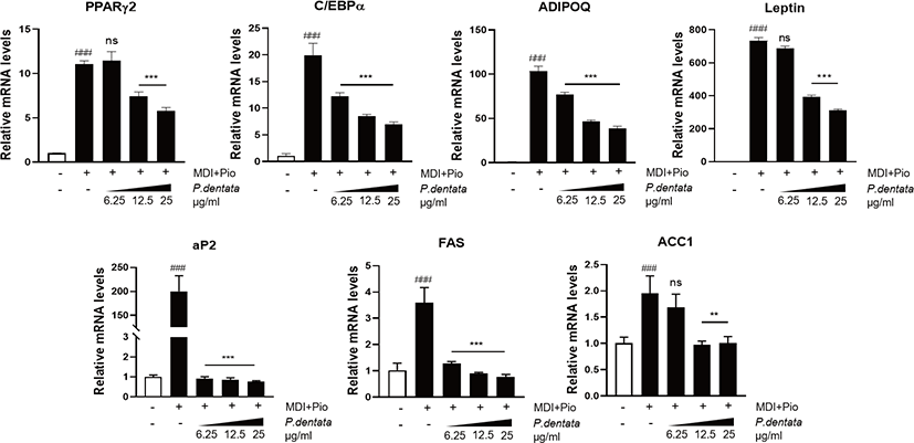
Next, we performed RT-qPCR analysis to examine the mRNA expression of adipogenic specific transcription factors such as PPAR-γ2, C/EBPα, and their target genes such as ADIPOQ, Leptin, FAS, aP2, and ACC1 after the treatment of P. dentata 50% MeOH extract. The extract decreased the PPAR-γ2, C/EBPα, as well as ADIPOQ, Leptin, FAS, aP2, and ACC1 mRNA expression. Gene expression of the PPAR-γ2, C/EBPα, and their adipogenic related genes following P. dentata treatment was significantly lower compared with that of differentiated control adipocytes treated MDI (methylisobutylxanthine, dexamethasone, insulin) plus pioglitazone. P. dentata 50% MeOH extract significantly downregulated the expression of adipogenesis associated genes in a dose-dependent manner (Fig. 3).

jast-62-6-854-g3
Fig. 3. Expression of adipogenic related genes in 3T3-L1 cells with Porphyra dentata 50% MeOH extract. The expression of adipogenic related genes which PPAR-γ2, C/EBPα (adipogenic transcription factors), ADIPOQ, leptin, aP2 (adipokine), FAS and ACC1 (lipogenic enzyme). 3T3-L1 cells cultured with various concentrations of P. dentata 50% MeOH extract (6.25, 12.5, and 25 μg/mL) with differentiation media were analyzed on day 5 by RT-qPCR. P. dentata 50% MeOH extract treatment decreased adipogenic related genes mRNA expression in a concentration-dependent manner on 5 days. Data were presented as mean and standard errors from three experiments. ###p < 0.001 vs. preadipocyte, ***p < 0.001, **p < 0.01 vs. MDI+pio. All data are presented as mean ± SD, and experiments were performed three times. PPARγ2, peroxisome proliferator-activated receptor γ2; MDI, methylxanthine, dexamethasone, insulin; C/EBPα, CCAAT/enhancer binding protein α; ADIPOQ, adiponectin; aP2, adipocyte protein 2; FAS, fatty acid synthase; ACC1, acetyl-coA carboxylase-1.
Download Original Figure

DISCUSSION

The 3T3-L1 mouse preadipocytes have been widely used for screening the effective agents to regulate the adipogenesis. The adipogenesis was determined by Oil Red O staining to show the amount of lipid droplets by specifically staining neutral triglycerides with high levels of adiopocyte-related genes expression in 3T3-L1 cells [22].

In the present study, we demonstrated that P. dentata extract dramatically inhibited lipid accumulation during adipocyte differentiation with decreasing PPARγ2, C/EBPα, ADIPOQ, leptin, FAS, aP2, and ACC1 expression. One of the alternative splicing forms of PPAR, PPARγ2, is a lipid-activated transcription factor which specifically expressed in adipose tissue [23]. In response to fatty acids, PPARγ2 leads to fat accumulation in adipocytes by modulating target genes involved in lipid metabolism [24]. However, PPARγ2 does not function alone but cooperatively with transcription factors in the C/EBP family to induce adipocyte differentiation [9,24]. The C/EBPs belong to the basic-leucine zipper class of transcription factors and has several forms including C/EBPα, C/EBPβ, C/EBPγ, and C/EBPδ [25]. The temporal expression of these factors during adipocyte differentiation indicates a cascade whereby early induction of C/EBPβ and C/EBPδ leads to induction of C/EBPα, which C/EBPα induces expression of many adipogenic related genes directly [26]. In this study, the mRNA expression of PPARγ2 and C/EBPα decreased significantly after treatment of P. dentata extract compared with that in differentiated control cells. It has been reported that PPARγ2 and C/EBPα cooperates to increase adipogenic genes using a positive feedback loop between them leading to adipogenesis [27]. ADIPOQ, leptin, and aP2 investigated in this study, are adipokine which cytokine secreted by adipose tissue and in obesity [28,29]. ADIPOQ is an adipocyte-specific factor, which adipocyte-derived hormone, it is abundantly produced and secreted by adipose tissues and widely recognized for its anti-inflammatory effects [30]. Leptin is also adipocyte-derived hormone that circulates in proportion to fat mass and acts as a negative regulator of energy homeostasis [31]. aP2 called fatty acid binding protein 4 (FABP4) is one of the only genes characterized by sufficient regulatory sequences to direct adipose-specific expression in vivo [32,33]. Having played an important role as adipocytes differentiation marker, as it can lead to the development of increasingly large adipocytes by leptin resistance and contribute to the accumulation of excessive fat masses found in obese states [34]. These adipokine levels were increased during differentiation from preadipocytes to maturation adipocytes [34,35]. The other adipogenic related genes, FAS and ACC1 are lipogenic enzymes. FAS is the key enzyme in lipogenesis, catalyzing the reactions for the synthesis of long-chain fatty acids [36]. ACC1 is a multi-subunit lipogenic enzyme that catalyzes the irreversible carboxylation of acetyl-CoA to produce malonyl-CoA for the biosynthesis of fatty acids [37]. Plants have been used as traditional natural medicines for healing many diseases [38] and many studies have shown that plant-derived foods have the potential to reduce obesity [39]. A plant belonging to the genus Porphyra, called laver, also consumed mainly as processed food or used a source of health-enhancing substances and this group have a unique active substances which provide health benefits [39,40]. Porphyra species contain biological active compounds, including polysaccharides, carotenoids, phenolic compounds, and MAAs. These compounds have been reported to have antioxidant [41], anti-inflammatory [42], anti-cancer [43,44], prevention of nervous system [45], and bone disease [46]. In particular, fucoidan, carotenoids, and phenolic compounds (EGCG) inhibited lipid accumulation in 3T3-L1 cells.

Previously, Kim et al. [22] indicated that Pyropia yezoensi (P. yezoensis) methanol extract, one types of laver contain high MAAs content (120 mg/g dried extract) reduces the contents of accumulation lipid determined by Oil Red O staining in a dose-dependent manner [22]. However, they demonstrated that treatment with high concentration (5 mg/mL) of the P. yezoensis methanol extract inhibited adipogenesis with decrease of preadipocytes proliferation via oxidative stress and proapoptotic effects. Our result indicates that P. dentata 50% MeOH extract at low concentration of ~25 μg/mL significantly suggest anti-adipogenesis in a dose-dependent manner with no cytotoxicity in 3T3-L1 cells. Hence, further study is needed to identify whether bioactive compounds (polysaccharides, carotenoids, phenolics, etc.) contained in laver contribute suppression of lipid accumulation in adipocyte.

In conclusion, P. dentata extract inhibited the accumulation of lipid droplets in concentration-dependent manner. Moreover, P. dentata extract inhibits the expression of adipogenic related genes involved in the adipogenesis from preadipocytes to mature adipocytes in 3T3-L1 cells. Especially, in all experiments, lipid droplets formation and gene expression are inhibited in a concentration-dependent manner (6.25, 12.5, and 25 μg/mL) of P. dentata extract. Thus, the result revealed that P. dentata has an effect of anti-obesity that inhibits adipogenesis. Since pharmacological effects of the Porphyra species are proven [47], and P. dentata belonging to that species is also consumed as food, it has the potential to be used as a dietary supplement and medicinal food item to suppress obesity.

Competing interests

No potential conflict of interest relevant to this article was reported.

Funding sources

This research was supported by Basic Science Research Program through the National Research Foundation of Korea (NRF) funded by the Ministry of Education, Science and Technology (no. NRF-2019R1I1A3A01059211).

Acknowledgements

We thank the members of the Kim laboratory for their discussions and help.

Availability of data and material

Upon reasonable request, the datasets of this study can be available from the corresponding author.

Authors’ contributions

Conceptualization: Cho JY, Kim SH.

Data curation: Cho JY, Kim SH.

Formal analysis: Choi SY, Lee SY, Cho JY, Kim SH.

Methodology: Choi SY, Lee SY, Jang Dh, Lee SJ.

Writing - original draft: Choi SY.

Writing - review & editing: Cho JY, Kim SH.

Ethics approval and consent to participate

This article does not require IRB/IACUC approval because there are no human and animal participants.

REFERENCES

1.

Heymsfield SB, Wadden TA. Mechanisms, pathophysiology, and management of obesity. N Engl J Med. 2017; 376:254-66

2.

Khandekar MJ, Cohen P, Spiegelman BM. Molecular mechanisms of cancer development in obesity. Nat Rev Cancer. 2011; 11:886-95

3.

Kim GC, Kim JS, Kim GM, Choi SY. Anti-adipogenic effects of Tropaeolum majus (nasturtium) ethanol extract on 3T3-L1 cells. Food Nutr Res. 2017; :61-1339555

4.

González-Castejón M, Rodriguez-Casado A. Dietary phytochemicals and their potential effects on obesity: a review. Pharmacol Res. 2011; :64-43855

5.

Hammarstedt A, Gogg S, Hedjazifar S, Nerstedt A, Smith U. Impaired adipogenesis and dysfunctional adipose tissue in human hypertrophic obesity. Physiol Rev. 2018; 98:1911-41

6.

Cao Y. Angiogenesis modulates adipogenesis and obesity. J Clin Invest. 2007; 117:2362-8

7.

Spiegelman BM, Flier JS. Adipogenesis and obesity: rounding out the big picture. Cell. 1996; 87:377-89

8.

Shao D, Lazar MA. Peroxisome proliferator activated receptor γ, CCAAT/enhancer-binding protein α, and cell cycle status regulate the commitment to adipocyte differentiation. J Biol Chem. 1997; 272:21473-8

9.

Auwerx J, Martin G, Guerre-Millo M, Staels B. Transcription, adipocyte differentiation, and obesity. J Mol Med. 1996; 74:347-52

10.

Moseti D, Regassa A, Kim WK. Molecular regulation of adipogenesis and potential anti-adipogenic bioactive molecules. Int J Mol Sci. 2016; :17-124

11.

Holdt SL, Kraan S. Bioactive compounds in seaweed: functional food applications and legislation. J Appl Phycol. 2011; 23:543-97

12.

Yoshiki M, Tsuge K, Tsuruta Y, Yoshimura T, Koganemaru K, Sumi T, et al. Production of new antioxidant compound from mycosporine-like amino acid, porphyra-334 by heat treatment. Food Chem. 2009; 113:1127-32

13.

Rui Y, Zhaohui Z, Wenshan S, Bafang L, Hu H. Protective effect of MAAs extracted from Porphyra tenera against UV irradiation-induced photoaging in mouse skin. J Photochem Photobiol B. 2019; 192:26-33

14.

Isaka S, Cho K, Nakazono S, Abu R, Ueno M, Kim D, et al. Antioxidant and anti-inflammatory activities of porphyran isolated from discolored nori (Porphyra yezoensis). Int J Biol Macromol. 2015; 74:68-75

15.

Wang X, Zhang Z. The antitumor activity of a red alga polysaccharide complexes carrying 5-fluorouracil. Int J Biol Macromol. 2014; 69:542-5

16.

Wan-Loy C, Siew-Moi P. Marine algae as a potential source for anti-obesity agents. Mar Drugs. 2016; :14-222

17.

Gómez-Zorita S, González-Arceo M, Trepiana J, Eseberri I, Fernández-Quintela A, Milton-Laskibar I, et al. Anti-obesity effects of macroalgae. Nutrients. 2020; :12-2378

18.

Rodríguez-Pérez C, Segura-Carretero A, del Mar Contreras M. Phenolic compounds as natural and multifunctional anti-obesity agents: a review. Crit Rev Food Sci Nutr. 2019; 59:1212-29

19.

Kim KJ, Lee BY. Fucoidan from the sporophyll of Undaria pinnatifida suppresses adipocyte differentiation by inhibition of inflammation-related cytokines in 3T3-L1 cells. Nutr Res. 2012; 32:439-47

20.

Cho TJ, Rhee MS. Health functionality and quality control of laver (Porphyra, Pyropia): current issues and future perspectives as an edible seaweed. Mar Drugs. 2019; :18-14

21.

Kazłowska K, Hsu T, Hou CC, Yang WC, Tsai GJ. Anti-inflammatory properties of phenolic compounds and crude extract from Porphyra dentata. J Ethnopharmacol. 2010; 128:123-30

22.

Kim H, Lee Y, Han T, Choi EM. The micosporine-like amino acids-rich aqueous methanol extract of laver (Porphyra yezoensis) inhibits adipogenesis and induces apoptosis in 3T3-L1 adipocytes. Nutr Res Pract. 2015; 9:592-8

23.

de Sá PM, Richard AJ, Hang H, Stephens JM. Transcriptional regulation of adipogenesis. Compr Physiol. 2017; 7:635-74

24.

Tontonoz P, Hu E, Graves RA, Budavari AI, Spiegelman BM. mPPARγ2: tissue-specific regulator of an adipocyte enhancer. Genes Dev. 1994; 8:1224-34

25.

Rosen ED, Walkey CJ, Puigserver P, Spiegelman BM. Transcriptional regulation of adipogenesis. Genes Dev. 2000; 14:1293-307

26.

Rosen ED, MacDougald OA. Adipocyte differentiation from the inside out. Nat Rev Mol Cell Biol. 2006; 7:885-96

27.

Wu Z, Rosen ED, Brun R, Hauser S, Adelmant G, Troy AE, et al. Cross-regulation of C/EBPα and PPARγ controls the transcriptional pathway of adipogenesis and insulin sensitivity. Mol Cell. 1999; 3:151-8

28.

Ghantous CM, Azrak Z, Hanache S, Abou-Kheir W, Zeidan A. Differential role of leptin and adiponectin in cardiovascular system. Int J Endocrinol. 2015; :2015-534320

29.

Li Y, Rong Y, Bao L, Nie B, Ren G, Zheng C, et al. Suppression of adipocyte differentiation and lipid accumulation by stearidonic acid (SDA) in 3T3-L1 cells. Lipids Health Dis. 2017; :16-181

30.

Fang H, Judd RL. Adiponectin regulation and function. Compr Physiol. 2018; 8:1031-63

31.

Bell BB, Rahmouni K. Leptin as a mediator of obesity-induced hypertension. Curr Obes Rep. 2016; 5:397-404

32.

Cao H, Sekiya M, Ertunc ME, Burak MF, Mayers JR, White A, et al. Adipocyte lipid chaperone aP2 is a secreted adipokine regulating hepatic glucose production. Cell Metab. 2013; 17:768-78

33.

Tontonoz P, Hu E, Spiegelman BM. Stimulation of adipogenesis in fibroblasts by PPARγ2, a lipid-activated transcription factor. Cell. 1994; 79:1147-56

34.

Sáinz N, Barrenetxe J, Moreno-Aliaga MJ, Martínez JA. Leptin resistance and diet-induced obesity: central and peripheral actions of leptin. Metabolism. 2015; 64:35-46

35.

Simons PJ, van den Pangaart PS, van Roomen CPAA, Aerts JMFG, Boon L. Cytokine-mediated modulation of leptin and adiponectin secretion during in vitro adipogenesis: evidence that tumor necrosis factor-α- and interleukin-1β-treated human preadipocytes are potent leptin producers. Cytokine. 2005; 32:94-103

36.

Hogan JC, Stephens JM. The regulation of fatty acid synthase by STAT5A. Diabetes. 2005; 54:1968-75

37.

Yang SM, Park YK, Kim JI, Lee YH, Lee TY, Jang BC. LY3009120, a pan-Raf kinase inhibitor, inhibits adipogenesis of 3T3-L1 cells by controlling the expression and phosphorylation of C/EBP-α, PPAR-γ, STAT-3, FAS, ACC, perilipin A and AMPK. Int J Mol Med. 2018; 42:3477-84

38.

Roh C, Jung U. Screening of crude plant extracts with anti-obesity activity. Int J Mol Sci. 2012; 13:1710-9

39.

Boeing H, Bechthold A, Bub A, Ellinger S, Haller D, Kroke A, et al. Critical review: vegetables and fruit in the prevention of chronic diseases. Eur J Nutr. 2012; 51:637-63

40.

Rajapakse N, Kim SK. Nutritional and digestive health benefits of seaweed. Adv Food Nutr Res. 2011; 64:17-28

41.

Zhang Z, Zhang Q, Wang J, Zhang H, Niu X, Li P. Preparation of the different derivatives of the low-molecular-weight porphyran from Porphyra haitanensis and their antioxidant activities in vitro. Int J Biol Macromol. 2009; 45:22-6

42.

Yanagido A, Ueno M, Jiang Z, Cho K, Yamaguchi K, Kim D, et al. Increase in anti-inflammatory activities of radical-degraded porphyrans isolated from discolored nori (Pyropia yezoensis). Int J Biol Macromol. 2018; 117:78-86

43.

Yu X, Zhou C, Yang H, Huang X, Ma H, Qin X, et al. Effect of ultrasonic treatment on the degradation and inhibition cancer cell lines of polysaccharides from Porphyra yezoensis. Carbohydr Polym. 2015; 117:650-6

44.

Noda H, Amano H, Arashima K, Nisizawa K. Antitumor activity of marine algae. Hydrobiologia. 1990; 204:577-84

45.

Mohibbullah M, Bhuiyan MMH, Hannan MA, Getachew P, Hong YK, Choi JS, et al. The edible red alga Porphyra yezoensis promotes neuronal survival and cytoarchitecture in primary hippocampal neurons. Cell Mol Neurobiol. 2016; 36:669-82

46.

Ueno M, Cho K, Isaka S, Nishiguchi T, Yamaguchi K, Kim D, et al. Inhibitory effect of sulphated polysaccharide porphyran (isolated from Porphyra yezoensis) on RANKL-induced differentiation of RAW264.7 cells into osteoclasts. Phytother Res. 2018; 32:452-8

47.

Venkatraman KL, Mehta A. Health benefits and pharmacological effects of Porphyra species. Plant Foods Hum Nutr. 2019; 74:10-7