Journal of Animal Science and Technology
Korean Society of Animal Sciences and Technology
RESEARCH ARTICLE

Effect of hyperthermia on cell viability, amino acid transfer, and milk protein synthesis in bovine mammary epithelial cells

Jia Zhou1https://orcid.org/0000-0001-8293-105X, Sungming Yue2https://orcid.org/0000-0002-2151-6511, Benchu Xue1https://orcid.org/0000-0002-7163-3812, Zhisheng Wang1https://orcid.org/0000-0002-2520-1912, Lizhi Wang1https://orcid.org/0000-0002-4915-4225, Quanhui Peng1https://orcid.org/0000-0003-1421-9145, Rui Hu1https://orcid.org/0000-0003-0961-6760, Bai Xue1,*https://orcid.org/0000-0001-8008-8425
1Animal Nutrition Institute, Sichuan Agricultural University, Chengdu 611130, China
2Department of Bioengineering, Sichuan Water Conservancy Vocation College, Chengdu 611845, China
*Corresponding author: Bai Xue, Animal Nutrition Institute, Sichuan Agricultural University, Chengdu 611130, China. Tel: +86-28-86291781, E-mail: xuebai@sicau.edu.cn

© Copyright 2022 Korean Society of Animal Science and Technology. This is an Open-Access article distributed under the terms of the Creative Commons Attribution Non-Commercial License (http://creativecommons.org/licenses/by-nc/4.0/) which permits unrestricted non-commercial use, distribution, and reproduction in any medium, provided the original work is properly cited.

Received: Sep 19, 2021; Revised: Nov 08, 2021; Accepted: Nov 16, 2021

Published Online: Jan 31, 2022

Abstract

The reduction of milk yield caused by heat stress in summer is the main condition restricting the economic benefits of dairy farms. To examine the impact of hyperthermia on bovine mammary epithelial (MAC-T) cells, we incubated the MAC-T cells at thermal-neutral (37°C, CON group) and hyperthermic (42°C, HS group) temperatures for 6 h. Subsequently, the cell viability and apoptotic rate of MAC-T cells, apoptosis-related genes expression, casein and amino acid transporter genes, and the expression of the apoptosis-related proteins were examined. Compared with the CON group, hyperthermia significantly decreased the cell viability (p < 0.05) and elevated the apoptotic rate (p < 0.05) of MAC-T cells. Moreover, the expression of heat shock protein (HSP)70, HSP90B1, Bcl-2-associated X protein (BAX), Caspase-9, and Caspase-3 genes was upregulated (p < 0.05). The expression of HSP70 and BAX (pro-apoptotic) proteins was upregulated (p < 0.05) while that of B-cell lymphoma (BCL)2 (antiapoptotic) protein was downregulated (p < 0.05) by hyperthermia. Decreased mRNA expression of mechanistic target of rapamycin (mTOR) signaling pathway-related genes, amino acid transporter genes (SLC7A5, SLC38A3, SLC38A2, and SLC38A9), and casein genes (CSNS1, CSN2, and CSN3) was found in the heat stress (HS) group (p < 0.05) in contrast with the CON group. These findings illustrated that hyperthermia promoted cell apoptosis and reduced the transport of amino acids into cells, which inhibited the milk proteins synthesis in MAC-T cells.

Keywords: Hyperthermia; Heat stress; Apoptosis; Milk protein synthesis; Amino acid transport

INTRODUCTION

The rise in global temperature in recent decades has negatively affected agriculture and food supply. More than half of dairy cows live in subtropical and tropical areas that have a temperature-humidity index (THI) which tends to reach 68 or more, and the risk of heat stress in dairy cows is inevitable [1]. Heat stress leads to a 10%–35% decline in milk yield [2] and an estimated 5.4% loss in the monthly income of dairy farmers during summer [3]. Therefore, understanding the mechanism by which heat stress induces a decrease in milk protein synthesis is crucial to improve the milk production potential of dairy cattle during summer.

Heat stress, which reduces the dry matter intake (DMI) of dairy cows, has been traditionally considered as the major cause for the decreased milk production potential under hyperthermic environments [4,5]. However, for the past few years, the experiment of pair-fed to non-heat-stress cows confirmed that the decrease in milk yield and milk composition was only partly caused by the reduction of DMI [6,7]. Further studies indicated that the decrease in the production of milk protein induced by heat stress was specifically caused by a decline in the activity of mammary protein synthesis rather than a decrease in milk yield [6,8]. Heat stress promotes the consumption of extra-mammary amino acids, including urinary nitrogen excretion and rumen microbial protein synthesis, in dairy cows, which reduces the amount of amino acids available to the mammary gland for the synthesis of milk proteins [9]. Transcriptome analysis indicated that heat stress strongly inhibited the amino acids metabolic activity in the mammary tissue, and the data suggested that the decreased availability of amino acids resulted in a decreased synthesis of milk proteins [10]. In addition, hyperthermia reduced cell viability in bovine mammary epithelial cells (BMECs) [11] and alveoli number in the lactating mammary gland [12]. Hyperthermia negatively regulates the number and activity of mammary gland cells, thereby contributing to a decrease in milk production under high-temperature stress [13]. However, to our knowledge, the influence of heat stress on the transport of amino acids in the mammary gland of lactating cows is insufficient in the existing literature.

As the precursor of the synthesis of milk proteins, amino acids perform critical functions in the regulation of physiological functions [14]. For instance, the branched-chain amino acids, such as isoleucine, valine, and leucine regulate nutrition metabolism, immunity, and energy homeostasis in mammals [15]. Methionine (Met) and arginine (Arg) may stimulate the mammalian target of rapamycin complex 1 (mTORC1) and promote protein synthesis [16]. Dietary supplementation of Met could increase the milk protein concentration and improve milk production in dairy cows [17,18]. Under hyperthermic conditions, enhanced supply of Met and Arg had a positive effect on milk protein synthesis in heat-stressed BMECs [19], and supplementation of Met helped maintain milk composition in heat-stressed lactating Holstein cows [20]. We hypothesized that the heat stress-induced reduction in milk protein synthesis was due to the decrease in the uptake of amino acids by mammary cells. Heat stress refers to a sequence of non-specific physiological responses to maintain a constant body temperature [4]. In vitro, apoptosis induced by hyperthermia is also considered a response to heat stress [21,22]. Bovine mammary epithelial (MAC-T) cells are well-known mammary epithelial cell line and retain the phenotypic characteristics of BMECs [23,24], have been used extensively to study apoptosis in the immune response or oxidative stress [25,26], milk protein synthesis, and mammalian lactation [27,28]. Thus, we primarily aimed to examine the impact of heat stress on the synthesis of milk protein by incubating MAC-T cells at a hyperthermic temperature (42°C).

MATERIALS AND METHODS

Cell culture and experimental design

Frozen bovine MAC-T cells were recovered and allowed to grow in 75 cm2 cell culture flasks at a temperature of 37°C and 5% CO2 concentration to obtain enough biological material for subsequent analysis. Cells at 80%–90% confluency were transferred into 6-well plates (1.2~1.5×10⁵ cells per well, Thermo Scientific, Waltham, MA, USA). To culture MAC-T cells, we utilized the complete medium consist of Dulbecco’s modified Eagle’s medium (DMEM,Thermo Scientific) accompanied by 10 percent fetal bovine serum (FBS; Thermo Scientific), 100 μg/mL streptomycin, and 100 IU/mL penicillin G (Sigma Aldrich, St Louis, MO, USA). After every 48 hours, the culture medium was replaced. The cells were washed using phosphate-buffered saline (PBS, Thermo Scientific) three times and the medium was changed until the confluency was 80% to 90%. Then, MAC-T cells were divided into two groups (n=6 replicas for each treatment) and subjected to incubation at 37°C (CON) or 42°C (heat stress [HS]) for 6 h, respectively. The incubation temperature and time were set at 42°C and 6 h, based on a previous study by Collier et al. [29] where the mRNA concentration of heat shock protein 70 (HSP70) was considerably elevated in BMECs within 1 and 2 h, and it attained a peak after 4 hours following the exposure of the cells to 42°C.

Cell viability and apoptosis assays

The viability of the cells was assessed utilizing an MTT test kit in accordance with the instructions stipulated by the manufacturer. Briefly, 100 μL medium containing MAC-T cells (2×10⁴/mL) were transferred into 96-well culture plates, followed by treatment at 37°C or 42°C for 6 hours. Afterward, they were subjected to incubation for 16 hours at 37°C after being incubated for 4 hours with 10 μL of MTT staining solution within every well plate. Subsequently, 100 μL of the formazan crystals were added in all the wells at 37°C for 4 h until completely dissolved, and a microplate reader (Bio-Rad, Hercules, CA, USA) was utilized to determine the optical density (OD) at 570 nm. Cell apoptosis rate was determined utilizing an Annexin V-FITC/PI apoptosis detection kit (4A Biotech, Beijing, China) in compliance with the protocols provided by the manufacturer. The excitation wavelength was 525 nm (Annexin V-FITC, green fluorescence), and the emission wavelength was (595 nm PI, red fluorescence). The results were evaluated utilizing the Cell-Quest software (BD Biosciences, Franklin Lakes, NJ, USA).

Isolation of RNA and quantitative reverse transcription-polymerase chain reaction (qRT-PCR)

The Steady Pure Universal RNA Extraction Kit (Accurate Bio, Hunan, China) was utilized to extract and purify total RNA from MAC-T cells according to the instructions provided by the manufacturer. The NanoDrop 2000 spectrophotometer (Thermo Scientific) was utilized to determine the purity as well as the concentration of the isolated RNA. Additionally, the integrity of the RNA was examined utilizing an Agilent 2100 Bioanalyzer (Agilent Technologies, Santa Clara, CA, USA). The samples that had an RNA Integrity Number (RIN) ≥ 7.0 underwent dilution to 100 ng/μL with RNase-free water. The reverse transcription of the diluted RNA samples to cDNA was performed utilizing the Prime-Script™ RT-PCR reagent Kit with gDNA Eraser (Takara, Tokyo, Japan) in accordance with the protocols stipulated by the manufacturer. For additional analysis, RNase-free water was utilized to dilute the cDNA at a ratio of 1: 5.

With the aid of SYBR Premix Ex Taq reagents (TaKaRa, Dalian, China), we conducted qRT-PCR in an ABI 7500 real-time thermocycler (Applied Biosystems, Foster City, CA, USA), as earlier described [30,31]. To normalize the target gene expression, the reference gene utilized was glyceraldehyde-3-phosphate dehydrogenase (GAPDH) [31]. Table 1 contains a list of sequences of all primers, which were commercially manufactured by Invitrogen (Shanghai, China). The relative mRNA target genes expression was computed utilizing the comparative cycle threshold (2−ΔΔCt) method [32]. Each of the biological samples was replicated three times on a 96-well real-time PCR plate (Applied Biosystems).

Table 1. The primer sequences of genes
Gene Primer sequence (5’ -3’) Accession
GAPDH F: CTGGCAAAGTGGACATCGTC NM_001034034.2
R: GCCAGTAGAAGCAGGGATGA
BCL-2 F: AGATGTCTTCCCTGCTCCCT XM_010815066.3
R: TGCGGGACCCTGTAATTCTG
BAX F: AGAGGATGATCGCAGCTGTG XM_015458140.2
R: GAAGTCCAATGTCCAGCCCA
Caspase-3 F: TGGTACAGACGTGGATGCAG XM_010820245.3
R: TCCCCTCTGAAGAAACTTGCT
Caspase-9 F: GGCCAGGCAGCTAATCCTAG XM_024975972.1
R: TTCCTTGGCTCGGCTTTGAT
HSP70 F: TGCATATTCATCTCCGGCCC XM_005225768.4
R: CTCCTTCCCATCGCCTCATC
HSP90B1 F: AGAACCTGCTGCATGTCACA NM_174700.2
R: ACCAACACCAAACTGACCGA
CSN1S1 F: ATCAAGCACCAAGGACTCCC XM_024993016.1
R: GCTCAGGGTAGAAGTAGGCC
CSN2 F: TCCATTCAGCTCCTCCTTCAC XM_015471671.2
R: GGGAGGCTGTTATGGATGGG
CSN3 F: CCCAGGAGCAAAACCAAGAAC NM_174294.2
R: TGAAGAATTTGGGCAGGTGAC
SLC7A5 F: CGTCCTCCAGTGCATCATGA NM_174613.2
R: TAGAAACTTGATGGGCCGCT
AKT1 F: GCGCCACCATGAAGACTTTC XM_024981593.1
R: CCTGGTGTCCGTCTCAGATG
mTOR F: AGGGCATGAATCGGGATGAC XM_002694043.6
R: GTGAAGGCAGAAGGTCGGAA
RPS6 F: CCAGAAGCTCATTGAAGTGGA NM_001015548.2
R: GCTGAATCTTGGGTGCTTTAGT
RPS6KB1 F: GGGCCCCTGAGATCTTGATG NM_205816.1
R: CGTGAGGTAGGGAGGCAAAT
SLC38A3 F: GCTGCCCCTTGTCATACAGA XM_024982409.1
R: CGTAGAAGGTGAGGTAGCCG
SLC38A9 F: TTGGGCAGTGGTCAAGTCTC XM_024981327.1
R: CGAATAGCCTTCCAAGTGACG
SLC38A2 F: GGAGATGGTTGGGAAGCTCA XM_024991403.1
R: CATCATTCTTCGACGGCTGC

GAPDH, glyceraldehyde-3-phosphate dehydrogenase; BCL-2, B cell leukemia/lymphoma 2; BAX, Bcl-2-associated X protein; Caspase-3, cysteinyl aspartate specific proteinase-3; Caspase-9, cysteinyl aspartate specific proteinase-9; HSP70, heat shock protein 70; HSP90B1, heat shock protein 90B1; CSN1S1, casein alpha s1; CSN2, casein beta; CSN3, casein kappa; SLC7A5, solute carrier family 7, member 5; AKT1, serine-threonine protein kinas 1; mTOR, mammalian target of rapamycin; RPS6, ribosomal protein S6; RPS6KB1, ribosomal protein S6 kinase B1; SLC38A3, solute carrier family 38, member 3; SLC38A9, solute carrier family 38, member 9; SLC38A2, solute carrier family 38, member 2.

Download Excel Table
Western blot

The Western blot analysis was conducted in the same way as previously reported [33]. Briefly, MAC-T cells were solubilized in radioimmunoprecipitation assay (RIPA) Lysis and Extraction Buffer (Invitrogen, Waltham, MA, USA) to obtain total protein. After denaturation at high temperature, the protein samples extracted from cells were isolated utilizing sodium dodecyl sulfate-polyacrylamide gel electrophoresis (SDS-PAGE) and subsequently loaded onto a nitrocellulose membrane. Blocking of the membrane was carried out using 5% skimmed milk generated in Tris-buffer, followed by incubation using primary antibodies (Complete details are listed in Table 2) over the night at 4°C. Subsequently, incubation of the membrane was conducted using horseradish peroxidase (HRP)-conjugated anti-rabbit IgG secondary antibody (Complete details are listed in Table 2) at ambient temperature for 4 hours. Finally, detection of the blot was done utilizing ECL™ Western Blotting Detection Reagent (GE Healthcare, Piscataway, NJ, USA) and visualization of the proteins was achieved using enhanced chemiluminescence (Amersham Biosciences, Piscataway, NJ, USA). The intensity of β-actin was utilized as an endogenous control.

Table 2. The antibodies used for Western Bloting
Antibody Category Source INC
BCL2 Primary antibody Rabbit Cell Signaling Technology
BAX Primary antibody Rabbit Santa Cruz
HSP70 Primary antibody Mouse Cell Signaling Technology
β-Actin Primary antibody Rabbit Cell Signaling Technology
Goat Anti-Rabbit IgG H&L (HRP) Secondary antibody Goat Abcam

BCL-2, B cell leukemia/lymphoma 2; BAX, Bcl-2-associated X protein; HSP70, heat shock protein 70.

Download Excel Table
Statistical analysis

The independent two-sample t-test was utilized to examine all of the data with the aid of the SPSS 17.0 package (SPSS, Chicago, IL, USA). The data were expressed as the mean ± SD. p < 0.05 was considered significant.

RESULTS

Impacts of heat stress on apoptosis and viability of MAC-T cells

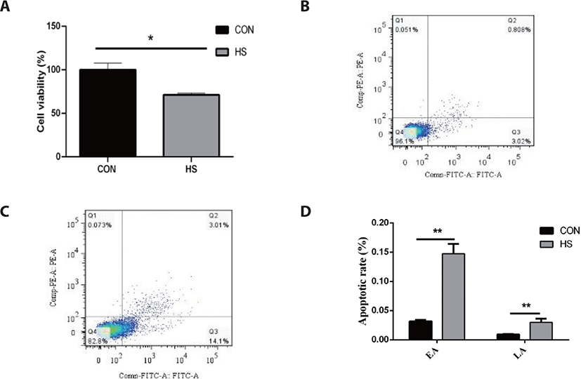
As illustrated in Fig. 1, MAC-T cells thermally treated at 42°C for 6 h showed a 28.81% decrease in cell viability as opposed to the CON group (p < 0.05). Furthermore, heat stress considerably elevated the early apoptotic rate (14.72% vs. 3.18%) and late apoptotic rate (3.01% vs. 0.91%) of MAC-T cells (p < 0.05).

jast-64-1-110-g1
Fig. 1. Impacts of heat stress on the apoptosis and cell viability of MAC-T cells. (A) The MAC-T cells viability was evaluated after being thermal treatment at 42°C for 6 h; (B) Populations of early and late apoptotic MAC-T cells cultured at 37°C for 6 h, as determined by flow cytometry; (C) Populations of early and late apoptotic MAC-T cells cultured at 42°C for 6 h, as determined by flow cytometry. (D) The early apoptotic (EA) and late apoptotic (LA) rates of MAC-T cells after being treated for 6 h. * p < 0.05, ** p < 0.01. HS, heat stress.
Download Original Figure
Impacts impacts of heat stress on the expression of heat shock and apoptosis-related genes of MAC-T cells

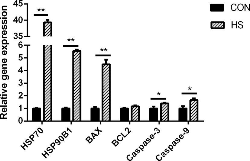
Heat stress greatly elevated the gene expression of HSP90B) and HSP70 (p < 0.01) in MAC-T cells (p < 0.01; Fig. 2). Bcl-2-associated X protein (BAX) gene expression was greatly elevated in response to heat stress (p < 0.01), while the B-cell lymphoma 2 (BCL2) gene expression was not affected. In addition, heat stress considerably elevated the caspase-9 and caspase-3 gene expressions (both p < 0.05).

jast-64-1-110-g2
Fig. 2. Impacts of heat stress on the expression of heat shock and apoptosis-related genes. * p < 0.05, ** p < 0.01. HSP70, heat shock protein 70; HSP90B1, heat shock protein 90B1; BAX, Bcl-2-associated X protein; BCL2, B-cell lymphoma 2; HS, heat stress.
Download Original Figure
Impacts of heat stress on the expression of heat shock and apoptosis-related proteins in MAC-T cells

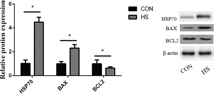
The HSP70 protein expression was substantially elevated in MAC-T cells upon exposure to heat stress (p < 0.01; Fig. 3). Heat stress substantially elevated the BAX protein expression (p < 0.01) while decreasing that of BCL2 (p < 0.05).

jast-64-1-110-g3
Fig. 3. Impacts of heat stress on the expression of heat shock and apoptosis-related proteins. * p < 0.05.HSP70, heat shock protein 70; BAX, Bcl-2-associated X protein; BCL2, B-cell lymphoma 2; HS, heat stress.
Download Original Figure
Impacts of heat stress on the expression of mTOR signaling pathway-related genes in MAC-T cells

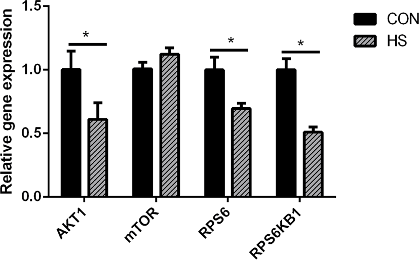
Heat stress considerably reduced the gene expression of ribosomal protein S6 (RPS6, p < 0.05), AKT serine/threonine kinase 1 (AKT1, p < 0.05), and ribosomal protein S6 kinase B1 (RPS6KB1, p < 0.05) (Fig. 4).

jast-64-1-110-g4
Fig. 4. Impacts of heat stress on the expression of mTOR signaling pathway-related genes. * p < 0.05. AKT1, serine/threonine kinase 1; mTOR, mechanistic target of rapamycin kinase; RPS6, ribosomal protein S6; RPS6KB1, ribosomal protein S6 kinase B1; HS, heat stress.
Download Original Figure
Impacts of heat stress on the expression of casein and amino acid transporter genes in MAC-T cells

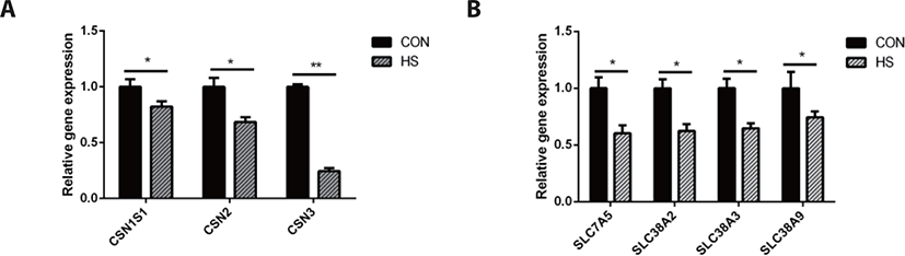
Heat stress significantly downregulated the gene expression of casein kappa (CSN3, p < 0.01) and casein alpha s1 (CSN1S1, p < 0.05), casein beta (CSN2, p < 0.05). Moreover, heat stress downregulated the gene expression of solute carrier family 38 member 2 (SLC38A2, p < 0.05), SLC38A9 (p <0.05), SLC38A3 (p < 0.05), and SLC7A5 ( p < 0.05) as shown in Fig. 5.

jast-64-1-110-g5
Fig. 5. Impacts of heat stress on the expression of casein and amino acid transporter genes. (A) The expression of casein genes; (B) The expression of amino acid transporter genes. * p < 0.05, ** p < 0.01. CSN1S1, casein alpha s1; CSN2, casein beta; CSN3, casein kappa; SLC7A5, solute carrier family 7 member 5; SLC38A2, solute carrier family 38 member 2; SLC38A3, solute carrier family 38 member 3; SLC38A9, solute carrier family 38 member 9; HS, heat stress.
Download Original Figure

DISCUSSION

High temperature can induce DNA damage, mitochondrial dysfunction, and abnormal gene expression and protein synthesis, eventually leading to cell death [34-36]. Liu et al. [37] showed that heat-stressed BMECs were characterized by the presence of condensed nuclei and cytoplasmic vacuoles. Moreover, they found that cells released a large number of cellular fragments into the medium and exhibited cytolysis and disorganization [37]. Hyperpyrexia could cause a decrease in the total number and activity of BMECs by inducing apoptosis [38]. During heat stress, cells mount a series of regulatory stress responses to maintain cell homeostasis [39]. For instance, as an adaptive cellular response to heat stress, cells rapidly upregulate the transcription and translation of HSPs to protect against protein aggregation and degradation [40], thereby restoring the normal function of the mammary gland. Both HSP90 and HSP70 perform mostly anti-apoptotic functions [41]. However, heat stress also induces the expression of pro-and anti-apoptotic members of the Bcl-2 protein family, which are known to regulate cell death [42]. Through the interaction of these proteins, the binding of cytochrome c released from mitochondria to cytosolic Apaf-1 results in the formation of a caspase-activating complex known as apoptosome [42]. The dimerization of caspase-9 within the apoptosome complex activates caspase-3, which results in apoptotic body formation and cellular inactivation through the cleavage specific proteins [43]. During this process, Bak and Bax, the pro-apoptotic Bcl-2 family members, perform a function of positively modulating the cytochrome c release from mitochondria [44], while the antiapoptotic Bcl-2 family members, Bcl-2 and Bcl-xL, suppress its release [45]. In this study, we found that hyperthermia decreased the viability and increased the apoptotic rate of MAC-T cells. The protein and gene expression of BAX was upregulated in the HS group, which is considered a crucial step in the mitochondrial apoptotic pathway [46]. Moreover, the higher expression of HSP70, HSP90B1, caspase-9 and caspase-3 genes and HSP70 protein was observed in the HS group. These results suggested that MAC-T cells underwent apoptosis after incubation at 42°C for 6 h, which might have resulted in a decrease in milk protein synthesis.

As the MAC-T cell line is incapable of secreting milk components, milk protein content could not be detected directly in this study. The CSN2 gene expression is positively associated with milk yield [47]. Hence, the expression of casein genes may be used to evaluate milk yield as an alternative to the evaluation of casein protein synthesis in MAC-T cells [48]. We compared the CSN1S1, CSN2, and CSN3 genes expression, which are the most highly expressed casein genes in milk protein [49], between the HS and CON groups and found that their expressions were significantly decreased in the HS group. These findings corroborate an earlier research report on the mammary gland tissue of heat-stressed lactating dairy cows [50] and another study on heat-stressed BMECs [51]. Therefore, heat stress could directly inhibit the synthesis of casein proteins, and the decrease in the DMI may be partly responsible for the decrease in the synthesis of milk protein in lactating cows under heat stress. Heat stress destroyed the cytoskeleton of BMECs, inhibited the cell cycle [52], and substantially reduced the mTOR signaling pathway activity [53], which is known as the regulator of protein synthesis. As a key upstream modulator of the mTOR signaling pathway, AKT performs a vital function in the maintenance of cell survival and depletion before the induction of apoptosis in fibroblast cells exposed to heat stress for a long term [54]. Hyperthermia decreased the phosphorylation state of AKT, RPS6K1, and RPS6, which are regarded as the upstream and downstream protein factors of the mTOR signaling pathway in MAC-T cells [54]. The suppression of the mTOR signaling pathway may be attributed the reduction of milk protein synthesis.

Amino acids are nutrients essential for the survival of all cell types. They not only serve as the precursor molecules for protein synthesis but can also regulate cellular function. For example, leucine (Leu), glutamine (Gln), and Arg function as signaling factors in the mTOR signaling pathway; serine (Ser), Glu, glycine (Gly), and aspartate (Asp) are necessary for nucleotide synthesis [55,56]. Thus, the normal function of mammary cells depends on the intracellular amino acid supply modulated by amino acid transporters. Interestingly, we also found that the gene expression of amino acid transporters was downregulated by hyperthermia. Amino acid transporters are membrane transporters and the majority of them belong to the solute carrier family of membrane transport proteins. SLC7A5 is a systemic L-type amino acid transporter (LAT1) that exclusively transports essential amino acids [57]. In many cells, the SLC7A5-mediated import of amino acids is essential to maintain mTOR activity [58]. One of the main functions of mTOR is to speed up the translation of mRNA, where amino acids are required as precursors [58]. Thus, the hyperthermia-induced decrease in SLC7A5 gene expression could have caused the decrease in amino acid transport, which inhibited the mTOR signaling pathway activity, eventually resulting in the reduction of lactoprotein synthesis in heat-stressed MAC-T cells. The inhibition of the mTOR signaling pathway significantly reduced the expression of β-casein and LAT1 (encoded by SLC7A5) [59]. The transporters classified as SLC38 family are known as sodium neutral amino acid transporters, which can perform the net transport of neutral amino acids [60]. This family of proteins contributes to maintaining the homeostatic pool of extracellular and intracellular amino acids [61]. These results suggest that the mTOR signaling pathway and amino acid transporters regulate each other to regulate the synthesis of milk protein in mammary cells of dairy cows. In contrast, a reduction in the supply of amino acids may also result in a decline in milk protein synthesis due to the shortage of essential substrates. In this research, the decreased expression of amino acid transporter genes in heat-stressed MAC-T cells might be linked to the decreased synthesis of milk proteins.

CONCLUSION

Hyperthermia induced apoptosis and lowered the expression of mTOR signaling pathway-related genes in MAC-T cells. Additionally, hyperthermia downregulated the expression of amino acid transporter genes, which might decrease the supply of amino acids available to MAC-T cells. Subsequently, the deficiency of amino acids was the root cause for the decreased rate of protein synthesis in MAC-T cells under heat stress. The results from this research may offer novel directions for the development of strategies to alleviate the loss of milk production induced by heat stress.

Competing interests

The authors declare no conflict of interest.

Funding sources

This research was funded by the Program for Doctoral Workstation of Nutrition and Health of High-Yield Dairy Cows established by Sichuan Agricultural University (Sichuan, China) and Menon Animal Nutrition Technology (Shanghai, China), and the Science and Technology Supporting Program of the National Science and Tech-nology Ministry, China, grant number 2012BAD12B02.

Acknowledgements

We acknowledge Menon Animal Nutrition Technology, Shanghai, China, for providing financial support in this study. We gratefully thank our professors and students for their help in this research.

Availability of data and material

Upon reasonable request, the datasets of this study can be available from the corresponding author.

Authors’ contributions

Conceptualization: Zhou J, Yue S, Xue Bai.

Formal analysis: Zhou J, Yue S, Xue Benchu.

Methodology: Yue S, Xue Benchu, Xue Bai.

Validation: Hu R.

Investigation: Wang Z, Wang L, Peng Q, Xue Bai.

Writing - original draft: Zhou J.

Writing - review & editing: Zhou J, Yue S, Xue Benchu, Wang Z, Wang L, Peng Q, Hu R, Xue Bai.

Ethics approval and consent to participate

This article does not require IRB/IACUC approval because there are no human and animal participants.

REFERENCES

1.

Baruselli PS, Ferreira RM, Vieira LM, Souza AH, Bó GA, Rodrigues CA. Use of embryo transfer to alleviate infertility caused by heat stress. Theriogenology. 2020; 155:1-11

2.

St-Pierre NR, Cobanov B, Schnitkey G. Economic losses from heat stress by US livestock industries. J Dairy Sci. 2003; 86:E52-77

3.

Hempel S, Menz C, Pinto S, Galán E, Janke D, Estellés F, et al. Heat stress risk in European dairy cattle husbandry under different climate change scenarios – uncertainties and potential impacts. Earth Syst Dyn. 2019; 10:859-84

4.

West JW. Effects of heat-stress on production in dairy cattle. J Dairy Sci. 2003; 86:2131-44

5.

Beede DK, Collier RJ. Potential nutritional strategies for intensively managed cattle during thermal stress. J Anim Sci. 1986; 62:543-54

6.

Cowley FC, Barber DG, Houlihan AV, Poppi DP. Immediate and residual effects of heat stress and restricted intake on milk protein and casein composition and energy metabolism. J Dairy Sci. 2015; 98:2356-68

7.

Wheelock JB, Rhoads RP, VanBaale MJ, Sanders SR, Baumgard LH. Effects of heat stress on energetic metabolism in lactating Holstein cows. J Dairy Sci. 2010; 93:644-55

8.

Cai M, Hu Y, Zheng T, He H, Xiao W, Liu B, et al. MicroRNA-216b inhibits heat stress-induced cell apoptosis by targeting Fas in bovine mammary epithelial cells. Cell Stress Chaperones. 2018; 23:921-31

9.

Gao ST, Guo J, Quan SY, Nan XM, Fernandez MVS, Baumgard LH, et al. The effects of heat stress on protein metabolism in lactating Holstein cows. J Dairy Sci. 2017; 100:5040-9

10.

Gao ST, Ma L, Zhou Z, Zhou ZK, Baumgard LH, Jiang D, et al. Heat stress negatively affects the transcriptome related to overall metabolism and milk protein synthesis in mammary tissue of midlactating dairy cows. Physiol Genomics. 2019; 51:400-9

11.

Zou Y, Shao J, Li Y, Zhao FQ, Liu JX, Liu H. Protective effects of inorganic and organic selenium on heat stress in bovine mammary epithelial cells. Oxid Med Cell Longev. 2019; 2019:1503478

12.

Dado-Senn B, Skibiel AL, Fabris TF, Dahl GE, Laporta J. Dry period heat stress induces microstructural changes in the lactating mammary gland. PLOS ON. 2019; 14e0222120

13.

Guo Z, Gao S, Ouyang J, Ma L, Bu D. Impacts of heat stress-induced oxidative stress on the milk protein biosynthesis of dairy cows. Animals. 2021; 11:726

14.

Liu Z, Ezernieks V, Wang J, Arachchillage NW, Garner JB, Wales WJ, et al. Heat stress in dairy cattle alters lipid composition of milk. Sci Rep. 2017; 7:961

15.

Nie C, He T, Zhang W, Zhang G, Ma X. Branched chain amino acids: beyond nutrition metabolism. Int J Mol Sci. 2018; 19:954

16.

Hu L, Chen Y, Cortes IM, Coleman DN, Dai H, Liang Y, et al. Supply of methionine and arginine alters phosphorylation of mechanistic target of rapamycin (mTOR), circadian clock proteins, and α-s1-casein abundance in bovine mammary epithelial cells. Food Funct. 2020; 11:883-94

17.

Wang C, Liu HY, Wang YM, Yang ZQ, Liu JX, Wu YM, et al. Effects of dietary supplementation of methionine and lysine on milk production and nitrogen utilization in dairy cows. J Dairy Sci. 2010; 93:3661-70

18.

Noftsger S, St-Pierre NR. Supplementation of methionine and selection of highly digestible rumen undegradable protein to improve nitrogen efficiency for milk production. J Dairy Sci. 2003; 86:958-69

19.

Salama AAK, Duque M, Wang L, Shahzad K, Olivera M, Loor JJ. Enhanced supply of methionine or arginine alters mechanistic target of rapamycin signaling proteins, messenger RNA, and microRNA abundance in heat-stressed bovine mammary epithelial cells in vitro. J Dairy Sci. 2019; 102:2469-80

20.

Pate RT, Luchini D, Murphy MR, Cardoso FC. Effects of rumen-protected methionine on lactation performance and physiological variables during a heat stress challenge in lactating Holstein cows. J Dairy Sci. 2020; 103:2800-13

21.

Xiong Y, Yin Q, Jin E, Chen H, He S. Selenium attenuates chronic heat stress-induced apoptosis via the inhibition of endoplasmic reticulum stress in mouse granulosa cells. Molecules. 2020; 25:557

22.

Wang Y, Yang C, Elsheikh NAH, Li C, Yang F, Wang G, et al. HO-1 reduces heat stress-induced apoptosis in bovine granulosa cells by suppressing oxidative stress. Aging (Albany NY). 2019; 11:5535-47

23.

Huynh HT, Robitaille G, Turner JD. Establishment of bovine mammary epithelial cells (MAC-T): an in vitro model for bovine lactation. Exp Cell Res. 1991; 197:191-9

24.

Lee HY, Heo YT, Lee SE, Hwang KC, Lee HG, Choi SH, et al. Short communication: retinoic acid plus prolactin to synergistically increase specific casein gene expression in MAC-T cells. J Dairy Sci. 2013; 96:3835-9

25.

Wang J, Jin Y, Wu S, Yu H, Zhao Y, Fang H, et al. Deoxynivalenol induces oxidative stress, inflammatory response and apoptosis in bovine mammary epithelial cells. J Anim Physiol Anim Nutr. 2019; 103:1663-74

26.

Li C, Wang Y, Li L, Han Z, Mao S, Wang G. Betaine protects against heat exposure–induced oxidative stress and apoptosis in bovine mammary epithelial cells via regulation of ROS production. Cell Stress Chaperones. 2019; 24:453-60

27.

Liao XD, Zhou CH, Zhang J, Shen JL, Wang YJ, Jin YC, et al. Effect of all-trans retinoic acid on casein and fatty acid synthesis in MAC-T cells. Asian-Australas J Anim Sci. 2020; 33:1012-22

28.

Zhong W, Shen J, Liao X, Liu X, Zhang J, Zhou C, et al. Camellia (Camellia oleifera Abel.) seed oil promotes milk fat and protein synthesis-related gene expression in bovine mammary epithelial cells. Food Sci Nutr. 2020; 8:419-27

29.

Collier RJ, Stiening CM, Pollard BC, VanBaale MJ, Baumgard LH, Gentry PC, et al. Use of gene expression microarrays for evaluating environmental stress tolerance at the cellular level in cattle. J Anim Sci. 2006; 84:E1-13

30.

Hua L, Zhuo Y, Jiang D, Li J, Huang X, Zhu Y, et al. Identification of hepatic fibroblast growth factor 21 as a mediator in 17β-estradiol-induced white adipose tissue browning. FASEB J. 2018; 32:5602-11

31.

Zhuo Y, Hua L, Feng B, Jiang X, Li J, Jiang D, et al. Fibroblast growth factor 21 coordinates adiponectin to mediate the beneficial effects of low-protein diet on primordial follicle reserve. EBioMedicine. 2019; 41:623-35

32.

Livak KJ, Schmittgen TD. Analysis of relative gene expression data using real-time quantitative PCR and the 2−ΔΔCT method. Methods. 2001; 25:402-8

33.

Hirano S. Western blot analysis. Methods Mol Biol. 2012; 926:87-97

34.

Lord-Fontaine S, Averill-Bates DA. Heat shock inactivates cellular antioxidant defenses against hydrogen peroxide: protection by glucose. Free Radic Biol Med. 2002; 32:752-65

35.

Roth Z. Physiology and endocrinology symposium: cellular and molecular mechanisms of heat stress related to bovine ovarian function. J Anim Sci. 2015; 93:2034-44

36.

Sakatani M. Effects of heat stress on bovine preimplantation embryos produced in vitro. J Reprod Dev. 2017; 63:347-52

37.

Liu HY, Zhao K, Zhou MM, Wang C, Ye JA, Liu JX. Cytoprotection of vitamin E on hyperthermia-induced damage in bovine mammary epithelial cells. J Therm Biol. 2010; 35:250-3

38.

Hu H, Wang J, Gao H, Li S, Zhang Y, Zheng N. Heat-induced apoptosis and gene expression in bovine mammary epithelial cells. Anim Prod Sci. 2016; 56:918-26

39.

Kumsta C, Chang JT, Schmalz J, Hansen M. Hormetic heat stress and HSF-1 induce autophagy to improve survival and proteostasis in C. elegans. Nat Commun. 2017; 8:14337

40.

Richter K, Haslbeck M, Buchner J. The heat shock response: life on the verge of death. Mol Cell. 2010; 40:253-66

41.

Arya R, Mallik M, Lakhotia SC. Heat shock genes — integrating cell survival and death. J Biosci. 2007; 32:595-610

42.

Stankiewicz AR, Livingstone AM, Mohseni N, Mosser DD. Regulation of heat-induced apoptosis by Mcl-1 degradation and its inhibition by Hsp70. Cell Death Differ. 2009; 16:638-47

43.

Taylor RC, Cullen SP, Martin SJ. Apoptosis: controlled demolition at the cellular level. Nat Rev Mol Cell Biol. 2008; 9:231-41

44.

Antonsson B, Montessuit S, Sanchez B, Martinou JC. Bax is present as a high molecular weight oligomer/complex in the mitochondrial membrane of apoptotic cells. J Biol Chem. 2001; 276:11615-23

45.

Leber B, Lin J, Andrews DW. Embedded together: the life and death consequences of interaction of the Bcl-2 family with membranes. Apoptosis. 2007; 12:897-911

46.

Stankiewicz AR, Lachapelle G, Foo CPZ, Radicioni SM, Mosser DD. Hsp70 inhibits heat-induced apoptosis upstream of mitochondria by preventing Bax translocation. J Biol Chem. 2005; 280:38729-39

47.

Tsiplakou E, Flemetakis E, Kouri ED, Karalias G, Sotirakoglou K, Zervas G. The effect of long-term under- and overfeeding on the expression of six major milk proteins’ genes in the mammary tissue of goats. J Anim Physiol Anim Nutr. 2016; 100:422-30

48.

Dong X, Zhou Z, Saremi B, Helmbrecht A, Wang Z, Loor JJ. Varying the ratio of Lys: Met while maintaining the ratios of Thr: Phe, Lys: Thr, Lys: His, and Lys: Val alters mammary cellular metabolites, mammalian target of rapamycin signaling, and gene transcription. J Dairy Sci. 2018; 101:1708-18

49.

Riley LG, Gardiner-Garden M, Thomson PC, Wynn PC, Williamson P, Raadsma HW, et al. The influence of extracellular matrix and prolactin on global gene expression profiles of primary bovine mammary epithelial cells in vitro. Anim Genet. 2010; 41:55-63

50.

Yue S, Wang Z, Wang L, Peng Q, Xue B. Transcriptome functional analysis of mammary gland of cows in heat stress and thermoneutral condition. Animals. 2020; 10:1015

51.

Li L, Wang Y, Li C, Wang G. Proteomic analysis to unravel the effect of heat stress on gene expression and milk synthesis in bovine mammary epithelial cells. Anim Sci J. 2017; 88:2090-9

52.

Li L, Sun Y, Wu J, Li X, Luo M, Wang G. The global effect of heat on gene expression in cultured bovine mammary epithelial cells. Cell Stress Chaperones. 2015; 20:381-9

53.

Ríus AG. Invited review: adaptations of protein and amino acid metabolism to heat stress in dairy cows and other livestock species. Appl Anim Sci. 2019; 35:39-48

54.

Galadari S, Thayyullathil F, Hago A, Patel M, Chathoth S. Akt depletion is an important determinant of L929 cell death following heat stress. Ann N Y Acad Sci. 2008; 1138:385-92

55.

Wise DR, Thompson CB. Glutamine addiction: a new therapeutic target in cancer. Trends Biochem Sci. 2010; 35:427-33

56.

Locasale JW. Serine, glycine and one-carbon units: cancer metabolism in full circle. Nat Rev Cancer. 2013; 13:572-83

57.

Scalise M, Galluccio M, Console L, Pochini L, Indiveri C. The human SLC7A5 (LAT1): the intriguing histidine/large neutral amino acid transporter and its relevance to human health. Front Chem. 2018; 6:243

58.

Sokolov AM, Holmberg JC, Feliciano DM. The amino acid transporter Slc7a5 regulates the mTOR pathway and is required for granule cell development. Hum Mol Genet. 2020; 29:3003-13

59.

Lin Y, Duan X, Lv H, Yang Y, Liu Y, Gao X, et al. The effects of L-type amino acid transporter 1 on milk protein synthesis in mammary glands of dairy cows. J Dairy Sci. 2018; 101:1687-96

60.

Mackenzie B, Erickson JD. Sodium-coupled neutral amino acid (system N/A) transporters of the SLC38 gene family. Pflugers Arch. 2004; 447:784-95

61.

Bröer S, Palacín M. The role of amino acid transporters in inherited and acquired diseases. Biochem J. 2011; 436:193-211