Journal of Animal Science and Technology
Korean Society of Animal Sciences and Technology
REVIEW

The roles of growth factors and hormones in the regulation of muscle satellite cells for cultured meat production

Syed Sayeed Ahmad1,2,#https://orcid.org/0000-0002-2829-2768, Hee Jin Chun1,#https://orcid.org/0000-0002-4171-7062, Khurshid Ahmad1,2https://orcid.org/0000-0002-1095-8445, Sibhghatulla Shaikh1,2https://orcid.org/0000-0002-7489-2393, Jeong Ho Lim1,2https://orcid.org/0000-0002-0375-8170, Shahid Ali1,2https://orcid.org/0000-0002-4724-5086, Sung Soo Han2,3https://orcid.org/0000-0003-0773-2661, Sun Jin Hur4https://orcid.org/0000-0001-9386-5852, Jung Hoon Sohn5https://orcid.org/0000-0002-5311-6424, Eun Ju Lee1,2,*https://orcid.org/0000-0003-2496-0463, Inho Choi1,2,*https://orcid.org/0000-0002-0884-5994
1Department of Medical Biotechnology, Yeungnam University, Gyeongsan 38541, Korea
2Research Institute of Cell Culture, Yeungnam University, Gyeongsan 38541, Korea
3School of Chemical Engineering, Yeungnam University, Gyeongsan 38541, Korea
4Department of Animal Science and Technology, Chung-Ang University, Anseong 17546, Korea
5Synthetic Biology and Bioengineering Research Center, Korea Research Institute of Bioscience and Biotechnology (KRIBB), Daejeon 34141, Korea

# These authors contributed equally to this work.

*Corresponding author: Eun Ju Lee, Department of Medical Biotechnology, Yeungnam University, Gyeongsan 38541, Korea. Tel: +82-53-810-3589, E-mail: gorapadoc0315@hanmail.net
*Corresponding author: Inho Choi, Department of Medical Biotechnology, Yeungnam University, Gyeongsan 38541, Korea. Tel: +82-53-810-3024, E-mail: inhochoi@ynu.ac.kr

© Copyright 2023 Korean Society of Animal Science and Technology. This is an Open-Access article distributed under the terms of the Creative Commons Attribution Non-Commercial License (http://creativecommons.org/licenses/by-nc/4.0/) which permits unrestricted non-commercial use, distribution, and reproduction in any medium, provided the original work is properly cited.

Received: Oct 31, 2022; Revised: Nov 25, 2022; Accepted: Nov 27, 2022

Published Online: Jan 31, 2023

Abstract

Cultured meat is a potential sustainable food generated by the in vitro myogenesis of muscle satellite (stem) cells (MSCs). The self-renewal and differentiation properties of MSCs are of primary interest for cultured meat production. MSC proliferation and differentiation are influenced by a variety of growth factors such as insulin-like growth factors (IGF-1 and IGF-2), transforming growth factor beta (TGF-β), fibroblast growth factors (FGF-2 and FGF-21), platelet-derived growth factor (PDGF) and hepatocyte growth factor (HGF) and by hormones like insulin, testosterone, glucocorticoids, and thyroid hormones. In this review, we investigated the roles of growth factors and hormones during cultured meat production because these factors provide signals for MSC growth and structural stability. The aim of this article is to provide the important idea about different growth factors such as FGF (enhance the cell proliferation and differentiation), IGF-1 (increase the number of myoblasts), PDGF (myoblast proliferation), TGF-β1 (muscle repair) and hormones such as insulin (cell survival and growth), testosterone (muscle fiber size), dexamethasone (myoblast proliferation and differentiation), and thyroid hormones (amount and diameter of muscle fibers and determine the usual pattern of fiber distributions) as media components during myogenesis for cultured meat production.

Keywords: Muscle satellite cells; Growth factors; Hormones; Myogenesis; Cultured meat

INTRODUCTION

In vitro cultured meat production is an innovative concept based on tissue engineering and myogenesis without slaughtering of livestock [13]. These meats are produced from muscle satellite (stem) cells (MSCs), which are being widely used for this purpose because they proliferate and differentiate efficiently and are primary generators of new myonuclei [4]. MSCs are important for the maintenance of skeletal muscle (SM) structure and function, and meat is largely composed of SM. The SM contains 90% muscle fibers and 10% connective and fat tissues [58]. Furthermore, meat produced using MSCs represents an excellent new source of high-quality protein [911]. MSC self-renewal capacity is important for maintaining MSC populations and generating enormous numbers of myogenic cells, which proliferate, divide, fuse, and contribute to the synthesis of new myofibers [12]. Cultured meat was initially produced using bovine MSCs [13], and considerable research has been undertaken in recent years to produce functional meat products [14].

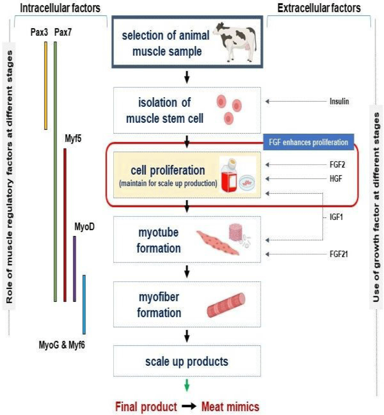
Myogenesis is a term used for the process leading from the development to the formation of SM tissue and involves MSC activation and proliferation (due to the expressions of muscle-specific genes and cell cycle-related factors) and the fusion of differentiating myoblasts into mature myofibers (under the direction of muscle regulatory factors). Myogenesis is mediated by MSCs and is responsible for the normal growth and repair of SM. This process is regulated by myogenic regulatory factors such as Pax3, Pax7, Myf5, MyoD, MyoG, and Myf6 [15,16] (Fig. 1).

jast-65-1-16-g1
Fig. 1. Cultured meat production based on myogenesis. MSC isolated from selected animals through biopsy or from animal tissues have different proliferation and differentiation properties. The roles of different growth and muscle regulatory factors during myogenesis are also included in the figure. FGF2, HGF, and IGF-1 enhance cell proliferation, and FGF21 aids myotube formation. FGF, fibroblast growth factor; HGF, hepatocyte growth factor; IGF, insulin-like growth factors; MSC, muscle satellite (stem) cell.
Download Original Figure

Furthermore, MSC culture [17,18] and the directed differentiation of pluripotent MSC [19,20] provide novel approaches to in vitro myogenesis. Several extracellular matrix (ECM) proteins such as fibromodulin (FMOD) [7,21], matrix gla protein [22], and dermatopontin (DPT) [23] have been reported to regulate myogenesis, and ECM contains molecules such as collagen, integrin, decorin, biglycan, DPT, FMOD, fibronectin, glycosaminoglycan, laminin, and dystrophin that provide structural support, cellular communication, and contribute much to the architectural maintenance of SM [5,24].

Cultured meat is an attractive option compared with regular meat as it is free from environmental pollution, ethical issues, and food security challenges [2527]. However, several issues, such as color, texture, flavor, and nutritional value, which all depend on protein and intramuscular fat contents, remain to be resolved [28,29]. Cattle, chickens, pigs, and fish have been reported to be suitable target species for biopsy leading to cultured meat production [25], and it has been established that a single biopsy may replace the killing of 20 animals [30].

The different types of MSC, namely, adult, pluripotent (PSCs; embryonic MSC), and induced pluripotent MSC) are used to produce cultured meat [31]. MSCs and adipocytes are required for initiating cultured meat production and, in combination, may contribute to achieving an original meat flavor and texture [12,32,33]. Porcine SM multipotent progenitor cells have a greater doubling capacity than MSCs, but they require expensive recombinant growth factors (GFs) and do not develop into SM fibers as effectively as MSCs [34]. Accordingly, MSCs are the most common source used for cultured meat production, and PSCs (embryonic stem cell [ESC] and iPSC) are considered a second option.

Technical difficulties related to culture media, such as GF, hormones, identification of animals (age and health), sampling of MSCs, consumer acceptance, and religious standpoints, pose considerable challenges to the establishment of viable large-scale in vitro meat production systems, and thus, more research is required before this meat culturing system can be implemented at industrial levels.

MSC proliferation and differentiation are influenced by many GFs such as fibroblast growth factor (FGF), insulin-like growth factors (IGF-1 and 2), transforming growth factor-beta (TGF-β) [35], platelet-derived growth factor (PDGF), and hepatocyte growth factor (HGF). GFs can manage growth signal responses during development and are highly active during cell proliferation and differentiation [16,3638]. The roles of several GFs and hormones during myogenesis for cultured meat production are summarized in Table 1. Three key hormone types related to the physiological system, that is, testosterone (promotes cellular growth and repair), growth hormone, and glucocorticoids, participate in cultured meat production [39]. The roles of GFs and expressions of muscle regulatory factors at different myogenesis stages are shown in Fig. 1, and a schematic of the process from MSC isolation to muscle fiber development is provided in Fig. 1.

Table 1. Growth factors, hormones, and other supplements and their functions during myogenesis and cultured meat production. Several GFs (IGFs, insulin, TGF-β, FGF, PDGF, and HGF) and hormones (thyroid hormones, testosterone, and glucocorticoids) and other supplements (heme proteins, transferrin, BMP, and mTOR) and their functions during cultured meat production and myogenesis are mentioned
Supplements Function Embryonic MSC Myogenic progenitor cells Tissue engineering References
IGF-1 ▪ Induces the proliferation and differentiation of cultured myoblast and promotes tissue growth and development V V V [4044]
IGF-2 ▪ Stimulate both proliferation and differentiation of myoblasts and MSC. V V [42,45]
TGF-β ▪ Helps in collagen type I synthesis in the ECM, and supporting myofiber development V V [4648]
FGF2 ▪ Activate myogenic progenitor cells and promotes proliferation and growth of mouse myoblasts.
▪ Upregulates Myf5 and MyoD.
▪ Increases the MSC proliferation.
V V V [4952]
FGF21 ▪ Involved in cell proliferation and differentiation. V [53,54]
Insulin ▪ Promotes MSC survival, self-renewal, and myoblast growth.
▪ Regulators of muscle protein synthesis and hypertrophy.
V V [43,5558]
HGF ▪ Activation of quiescent MSC in vivo upon muscle injury. V V [52,59,60]
Testosterone ▪ Increase in muscle fiber size and MSC number. [6163]
Transferrin ▪ Stimulate myogenesis and terminal differentiation in fast chicken muscle during embryonic development. V V V [64,65]
Heme protein (Hemoglobin and myoglobin) ▪ Provides typical color of meat and slightly metallic taste of beef.
▪ Increase the iron content in the product.
[66,67]
BMP ▪ Preserve the progenitor pool by inhibiting the Myf5 and MyoD expression. [68]
mTOR ▪ Regulate MSC activity and myogenesis by upregulating the expression of Pax7, Myf5, MyoD, and myogenin. [69,70]
Creatine ▪ Reduces muscle damage by decreasing the inflammatory response and oxidative stress, and activating MSC. Helps in myopathies. [71,72]
PDGF ▪ Increases and improve MSC proliferation. V V [68,73,74]
Thyroxine ▪ Helps to myotube in maintaining their differentiated state for a longer period. V V [57]

GF, growth factor; IGF, insulin-like growth factors; TGF, transforming growth factor; FGF, fibroblast growth factor; PDGF, platelet-derived growth factors; HGF, hepatocyte growth factor; BMP, bone morphogenetic protein; mTOR, mechanistic target of rapamycin; MSC, muscle satellite (stem) cell; ECM, extracellular matrix.

Download Excel Table

Types of GFs and hormones in culture medium critically maintain cell proliferation and differentiation, and it has been demonstrated that in their absence, in vitro cultured MSCs lose their stemness [4]. Thus, understanding the properties of MSCs and the effects of GFs and hormones that control myogenesis is critical for the mass production of cultured meat. Previous studies have shown that GFs and hormones are involved in myogenesis, and thus, in this review, we focus on how GFs and hormones exert their influences throughout the stages of myogenesis. The objective of this article was to summarize the roles of different GFs and hormones during MSC proliferation and differentiation for in vitro cultured meat production.

ROLES OF GROWTH FACTORS IN MYOGENESIS FOR CULTURED MEAT PRODUCTION

Fibroblast growth factors

FGFs are peptide hormones that belong to a large family of polypeptides locally present in developing tissues [75] and are secreted by liver, adipocytes, pancreas, and SM [76]. The FGF family is composed of 22 ligands that interact with four different FGF receptors, and FGF/FGFR signaling governs cell survival, proliferation, migration, differentiation, and tissue repair/regeneration [77]. FGF-21 is actively involved in cell proliferation and differentiation [53]. On the other hand, FGF-2 (basic FGF) is secreted by fibroblasts and has been used extensively for in vitro MSC culture. FGF-2 supplementation increased mouse myoblast growth by 25%, but the removal of FGF-2 from culture media increased the differentiation of myoblast cells. Also, the endogenous secretion of FGF-1 (acidic FGF) by MSCs may trigger cell proliferation and muscle regeneration [78,79]. On the other hand, FGF enhances cell proliferation and differentiation, which play important roles during the initial stage of myogenesis. In addition, FGF-2 also promotes tissue formation and repair by interacting with heparin sulfate cofactor [80].

Platelet-derived growth factor

PDGFs increase the production of three SM basement membrane components, namely, laminin (70%), fibronectin (30%), and type IV collagen (70%). PDGF has four known isoforms (PDGF A, B, C, and D), which are stabilized by intermolecular disulfide bonds. All four PDGF isoforms contain a highly conserved 100 amino acid GF domain [81]. PDGFs are powerful mitogens and affect a wide range of cells, including fibroblasts, smooth muscle cells, connective tissue, bone, and cartilage cells, and have been demonstrated to boost mouse myoblast proliferation. Thus, PDGFs are important components during the production of cell-cultured meat [82].

Insulin-like growth factors

IGFs play important roles in myogenesis and muscle regeneration by activating MSC proliferation, increasing protein synthesis, and promoting differentiation [83-86]. There are two IGF types, that is, IGF-1 and IGF-2. IGF-1 is a 70 aa long polypeptide that shares ~60% similarity with IGF-2 [87]. IGF-1 was reported to be actively involved in the proliferation and differentiation of myoblast cells [83,88]. The negative effect of myostatin (MSTN) was found to be compensated by the positive effect of IGF-1 during myogenesis [85]. IGF-1 promotes tissue growth and development, stimulates cell proliferation, and has anti-aging, anti-inflammatory, and antioxidant properties [41]. IGF-1 also has direct and indirect glucose-lowering effects, enhances free fatty acid oxidation in muscles, reduces the accumulation of free fatty acid in liver, and insulin signaling, reduces hepatic glucose output, and improves insulin sensitivity [41].

IGF-1 and 2 were reported to be equally effective at promoting chick embryonic myoblast differentiation and fusion [42], whereas IGF-2 was more effective than IGF-1 during turkey embryonic myoblast proliferation and differentiation. IGFs were found to stimulate in chicken and turkey MSC proliferation [89, 90], and treatment with IGF-1 and IGF-2 enhanced MSC growth.

When the effect of IGF-1 on chicken myoblast proliferation was examined IGF-1 dose-dependently increased myoblast numbers. Supplementation with 10, 100, or 1,000 ng/mL of IGF-1 for 24 h increased numbers by 9.5%, 63.1%, and 66%, respectively. The optimal concentration of IGF-1 for stimulating proliferation was found to be 100 ng/mL [40].

Myostatin

Myostatin (growth and development factor-8, MSTN) is present in SM, and muscle mass in MSTN-null mice was reported to be dramatically enhanced due to increased muscle fiber levels (hyperplasia) and sizes (hypertrophy) [91,92]. Furthermore, the double-muscled phenotype in cattle has been linked to the MSTN gene. Muscular hypertrophy in Belgian Blue double-muscled (BBDM) cattle is caused by an 11-base-pare deletion, and this naturally occurring mutation inactivates the MSTN gene and results in a huge increase in SM mass [93]. Cell culture experiments have also revealed that MSTN inhibits the proliferation and differentiation of cultured muscle cell lines and primary myogenic cells [93], which suggests siRNA1 and siRNA5 might reduce MSTN gene expression and boost sheep meat yield [94]. Some natural compounds [9597] and inhibitory peptides targeting MSTN [21,98], such as Ac-MIF1 and Ac-MIF2-NH2, have been reported to inhibit the effect of MSTN significantly and enhance cell proliferation [99]. Additionally, in vivo and in vitro data showed that FMOD prevents muscle aging by decreasing the activity of MSTN protein [92]. These findings indicate that MSTN gene sequence modification and MSTN inhibition are good strategies for the mass production of cultured meat from agricultural animals.

Transforming growth factor β

TGF-β1 is involved in muscle repair through MSC activation, connective tissue development, and immune response regulation [100]. TGF-β1 also attenuated MSC activation when used with HGF and when administered during early MSC activation (0–48 h) or the proliferative phase (48–96 h) TGF-β1 maintained or induced MSC quiescence, respectively, based on MyoD protein levels [101]. TGF-β1 can influence MSC proliferation and myogenesis, enhance mature SM mass, and regulate intramuscular fibrogenesis; it also plays critical roles in muscle formation. TGF-β1 was also demonstrated to restrict myogenesis in 2D cultures while increasing myogenesis in 3D cultures, which makes it a useful regulator of in vitro muscle tissue formation [102,103].

ROLE OF HORMONES IN MYOGENESIS FOR CULTURED MEAT PRODUCTION

Insulin

Insulin is a hormone that facilitates MSC survival and self-renewal in vitro [55]. Insulin maintains the differentiation power of myoblasts in vitro[104], and in combination with IGF-2, insulin augments the self-renewal property of MSCs [105], which is necessary for the initiation of culture meat production. Insulin works by activating its receptor and the PI3K/AKT cascade to promote cell survival and also activates integrin, which is required for cell survival during niche reformation after passage. In addition, insulin is needed for human ESC survival on E-cadherin-coated surfaces and in suspension, which suggests its involvement in cell-to-cell adhesion [55]. Insulin is a potent mitogen that promotes DNA synthesis and MSC proliferation and has a dose-dependent stimulatory impact on MSC differentiation and fusion. Insulin appears to promote myogenesis and differentiation by boosting the production of myogenin and myosin heavy chain (MHC) isoforms, and insulin administration thickens myotubes, which suggests a role in insulin-induced hypertrophy [106]. Accordingly, insulin is an important factor for cell survival and growth during the initial stage of cultured meat.

Testosterone

Testosterone primarily interacts with androgen receptors in SM to promote muscle growth. In SM, testosterone has a variety of ergogenic, anabolic, and anti-catabolic functions that dose-dependently contribute to muscle strength and hypertrophy [107,108]. In muscle, testosterone promotes muscle hypertrophy by stimulating protein synthesis and inhibiting protein degradation, and when these effects merge, testosterone stimulates muscle hypertrophy. Furthermore, the interaction between testosterone and intracellular androgen receptor modulates specific physiological signals [39,108]. Testosterone dose-dependently increases MSC numbers and muscle fiber sizes [61,62]. A significant increase in myoblasts numbers, which generated bigger myofibers, was observed when MSCs were treated with testosterone. However, the effect of testosterone on myogenesis in chickens and the mechanism by which testosterone regulates SM development remain unclear. In addition, testosterone significantly increased the cross-sectional area and density of myofibers in muscle [62].

Androgens and estrogens also significantly influence muscle physiology and metabolism and are involved in muscle mass development, maintenance, and repair [109]. Although estrogens and androgens both have favorable effects on muscle, androgens play a major role in the control of muscle physiology and also influence muscle mass by decreasing protein breakdown and autophagy [110]. Overall, estrogens and androgens regulate muscle formation and maintenance.

Glucocorticoids

Glucocorticoids, primarily cortisol, have substantial effects on human SM. Cortisol regulates SM energy homeostasis and metabolism [111,112]. Glucocorticoid receptors are present in the ECM of mammalian SM fibers and signal quickly through the mitogen-activated protein kinase pathway [113,114]. Dexamethasone is a synthetic glucocorticoid known for its anti-inflammatory and immunosuppressive properties and is used to suppress muscle degeneration and stabilize muscle strength [115]. At a concentration of 25 nM dexamethasone increased myogenic proliferation [116], and treatment with dexamethasone has also been reported to increase MSC myogenic differentiation marked by advanced sarcomere formation [105,116,117]. Also, the addition of dexamethasone to muscle cell cultures enhances myogenesis.

Thyroid hormones

SM is a primary target of thyroid hormones [118], which are important for stimulating MSC proliferation and differentiation and muscle recovery and myogenesis [119,120]. Thyroid hormones enhance the numbers and diameters of muscle fibers and also help determine muscle fiber distribution patterns [121], but high or low thyroid hormone levels promote muscle atrophy and impede muscle regeneration [121,122]. Peroxisome proliferator-activated receptor gamma coactivator 1-alpha (PPARGC-1α) is a major regulator of mitochondrial production and is significantly up-regulated by thyroid hormones. One study reported that the presence of thyroid hormones enhanced mitochondrial numbers, protein synthesis, and basal O2 consumption, adenosine triphosphate (ATP) turnover, and maximum respiratory capacity [118]. SM contains type 2 and 3 iodothyronine deiodinases (DIO2 and DIO3, respectively). DIO2 is a strictly controlled enzyme that catalyzes the outer-ring monodeiodination of the secreted prohormone tetraiodothyronine (T4) to produce active hormone tri-iodothyronine (T3), which can either stay in myocytes, signal through nuclear receptors, or exit cells and interact with the extracellular pool [121, 123]. Thyroid hormone signaling plays an important role in SM development and regeneration, and T3 signaling is crucial for SM development [123].

ANIMAL TISSUE CULTURE TECHNIQUES

Cell culture is a process whereby cells are grown in engineered environments, and animal cell culture is one of the most widely utilized research techniques in the biological sciences. Using tissue engineering techniques, in vitro meat production provides a unique means of producing meat without using animals [124]. The basic technology for in vitro meat production involves cultivating muscle tissue in a bioreactor. Starter cells for meat production can be obtained by live animal biopsy and then cultured in vitro [124,125]. The development of a basic culture medium has allowed the growth and differentiation of various cells studied under various conditions [126]. Interestingly, optimal in vitro culture temperature, pH, CO2, O2, osmolality, and nutrition requirements may not be identical to in vivo conditions. Furthermore, sterile cultivation conditions, a constant supply of nutrients, and complex incubation conditions are required [127]. Although nutrients, adhesion proteins, and GFs are abundant in fetal bovine serum (FBS), the exploration of alternative media and the creation of serum-free media formulations have attracted global interest due to shortcomings in in vitro data quality and repeatability and animal welfare issues. The majority of studies on the topic indicate that chemically specific serum-free mediums are the ultimate goal [128, 129]. Research efforts continue to be directed toward the development of optimal culture conditions that improve the efficacy of cultured meat production.

GFs are signaling molecules that stimulate growth, proliferation, and differentiation by triggering signaling cascades after binding to their receptors. GFs are important cell culture media components and provide signals for growth, structural stability, and cell-to-cell communication. However, GFs are expensive, and thus, some alternative means of synthesizing GFs are needed. Important GFs can be sourced from the genomes of different species. Similar sequences can be found by searching publicly available genome databases and synthesized or purchased from an available vendor. Recombinant DNA technology can then be used to insert sequences of interest into bacterial systems. Using this strategy, active GFs, such as FGF-2, IGFs, PDGF-BB, and TGF-β1 were effectively developed in Escherichia coli, and soluble GFs from cattle, chickens, and fish were produced [130].

CONCLUSION

GFs and hormones are important media components that regulate MSCs and improve cultured meat production. After binding to their receptors, these factors deliver the signals for growth, structural stability, cell-to-cell communication, proliferation, and differentiation. However, they are expensive media components, and thus, optimization and cheaper ways of synthesizing GFs and hormones are required. This review describes the importance of the regulatory roles played by different GFs (FGF-2, IGF-1, PDGF-BB, and TGF-1) and hormones (insulin, testosterone, glucocorticoids, dexamethasone, and thyroid hormones) during myogenesis and cultured meat production.

Competing interests

No potential conflict of interest relevant to this article was reported.

Funding sources

This work was supported by the Korea Institute of Planning and Evaluation for Technology in Food, Agriculture, Forestry (IPET) through High Value-added Food Technology Development Program funded by Ministry of Agriculture, Food and Rural Affairs (MAFRA)(321026-05 and 322008-5). This research was also supported by the Basic Science Research Program through the National Research Foundation of Korea (NRF) funded by the Ministry of Education (2020R1A6A1A03044512).

Acknowledgements

Not applicable.

Availability of data and material

Upon reasonable request, the datasets of this study can be available from the corresponding author.

Authors’ contributions

Conceptualization: Ahmad SS, Chun HJ.

Data curation: Ahmad K, Shaikh S, Lim JH, Ali S.

Formal analysis: Ahmad K, Shaikh S, Lim JH, Ali S, Han SS, Hur SJ, Sohn JH, Lee EJ, Choi I.

Writing - original draft: Lee EJ, Choi I.

Writing - review & editing: Ahmad SS, Chun HJ, Ahmad K, Shaikh S, Lim JH, Ali S, Han SS, Hur SJ, Sohn JH, Lee EJ, Choi I.

Ethics approval and consent to participate

This article does not require IRB/IACUC approval because there are no human and animal participants.

REFERENCES

1.

Chriki S, Hocquette JF. The myth of cultured meat: a review. Front Nutr. 2020; 7:7

2.

Handral HK, Tay SH, Chan WW, Choudhury D. 3D Printing of cultured meat products. Crit Rev Food Sci Nutr. 2022; 62:272-81

3.

Singh S, Yap WS, Ge XY, Min VLX, Choudhury D. Cultured meat production fuelled by fermentation. Trends Food Sci Technol. 2022; 120:48-58

4.

Machida S, Spangenburg EE, Booth FW. Primary rat muscle progenitor cells have decreased proliferation and myotube formation during passages. Cell Prolif. 2004; 37:267-77

5.

Ahmad K, Lim JH, Lee EJ, Chun HJ, Ali S, Ahmad SS, et al. Extracellular matrix and the production of cultured meat. Foods. 2021; 10:3116

6.

Piochi M, Micheloni M, Torri L. Effect of informative claims on the attitude of Italian consumers towards cultured meat and relationship among variables used in an explicit approach. Food Res Int. 2022; 151:110881

7.

Lee EJ, Jan AT, Baig MH, Ahmad K, Malik A, Rabbani G, et al. Fibromodulin and regulation of the intricate balance between myoblast differentiation to myocytes or adipocyte-like cells. FASEB J. 2018; 32:768-81

8.

Choi KH, Yoon JW, Kim M, Lee HJ, Jeong J, Ryu M, et al. Muscle stem cell isolation and in vitro culture for meat production: a methodological review. Compr Rev Food Sci Food Saf. 2021; 20:429-57

9.

Johnson SE. NC1184: molecular mechanisms regulating skeletal muscle growth and differentiation. J Anim Sci. 2022; 100:skac229

10.

Jan AT, Lee EJ, Ahmad S, Choi I. Meeting the meat: delineating the molecular machinery of muscle development. J Anim Sci Technol. 2016; 58:18

11.

Gastaldello A, Giampieri F, De Giuseppe R, Grosso G, Baroni L, Battino M. The rise of processed meat alternatives: a narrative review of the manufacturing, composition, nutritional profile and health effects of newer sources of protein, and their place in healthier diets. Trends Food Sci Technol. 2022; 127:263-71

12.

Shaikh S, Lee E, Ahmad K, Ahmad SS, Chun H, Lim J, et al. Cell types used for cultured meat production and the importance of myokines. Foods. 2021; 10:2318

13.

Verbruggen S, Luining D, van Essen A, Post MJ. Bovine myoblast cell production in a microcarriers-based system. Cytotechnology. 2018; 70:503-12

14.

Manassi CF, de Souza SS, Hassemer GS, Sartor S, Lima CMG, Miotto M, et al. Functional meat products: trends in pro-, pre-, syn-, para- and post-biotic use. Food Res Int. 2022; 154:111035

15.

Zuk PA, Benhaim P, Hedrick MH. Stem cells from adipose tissue.In In: Lanza R, Gearhart J, Hogan B, Melton D, Pedersen R, Thomson J, West M, editors.editors Handbook of stem cells. Amsterdam: Academic Press. 2004; p p. 425-47

16.

Ahmad SS, Ahmad K, Lee EJ, Lee YH, Choi I. Implications of insulin-like growth factor-1 in skeletal muscle and various diseases. Cells. 2020; 9:1773

17.

Ding S, Wang F, Liu Y, Li S, Zhou G, Hu P. Characterization and isolation of highly purified porcine satellite cells. Cell Death Discov. 2017; 3:17003

18.

Ding S, Swennen GNM, Messmer T, Gagliardi M, Molin DGM, Li C, et al. Maintaining bovine satellite cells stemness through p38 pathway. Sci Rep. 2018; 8:10808

19.

Bogliotti YS, Wu J, Vilarino M, Okamura D, Soto DA, Zhong C, et al. Efficient derivation of stable primed pluripotent embryonic stem cells from bovine blastocysts. Proc Natl Acad Sci USA. 2018; 115:2090-5

20.

Choi KH, Lee DK, Kim SW, Woo SH, Kim DY, Lee CK. Chemically defined media can maintain pig pluripotency network in vitro. Stem Cell Reports. 2019; 13:221-34

21.

Lee EJ, Jan AT, Baig MH, Ashraf JM, Nahm SS, Kim YW, et al. Fibromodulin: a master regulator of myostatin controlling progression of satellite cells through a myogenic program. FASEB J. 2016; 30:2708-19

22.

Ahmad S, Jan AT, Baig MH, Lee EJ, Choi I. Matrix gla protein: an extracellular matrix protein regulates myostatin expression in the muscle developmental program. Life Sci. 2017; 172:55-63

23.

Kim T, Ahmad K, Shaikh S, Jan AT, Seo MG, Lee EJ, et al. Dermatopontin in skeletal muscle extracellular matrix regulates myogenesis. Cells. 2019; 8:332

24.

Ahmad K, Lee EJ, Shaikh S, Kumar A, Rao KM, Park SY, et al. Targeting integrins for cancer management using nanotherapeutic approaches: recent advances and challenges. Semin Cancer Biol. 2021; 69:325-36

25.

Bomkamp C, Skaalure SC, Fernando GF, Ben-Arye T, Swartz EW, Specht EA. Scaffolding biomaterials for 3D cultivated meat: prospects and challenges. Adv Sci. 2022; 9:2102908

26.

Zhang J, Chen Q, Kaplan DL, Wang Q. High-moisture extruded protein fiber formation toward plant-based meat substitutes applications: science, technology, and prospect. Trends Food Sci Technol. 2022; 128:202-16

27.

Levi S, Yen FC, Baruch L, Machluf M. Scaffolding technologies for the engineering of cultured meat: towards a safe, sustainable, and scalable production. Trends Food Sci Technol. 2022; 126:13-25

28.

Listrat A, Lebret B, Louveau I, Astruc T, Bonnet M, Lefaucheur L, et al. How muscle structure and composition influence meat and flesh quality. Sci World J. 2016; 2016:3182746

29.

Fish KD, Rubio NR, Stout AJ, Yuen JSK, Kaplan DL. Prospects and challenges for cell-cultured fat as a novel food ingredient. Trends Food Sci Technol. 2020; 98:53-67

30.

Melzener L, Verzijden KE, Buijs AJ, Post MJ, Flack JE. Cultured beef: from small biopsy to substantial quantity. J Sci Food Agric. 2021; 101:7-14

31.

Reiss J, Robertson S, Suzuki M. Cell sources for cultivated meat: applications and considerations throughout the production workflow. Int J Mol Sci. 2021; 22:7513

32.

Brack AS, Rando TA. Tissue-specific stem cells: lessons from the skeletal muscle satellite cell. Cell Stem Cell. 2012; 10:504-14

33.

Yin H, Price F, Rudnicki MA. Satellite cells and the muscle stem cell niche. Physiol Rev. 2013; 93:23-67

34.

Wilschut KJ, Jaksani S, Van Den Dolder J, Haagsman HP, Roelen BAJ. Isolation and characterization of porcine adult muscle-derived progenitor cells. J Cell Biochem. 2008; 105:1228-39

35.

White TP. Satellite cell and growth factor involvement in skeletal muscle growth. Med Sci Sports Exerc. 1989; 21:S30

36.

Lee EJ, Pokharel S, Jan AT, Huh S, Galope R, Lim JH, et al. Transthyretin: a transporter protein essential for proliferation of myoblast in the myogenic program. Int J Mol Sci. 2017; 18:115

37.

Vlasova-St. Louis I, Bohjanen PR. Post-transcriptional regulation of cytokine and growth factor signaling in cancer. Cytokine Growth Factor Rev. 2017; 33:83-93

38.

Chen FM, Zhang M, Wu ZF. Toward delivery of multiple growth factors in tissue engineering. Biomaterials. 2010; 31:6279-308

39.

Kraemer WJ, Ratamess NA, Hymer WC, Nindl BC, Fragala MS. Growth hormone(s), testosterone, insulin-like growth factors, and cortisol: roles and integration for cellular development and growth with exercise. Front Endocrinol. 2020; 11:33

40.

Yu M, Wang H, Xu Y, Yu D, Li D, Liu X, et al. Insulin-like growth factor-1 (IGF-1) promotes myoblast proliferation and skeletal muscle growth of embryonic chickens via the PI3K/Akt signalling pathway. Cell Biol Int. 2015; 39:910-22

41.

Halmos T, Suba I. The physiological role of growth hormone and insulin-like growth factors. Orv Hetil. 2019; 160:1774-83

42.

Schmid C, Steiner T, Froesch ER. Preferential enhancement of myoblast differentiation by insulin-like growth factors (IGF I and IGF II) in primary cultures of chicken embryonic cells. FEBS Lett. 1983; 161:117-21

43.

Ewton DZ, Florini JR. Effects of the somatomedins and insulin on myoblast differentiation in vitro. Dev Biol. 1981; 86:31-9

44.

Huang YC, Dennis RG, Larkin L, Baar K. Rapid formation of functional muscle in vitro using fibrin gels. J Appl Physiol. 2005; 98:706-13

45.

Oksbjerg N, Gondret F, Vestergaard M. Basic principles of muscle development and growth in meat-producing mammals as affected by the insulin-like growth factor (IGF) system. Domest Anim Endocrinol. 2004; 27:219-40

46.

Massagué J, Cheifetz S, Endo T, Nadal-Ginard B. Type beta transforming growth factor is an inhibitor of myogenic differentiation. Proc Natl Acad Sci USA. 1986; 83:8206-10

47.

Weist MR, Wellington MS, Bermudez JE, Kostrominova TY, Mendias CL, Arruda EM, et al. TGF-β1 enhances contractility in engineered skeletal muscle. J Tissue Eng Regen Med. 2013; 7:562-71

48.

Amit M, Shariki C, Margulets V, Itskovitz-Eldor J. Feeder layer- and serum-free culture of human embryonic stem cells. Biol Reprod. 2004; 70:837-45

49.

Rando TA, Blau HM. Primary mouse myoblast purification, characterization, and transplantation for cell-mediated gene therapy. J Cell Biol. 1994; 125:1275-87

50.

Xu C, Rosler E, Jiang J, Lebkowski JS, Gold JD, O’Sullivan C, et al. Basic fibroblast growth factor supports undifferentiated human embryonic stem cell growth without conditioned medium. Stem Cells. 2005; 23:315-23

51.

Düsterhöft S, Pette D. Evidence that acidic fibroblast growth factor promotes maturation of rat satellite-cell-derived myotubes in vitro. Differentiation. 1999; 65:161-9

52.

Schiaffino S, Mammucari C. Regulation of skeletal muscle growth by the IGF1-Akt/PKB pathway: insights from genetic models. Skelet Muscle. 2011; 1:4

53.

Huang Z, Xu A, Cheung BMY. The potential role of fibroblast growth factor 21 in lipid metabolism and hypertension. Curr Hypertens Rep. 2017; 19:28

54.

Liu X, Wang Y, Zhao S, Li X. Fibroblast growth factor 21 promotes C2C12 cells myogenic differentiation by enhancing cell cycle exit. BioMed Res Int. 2017; 2017:1648715

55.

Godoy-Parejo C, Deng C, Liu W, Chen G. Insulin stimulates PI3K/AKT and cell adhesion to promote the survival of individualized human embryonic stem cells. Stem Cells. 2019; 37:1030-41

56.

Tipton KD, Wolfe RR. Exercise, protein metabolism, and muscle growth. Int J Sport Nutr Exerc Metab. 2001; 11:109-32

57.

Kumegawa M, Ikeda E, Hosoda S, Takuma T. In vitro effects of thyroxine and insulin on myoblasts from chick embryo skeletal muscle. Dev Biol. 1980; 79:493-9

58.

Florini JR, Ewton DZ. Insulin acts as a somatomedin analog in stimulating myoblast growth in serum-free medium. In Vitro. 1981; 17:763-8

59.

Miller KJ, Thaloor D, Matteson S, Pavlath GK. Hepatocyte growth factor affects satellite cell activation and differentiation in regenerating skeletal muscle. Am J Physiol Cell Physiol. 2000; 278:C174-81

60.

Allen RE, Sheehan SM, Taylor RG, Kendall TL, Rice GM. Hepatocyte growth factor activates quiescent skeletal muscle satellite cells in vitro. J Cell Physiol. 1995; 165:307-12

61.

Sinha-Hikim I, Roth SM, Lee MI, Bhasin S. Testosterone-induced muscle hypertrophy is associated with an increase in satellite cell number in healthy, young men. Am J Physiol Endocrinol Metab. 2003; 285:E197-205

62.

Herbst KL, Bhasin S. Testosterone action on skeletal muscle. Curr Opin Clin Nutr Metab Care. 2004; 7:271-7

63.

Kimura I, Hasegawa T, Ozawa E. Indispensability of iron-bound chick transferrin for chick myogenesis in vitro: (myogenesis/transfrrin/iron). Dev Growth Differ. 1982; 24:369-80

64.

Mescher AL, Munaim SI. Transferrin and the growth-promoting effect of nerves. Int Rev Cytol. 1988; 110:1-26

65.

Matsuda R, Spector D, Micou-Eastwood J, Strohman RC. There is selective accumulation of a growth factor in chicken skeletal muscle. II. Transferrin accumulation in dystrophic fast muscle. Dev Biol. 1984; 103:276-84

66.

Simsa R, Yuen J, Stout A, Rubio N, Fogelstrand P, Kaplan DL. Extracellular heme proteins influence bovine myosatellite cell proliferation and the color of cell-based meat. Foods. 2019; 8:521

67.

Suman SP, Joseph P. Myoglobin chemistry and meat color. Annu Rev Food Sci Technol. 2013; 4:79-99

68.

Syverud BC, VanDusen KW, Larkin LM. Growth factors for skeletal muscle tissue engineering. Cells Tissues Organs. 2016; 202:169-79

69.

Zhang P, Liang X, Shan T, Jiang Q, Deng C, Zheng R, et al. mTOR is necessary for proper satellite cell activity and skeletal muscle regeneration. Biochem Biophys Res Commun. 2015; 463:102-8

70.

Rion N, Castets P, Lin S, Enderle L, Reinhard JR, Eickhorst C, et al. mTOR controls embryonic and adult myogenesis via mTORC1. Development. 2019; 146:dev172460

71.

Gualano B, Roschel H, Lancha AH, Brightbill CE, Rawson ES. In sickness and in health: the widespread application of creatine supplementation. Amino Acids. 2012; 43:519-29

72.

Kim J, Lee J, Kim S, Yoon D, Kim J, Sung DJ. Role of creatine supplementation in exercise-induced muscle damage: a mini review. J Exerc Rehabil. 2015; 11:244-50

73.

Yablonka-Reuveni Z, Balestreri TM, Bowen-Pope DF. Regulation of proliferation and differentiation of myoblasts derived from adult mouse skeletal muscle by specific isoforms of PDGF. J Cell Biol. 1990; 111:1623-9

74.

Maley MAL, Davies MJ, Grounds MD. Extracellular matrix, growth factors, genetics: their influence on cell proliferation and myotube formation in primary cultures of adult mouse skeletal muscle. Exp Cell Res. 1995; 219:169-79

75.

McFarland DC. Influence of growth factors on poultry myogenic satellite cells. Poult Sci. 1999; 78:747-58

76.

Cheung BMY, Deng HB. Fibroblast growth factor 21: a promising therapeutic target in obesity-related diseases. Expert Rev Cardiovasc Ther. 2014; 12:659-66

77.

Mossahebi-Mohammadi M, Quan M, Zhang JS, Li X. FGF signaling pathway: a key regulator of stem cell pluripotency. Front Cell Dev Biol. 2020; 8:79

78.

Groux-Muscatelli B, Bassaglia Y, Barritault D, Caruelle JP, Gautron J. Proliferating satellite cells express acidic fibroblast growth factor during in vitro myogenesis. Dev Biol. 1990; 142:380-5

79.

Shahini A, Vydiam K, Choudhury D, Rajabian N, Nguyen T, Lei P, et al. Efficient and high yield isolation of myoblasts from skeletal muscle. Stem Cell Res. 2018; 30:122-9

80.

Koledova Z, Sumbal J, Rabata A, de La Bourdonnaye G, Chaloupkova R, Hrdlickova B, et al. Fibroblast growth factor 2 protein stability provides decreased dependence on heparin for induction of FGFR signaling and alters ERK signaling dynamics. Front Cell Dev Biol. 2019; 7:331

81.

Breitkopf K, Roeyen C, Sawitza I, Wickert L, Floege J, Gressner AM. Expression patterns of PDGF-A, -B, -C and -D and the PDGF-receptors α and β in activated rat hepatic stellate cells (HSC). Cytokine. 2005; 31:349-57

82.

Albrecht DE, Tidball JG. Platelet-derived growth factor-stimulated secretion of basement membrane proteins by skeletal muscle occurs by tyrosine kinase-dependent and -independent pathways. J Biol Chem. 1997; 272:2236-44

83.

Coleman ME, DeMayo F, Yin KC, Lee HM, Geske R, Montgomery C, et al. Myogenic vector expression of insulin-like growth factor I stimulates muscle cell differentiation and myofiber hypertrophy in transgenic mice. J Biol Chem. 1995; 270:12109-16

84.

Lawlor MA, Rotwein P. Insulin-like growth factor-mediated muscle cell survival: central roles for Akt and cyclin-dependent kinase inhibitor p21. Mol Cell Biol. 2000; 20:8983-95

85.

Valdés JA, Flores S, Fuentes EN, Osorio-Fuentealba C, Jaimovich E, Molina A. IGF-1 induces IP3-dependent calcium signal involved in the regulation of myostatin gene expression mediated by NFAT during myoblast differentiation. J Cell Physiol. 2013; 228:1452-63

86.

Zanou N, Gailly P. Skeletal muscle hypertrophy and regeneration: interplay between the myogenic regulatory factors (MRFs) and insulin-like growth factors (IGFs) pathways. Cell Mol Life Sci. 2013; 70:4117-30

87.

Le Roith D. Seminars in medicine of the Beth Israel Deaconess Medical Center. Insulin-like growth factors. N Engl J Med. 1997; 336:633-40

88.

Noguchi S. The biological function of insulin-like growth factor-I in myogenesis and its therapeutic effect on muscular dystrophy. Acta Myol. 2005; 24:115-8

89.

McFarland DC, Pesall JE, Gilkerson KK. The influence of growth factors on turkey embryonic myoblasts and satellite cells in vitro. Gen Comp Endocrinol. 1993; 89:415-24

90.

Duclos MJ, Wilkie RS, Goddard C. Stimulation of DNA synthesis in chicken muscle satellite cells by insulin and insulin-like growth factors: evidence for exclusive mediation by a type-I insulin-like growth factor receptor. J Endocrinol. 1991; 128:35-42

91.

McPherron AC, Lawler AM, Lee SJ. Regulation of skeletal muscle mass in mice by a new TGF-p superfamily member. Nature. 1997; 387:83-90

92.

Lee EJ, Ahmad SS, Lim JH, Ahmad K, Shaikh S, Lee YS, et al. Interaction of fibromodulin and myostatin to regulate skeletal muscle aging: an opposite regulation in muscle aging, diabetes, and intracellular lipid accumulation. Cells. 2021; 10:2083

93.

Grobet L, Martin LJR, Poncelet D, Pirottin D, Brouwers B, Riquet J, et al. A deletion in the bovine myostatin gene causes the double–muscled phenotype in cattle. Nat Genet. 1997; 17:71-4

94.

Lu J, Sun D, Xu L, Lu G, Zhao F, Wei C, et al. Selection of an effective small interference RNA to silence myostatin gene expression in sheep fibroblast cells. Biochem Genet. 2012; 50:838-47

95.

Ahmad SS, Ahmad K, Lee EJ, Shaikh S, Choi I. Computational identification of dithymoquinone as a potential inhibitor of myostatin and regulator of muscle mass. Molecules. 2021; 26:5407

96.

Ali S, Ahmad K, Shaikh S, Lim JH, Chun HJ, Ahmad SS, et al. Identification and evaluation of traditional Chinese medicine natural compounds as potential myostatin inhibitors: an in silico approach. Molecules. 2022; 27:4303

97.

Lee EJ, Shaikh S, Ahmad K, Ahmad SS, Lim JH, Park S, et al. Isolation and characterization of compounds from Glycyrrhiza uralensis as therapeutic agents for the muscle disorders. Int J Mol Sci. 2021; 22:876

98.

Baig MH, Ahmad K, Moon JS, Park SY, Ho Lim J, Chun HJ, et al. Myostatin and its regulation: a comprehensive review of myostatin inhibiting strategies. Front Physiol. 2022; 13:876078

99.

Lee EJ, Shaikh S, Baig MH, Park SY, Lim JH, Ahmad SS, et al. MIF1 and MIF2 myostatin peptide inhibitors as potent muscle mass regulators. Int J Mol Sci. 2022; 23:4222

100.

Delaney K, Kasprzycka P, Ciemerych MA, Zimowska M. The role of TGF-β1 during skeletal muscle regeneration. Cell Biol Int. 2017; 41:706-15

101.

Rathbone CR, Yamanouchi K, Chen XK, Nevoret-Bell CJ, Rhoads RP, Allen RE. Effects of transforming growth factor-beta (TGF-β1) on satellite cell activation and survival during oxidative stress. J Muscle Res Cell Motil. 2011; 32:99-109

102.

Krieger J, Park BW, Lambert CR, Malcuit C. 3D skeletal muscle fascicle engineering is improved with TGF-β1 treatment of myogenic cells and their co-culture with myofibroblasts. PeerJ. 2018; 6e4939

103.

Ben-Arye T, Levenberg S. Tissue engineering for clean meat production. Front Sustain Food Syst. 2019; 3:46

104.

Mandel JL, Pearson ML. Insulin stimulates myogenesis in a rat myoblast line. Nature. 1974; 251:618-20

105.

Dodson MV, Allen RE, Hossner KL. Ovine somatomedin, multiplication-stimulating activity, and insulin promote skeletal muscle satellite cell proliferation in vitro. Endocrinology. 1985; 117:2357-63

106.

Rhoads RP, Baumgard LH, El-Kadi SW, Zhao LD. PHYSIOLOGY AND ENDOCRINOLOGY SYMPOSIUM: roles for insulin-supported skeletal muscle growth. J Anim Sci. 2016; 94:1791-802

107.

Kraemer WJ, Ratamess NA, Nindl BC. Recovery responses of testosterone, growth hormone, and IGF-1 after resistance exercise. J Appl Physiol. 2017; 122:549-58

108.

Vingren JL, Kraemer WJ, Ratamess NA, Anderson JM, Volek JS, Maresh CM. Testosterone physiology in resistance exercise and training: the up-stream regulatory elements. Sports Med. 2010; 40:1037-53

109.

Carson JA, Manolagas SC. Effects of sex steroids on bones and muscles: similarities, parallels, and putative interactions in health and disease. Bone. 2015; 80:67-78

110.

Rossetti ML, Steiner JL, Gordon BS. Androgen-mediated regulation of skeletal muscle protein balance. Mol Cell Endocrinol. 2017; 447:35-44

111.

Munck A, Guyre PM, Holbrook NJ. Physiological functions of glucocorticoids in stress and their relation to pharmacological actions. Endocr Rev. 1984; 5:25-44

112.

Sheffield-Moore M, Urban RJ. An overview of the endocrinology of skeletal muscle. Trends Endocrinol Metab. 2004; 15:110-5

113.

Boncompagni S, Arthurton L, Akujuru E, Pearson T, Steverding D, Protasi F, et al. Membrane glucocorticoid receptors are localised in the extracellular matrix and signal through the MAPK pathway in mammalian skeletal muscle fibres. J Physiol. 2015; 593:2679-92

114.

Pérez MHA, Cormack J, Mallinson D, Mutungi G. A membrane glucocorticoid receptor mediates the rapid/non-genomic actions of glucocorticoids in mammalian skeletal muscle fibres. J Physiol. 2013; 591:5171-85

115.

Lin JW, Huang YM, Chen YQ, Chuang TY, Lan TY, Liu YW, et al. Dexamethasone accelerates muscle regeneration by modulating kinesin-1-mediated focal adhesion signals. Cell Death Discov. 2021; 7:35

116.

Syverud BC, VanDusen KW, Larkin LM. Effects of dexamethasone on satellite cells and tissue engineered skeletal muscle units. Tissue Eng Part A. 2016; 22:480-9

117.

Guerriero V, Florini JR. Dexamethasone effects on myoblast proliferation and differentiation. Endocrinology. 1980; 106:1198-202

118.

Lesmana R, Sinha RA, Singh BK, Zhou J, Ohba K, Wu Y, et al. Thyroid hormone stimulation of autophagy is essential for mitochondrial biogenesis and activity in skeletal muscle. Endocrinology. 2016; 157:23-38

119.

Bloise FF, Cordeiro A, Ortiga-Carvalho TM. Role of thyroid hormone in skeletal muscle physiology. J Endocrinol. 2018; 236:R57-68

120.

Bloise FF, Oliveira TS, Cordeiro A, Ortiga-Carvalho TM. Thyroid hormones play role in Sarcopenia and Myopathies. Front Physiol. 2018; 9:560

121.

Salvatore D, Simonides WS, Dentice M, Zavacki AM, Larsen PR. Thyroid hormones and skeletal muscle—new insights and potential implications. Nat Rev Endocrinol. 2014; 10:206-14

122.

Martín AI, Priego T, López-Calderón A. Hormones and muscle atrophy. Adv Exp Med Biol. 2018; 1088:207-33

123.

Lee EJ, Shaikh S, Choi D, Ahmad K, Baig MH, Lim JH, et al. Transthyretin maintains muscle homeostasis through the novel shuttle pathway of thyroid hormones during myoblast differentiation. Cells. 2019; 8:1565

124.

Bhat ZF, Kumar S, Bhat HF. In vitro meat: a future animal-free harvest. Crit Rev Food Sci Nutr. 2017; 57:782-9

125.

Bhat ZF, Bhat H. Animal-free meat biofabrication. Am J Food Technol. 2011; 6:441-59

126.

Bhatia S, Goli D. Introduction to pharmaceutical biotechnology, volume 1: basic techniques and concepts. Bristol: IOP. 2018

127.

Verma A. Animal tissue culture: principles and applications.In In: Verma AS, Singh A, editors.editors Animal biotechnology: models in discovery and translation. Amsterdam: Academic Press. 2014; p p. 211-31

128.

van der Valk J, Bieback K, Buta C, Cochrane B, Dirks WG, Fu J, et al. Fetal bovine serum (FBS): past – present – future. Altern Anim Exp. 2018; 35:99-118

129.

Bauman E, Granja PL, Barrias CC. Fetal bovine serum-free culture of endothelial progenitor cells—progress and challenges. J Tissue Eng Regen Med. 2018; 12:1567-78

130.

Venkatesan M, Semper C, Skrivergaar S, DiLeo R, Mesa N, Rasmussen MK, et al. Recombinant production of growth factors for application in cell culture. iScience. 2022; 25:105054