

INTRODUCTION
MIT-001, also known as NecroX-7, is a novel mitochondria-targeted reactive oxygen species (ROS) scavenger developed by MitoImmune Therapeutics Inc., and has been shown to protect mammalian cells from oxidative damage [1]. MIT-001 inhibits ROS generation and calcium accumulation in mitochondria, thereby suppressing the release of molecules associated with cell damage and apoptosis [2,3]. Until recently, the activation of ROS or mitochondrial protection by MIT-001 in vitrified-warmed bovine blastocysts had not been investigated.
Embryo cryopreservation offers flexibility in gamete management, which is crucial for livestock production and infertility treatment. It is especially advantageous for preserving and transporting livestock genetic resources, thereby improving reproductive potential, facilitating genetic enhancement, and contributing to biodiversity conservation [4]. However, cryopreserved embryos often exhibit reduced viability and quality compared with fresh embryos because of intracellular ice crystal formation during vitrification [5]. Additionally, the use of cryoprotectants prior to vitrification, along with optimal culture conditions (e.g., temperature), significantly affects blastocyst viability [6]. Post-warming re-expansion is a critical indicator of the recovery and survival of cryopreserved embryos [7].
During early embryonic development, mitochondrial function and quantity are tightly regulated by a quality control system that plays a crucial role in maintaining mitochondrial homeostasis. Mitochondria play a vital role in ROS balance during oxidative phosphorylation, and their dysfunction results in the harmful accumulation of superoxide anions within cells. Interestingly, vitrification and warming have been linked to elevated ROS levels and mitochondrial impairment in various species, including pigs, cattle, and humans. Mitochondrial dysfunction can have profound implications for embryo viability, as mitochondria are critical not only for ATP production but also for regulating apoptosis and controlling ROS generation [8]. Currently, there is no study on the protective effects of ROS removal by MIT-001 in vitrified-warmed bovine blastocysts, and recent research suggests that mitochondrial dysfunction and low membrane potential after vitrification/warming are hallmark features of cryoinjury [8,9]. Therefore, we investigated the effect of MIT-001, a potential antioxidant agent targeting mitochondria, on the survival rate of cryopreserved bovine blastocysts in vitro.
Additionally, mitochondrial fission is necessary for mitochondrial quality control, leading to blastocyst developmental competence in porcine embryos during in vitro culture (IVC) progression [10]. In mammals, it is triggered by dynamin-related protein 1 (DRP1), which forms ring-like structures around the constriction points of dividing mitochondria [11]. However, in contrast to its role in cell survival, DRP1-induced mitochondrial fragmentation contributes to the release of proapoptotic factors during apoptosis [12]. Indeed, excessive mitochondrial fission is linked to apoptosis, and modulation of DRP1 is a promising strategy for improving early embryonic development.
p38 mitogen-activated protein kinases (MAPK) are activated in response to various cellular stressors, including oxidative stress, cytokines, and DNA damage, often culminating in apoptosis and altered cell differentiation [13]. Cortical filamentous actin (F-actin) stabilization plays a critical role in blastocyst development and re-expansion after vitrification and warming. Studies on murine and porcine preimplantation embryos have indicated that p38 MAPK activation stabilizes F-actin and mitochondrial function, thereby improving embryo development and survival [14,15].
Given the effects of ROS production and mitochondrial dysfunction on cryoinjury, we evaluated MIT-001’s protective effects of against vitrification and warming. Specifically, we assessed the blastocyst viability, cytoskeletal integrity, mitochondrial function, re-expansion potential, and p38 MAPK activation. The goal was to determine whether MIT-001 could mitigate ROS-induced damage, preserve cytoskeletal structure, and improve overall blastocyst quality, offering new insights into enhancing cryopreservation outcomes in assisted reproductive technology and livestock genetic resource conservation.
MATERIALS AND METHODS
Unless otherwise stated, all chemicals utilized in this study were obtained from Sigma-Aldrich. Bovine ovaries were obtained from a local slaughterhouse (Gimhae, Gyeongnam, Korea). No experiments were performed on live animals.
Bovine embryos were produced using the methods described by Jeong et al. [16]. for embryo development and vitrification-warming. Blastocysts generated through the IVC process were classified into four stages based on their average diameter (Supplementary Fig. 1, Early: 150 μm, Mid: 161 μm, Late: 175 μm and Expanded: 222 μm), ranging from the earliest to the fully expanded stage. Survival was determined based on re-expansion rates after 24 h of post-warming culture. The survival of blastocysts after cryopreservation and post-warming was evaluated according to these criteria, including the average diameter of each group.
Bovine blastocysts (n = 180) were assigned to three groups based on the MIT-001 (1 µM, Soon Ha Kim, Mitoimmune Therapeutics) treatment period: (I) warming only (WARM), (II) vitrification only (VITR), and (III) both vitrification and warming (VITR-WARM), compared with the Non-treated control group. After MIT-001 treatment, surviving bovine blastocysts were used for experimental analysis (Supplementary Fig. 2).
Bovine ovaries were collected from a local slaughterhouse and transported to the lab in 0.9% saline solution, supplemented with 75 μg/mL potassium penicillin G sodium salt, maintained at 36°C–38°C. Cumulus-oocyte complexes (COCs) were aspirated from follicles measuring 3–6 mm in diameter using a 10 mL disposable syringe fitted with an 18-gauge needle. Approximately, 15 COCs were matured in 50 μL of in vitro maturation (IVM) medium in a 60 mm dish under paraffin oil for 22 h at 38.5°C in an atmosphere containing 5% CO2. The medium for oocyte maturation consisted of Medium 199 (Gibco-BRL) supplemented with 0.2 mM sodium pyruvate, 0.6 mM cysteine, 10 IU/mL pregnant mare serum gonadotropin (PMSG), 10 IU/mL human chorionic gonadotropin (hCG), 10 ng/mL EGF, 25 μg/mL gentamicin, 25 μM β-mercaptoethanol, 10% fetal bovine serum (FBS; Gibco-BRL), and 1 μg/mL estradiol-β. After IVM, 15 oocytes were fertilized using vitrified-warmed sperm at a concentration of 2 × 10⁶ cells/mL in 50 μL of fertilization medium (Fert-TALP). The fertilization medium contained 0.2 mM sodium pyruvate, 25 μg/mL gentamicin, and 0.6% bovine serum albumin (BSA). Additionally, 10 μg/mL heparin, 80 μM penicillamine, 4 μM hypotaurine, and 2 μM epinephrine (PHE) were added during sperm addition. After 22 h of insemination, cumulus-enclosed oocytes were stripped by vortexing and then transferred into Charles Rosekrans amino acid (CR1-aa) medium containing 0.4 mM sodium pyruvate, 1 mM glutamine, 0.3 mg/mL glutathione, 25 μg/mL gentamicin, and 0.3% BSA for IVC. After two days of culture, cleaved embryos were further cultured in 50 μL of CR1-aa medium supplemented with 0.4 mM sodium pyruvate, 1 mM glutamine, 0.3 mg/mL glutathione, 25 μg/mL gentamicin, and 10% FBS for five days at 38.5°C in 5% CO2.
Vitrification was performed using Cryotop (Kitazato), following a modified method of Jeong et al., [16]. Briefly, blastocysts were transferred to an equilibrated solution (ES) containing 7.5% ethylene glycol (EG) and 7.5% dimethyl sulfoxide (DMSO) in phosphate-buffered saline PBS (Gibco-BRL) with 20% FBS for 5 min at room temperature. Blastocysts were transferred to a vitrification solution (VS) containing 15% ethylene glycol (EG), 15% DMSO, and 0.5 M sucrose in PBS with 20% FBS. After 25 s, the samples were placed on the cryotop and immediately submerged in liquid nitrogen. The entire process, from VS exposure to immersion in liquid nitrogen, was maintained for 1 min. The vitrified blastocysts were warmed by immersing the Cryotop in a warming solution (1.0 M sucrose in PBS with 20% FBS) for 1 min, followed by immersion in a diluted solution (0.5 M sucrose in PBS with 20% FBS) for 3 min. They were then treated with a further diluted solution (0.25 M sucrose in PBS with 20% FBS) for 5 min at room temperature, followed by incubation in a washing medium (PBS containing 20% FBS) for 5 min. The survival of vitrified-warmed blastocysts was determined according to the rate of re-expansion after 24 h of recovery in the culture medium. The blastocysts were divided into four groups (Non-treated, WARM, VITR, VITR-WARM) depending on the presence of MIT-001 (1 μM) during the vitrification and/or warming steps.
Apoptotic cells in vitrified-warmed blastocysts were detected using a Terminal deoxynucleotidyl transferase dUTP nick end labeling (TUNEL) assay with an in situ Cell Death Detection Kit (Roche Diagnostics GmbH). Blastocysts were washed with 0.1% PVA in PBS and subsequently fixed in 3.7% formaldehyde in PBS for 2 h at 38.5°C. Blastocysts were permeabilized with 0.5% Triton X-100 for 30 minutes at 38.5°C. The fixed blastocysts were incubated in TUNEL reaction medium for 1 h at 38.5°C in the dark, followed by washing and mounting on glass slides with 1.5 μg/mL 4’,6-diamidino-2-phenylindole (DAPI; Vector Laboratories). Images were captured using an iRiSTM digital cell imaging system (Logos Biosystems). Nuclei were considered TUNEL-positive only if they exhibited light-green fluorescence against a blue DAPI background.
ROS levels in the vitrified-warmed blastocysts were measured using dihydroethidium (DHE; D11347, Invitrogen), dichlorodihydrofluorescein diacetate (H2DCF-DA), and Mito-SOX (M36008, Invitrogen). Vitrified-warmed blastocysts in CR1-aa medium (with 10% FBS) were washed three times with 0.1% PVA in PBS. Blastocysts were transferred into 1X PBS containing 10 μM DHE, 5 μM H2DCF-DA and 1 μM Mito-SOX for 30 min at 38.5°C. The intensities of H2DCF-DA and MitoSOX were measured using an LSM 800 confocal microscope (Zeiss). Fluorescence images were analyzed using ImageJ 1.46r software (The National Institutes of Health [NIH]).
Vitrified-warmed blastocysts were incubated in 1X PBS with 4 μM MitoTracker Orange (Invitrogen, M7514) for 30 minutes at 38.5°C. The incubated blastocysts were washed with 0.1% PVA in PBS. Stained blastocysts were fixed in 3.7% formaldehyde for 2 h at 38.5°C and washed three times with 0.1% PVA in PBS. The MitoTracker image intensity was captured using an LSM 800 confocal microscope (Zeiss). Mitochondrial activity was assessed by quantifying orange fluorescence using the ImageJ 1.46r software (NIH). All images were obtained at consistent intensities and exposure times.
Vitrified-warmed blastocysts were washed three times with 0.1% PVA in PBS and fixed in 3.7% formaldehyde in PBS. Fixed blastocysts were then washed three times in 0.1% PVA in PBS and permeabilized with 0.5% Triton X-100 for 30 min at 38.5°C. After permeabilization, blastocysts were blocked with a blocking solution (0.1% PVA in PBS containing 1% BSA, 0.1% Tween-20, and 0.1% Triton X-100). The blocked blastocysts were incubated with the following primary antibodies: 1:200 dilution of anti-phospho-p38 MAPK (Thr180/Tyr182; Cell Signaling) and 1:100 dilution of anti-DRP1 (Santa Cruz Biotechnology) overnight at 4°C. The blastocysts were then incubated with goat anti-rabbit IgG (Santa Cruz Biotechnology) diluted 1:200 in blocking solution for 2 h at room temperature. Image data were quantified using ImageJ 1.46r software (NIH). All images were obtained at the same intensity and exposure time.
Embryos were washed three times with 0.1% PVA in PBS and incubated IVC medium with JC-1 (100:1; Cayman Chemical) for 30 min at 38.5°C. Washed embryos were fixed in 3.7% formaldehyde in PBS for 2 h at 38.5°C. The aggregated form (J-aggregate) of mitochondria emitted red fluorescence, while the monomers (J-monomer) emitted green fluorescence.
Vitrified-warmed blastocysts were washed with 0.1% PVA in PBS and then fixed in 3.7% formaldehyde in PBS for 2 h at 38.5°C. Blastocysts were permeabilized with 0.5% Triton X-100 for 30 min at room temperature, and after washing were incubated with 5 μg/mL phalloidin-FITC for 1 h at 38.5°C. After washing, embryos were counterstained with 1.5 μg/mL of DAPI (Vector Laboratories) for 1 min and mounted on glass slides. Images were captured using a Zeiss LSM 800 confocal microscope, and the fluorescence intensity of F-actin in the embryos was quantified using the ImageJ 1.46r software (NIH).
All experiments were conducted in triplicates. All data obtained in this study are presented as the means ± SD. Percentage data were analyzed using the Student’s t-test and one-way ANOVA, followed by Tukey’s multiple comparison test. Image data (TUNEL assay, DHE, DCF-DA, Mito-SOX, Mito-Tracker, JC-1 staining, DRP1, phospho-P38 MAPK, and F-actin) were analyzed using GraphPad Prism 5.0 software. Densitometry histogram values were measured using ImageJ 1.46r software (NIH). Differences were considered significant at *p < 0.05, **p < 0.01, and ***p < 0.001.
RESULTS
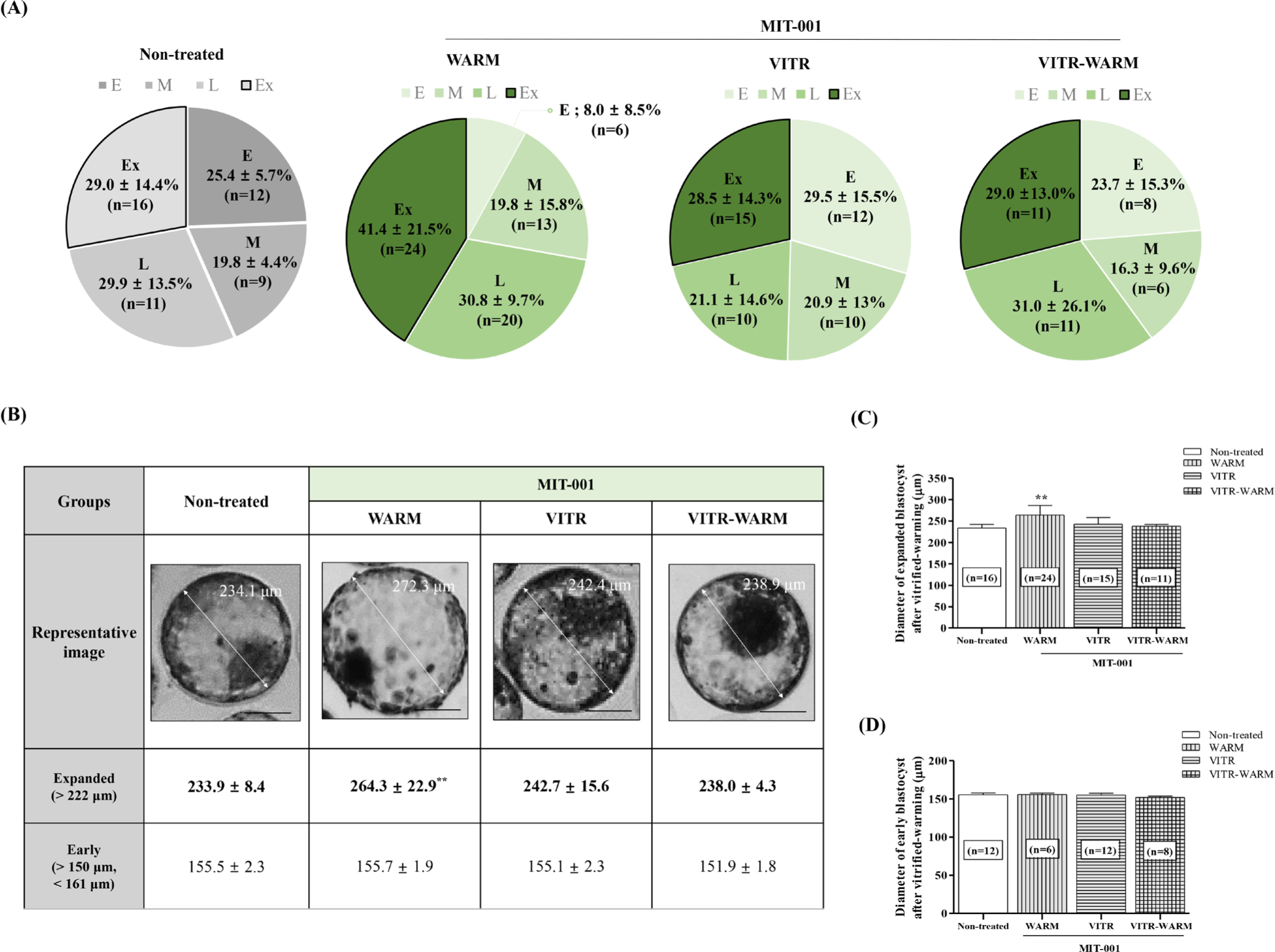
To investigate the effect of MIT-001 on cryopreservation survival, blastocysts were vitrified 7 days after IVC. After vitrification-warming, the blastocysts were classified according to their developmental stages and diameters. Blastocysts with a minimum diameter of 150 μm after post-warming were selected as the criterion for survival analysis (Supplementary Figs. 1 and 2). During the 24 h warming period after cryopreservation, the percentage of expanded blastocysts (Ex: 41.4 ± 21.5%, n = 24) following MIT-001 supplementation was similar to the results observed in IVC bovine embryos (IVC, Ex; 48.2 ± 19.2 %, n = 88) prior to cryopreservation (Fig. 1 and Supplementary Fig. 1). The results revealed an increase in the proportion of expanded blastocysts (Ex) in the WARM group compared to the other groups (Fig. 1A; Non-treated: 29.0 ± 14.4%. vs. WARM: 41.4 ± 21.5% vs. VITR: 28.5 ± 14.3% vs. VITR-WARM: 29.0 ± 13.0%), while the proportion of early blastocysts (E) was lower (Fig. 1A; Non-treated: 25.4 ± 5.7% vs. WARM: 8.0 ± 8.5% vs. VITR: 29.5 ± 15.5% vs. VITR-WARM: 23.7 ± 15.3%). Furthermore, the expanded blastocyst diameter was significantly improved (Non-treated: 233.9 ± 8.4 μm vs. WARM: 264.3 ± 22.9 μm vs. VITR: 242.7 ± 15.6 μm vs. VITR-WARM: 238.0 ± 4.3 μm) in the WARM group (Figs. 1B and 1C). However, the analysis of blastocyst diameter within each stage demonstrated that there was no significant difference in early blastocysts (Fig. 1D).
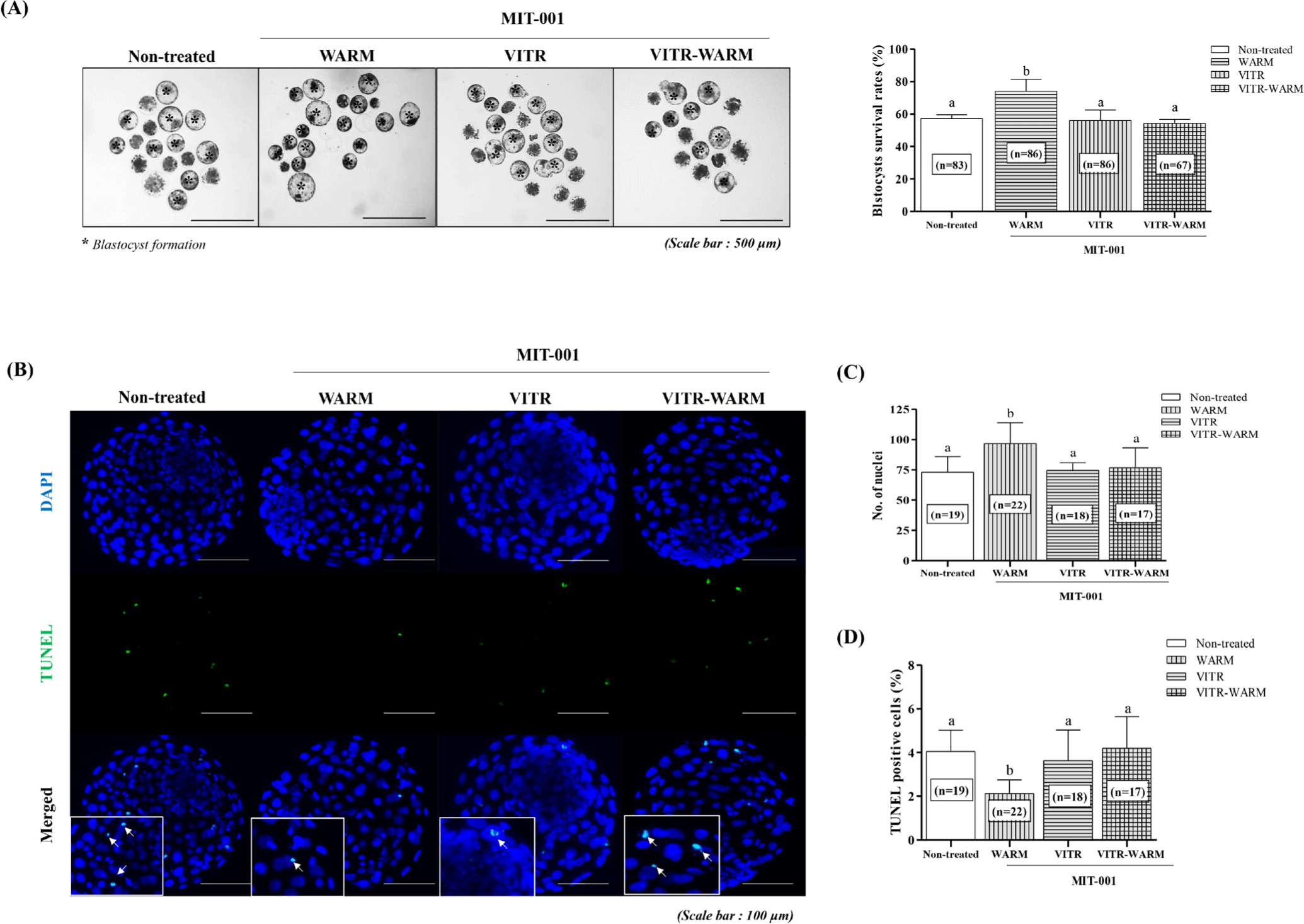
To evaluate the effects of MIT-001 on the cryopreservation of bovine blastocysts, we compared the survival rates of blastocysts exposed to various MIT-001 treatments during vitrification and warming (Table 1 and Fig. 2A). The results showed a significantly higher survival rate in the WARM group (p < 0.01, Non-treated: 57.3 ± 2.3% vs. WARM: 74.2 ± 7.3% vs. VITR: 56.2 ± 6.4% vs. VITR-WARM: 54.3 ± 5.6%), which received MIT-001 only during the warming process. To further assess the quality of vitrified-warmed blastocysts, we conducted DAPI/TUNEL staining to measure the total number of nuclei and DNA damage-induced apoptosis (Table 2 and Figs. 2B, 2C and 2D). We found that the WARM group exhibited a significantly higher number of nuclei (p < 0.01) and a lower percentage of TUNEL-positive cells, compared to the other groups (p < 0.01, Non-treated: 4.0 ± 1.0% vs. WARM: 2.2 ± 0.7% vs. VITR: 3.6 ± 1.4% vs. VITR-WARM: 4.2 ± 1.4%). These results suggest that MIT-001 supplementation during the warming process effectively reduced apoptosis in vitrified-warmed bovine blastocysts, thereby enhancing their overall viability.
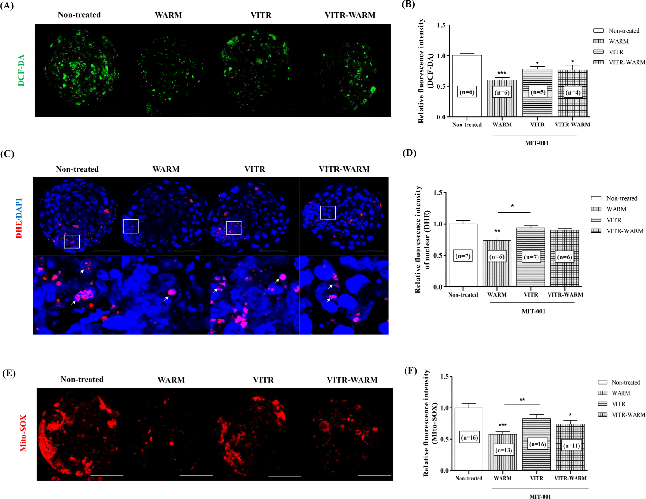
To assess intracellular ROS and superoxide in the nucleus and mitochondria, which are representative of cryoinjury, fluorescence expression was analyzed in surviving blastocysts from the MIT-001-treated groups during vitrification and warming (Fig. 3). The results revealed that the WARM group exhibited significantly lower (p < 0.001) intracellular ROS levels, as determined by DCF-DA staining (Figs. 4A and 4B). Nuclear superoxide levels, as assessed by DHE staining, were significantly lower in the WARM group compared with the non-treated (p < 0.01) and VITR groups (Figs. 3C and 3D). The patterns of mitochondria-specific superoxide production were similar (Figs. 3E and 3F). These findings suggest that the addition of MIT-001 during the warming process effectively mitigated oxidative stress induced by intracellular ROS, and excessive superoxide generation in the nuclei and mitochondria of vitrified-warmed bovine blastocysts.
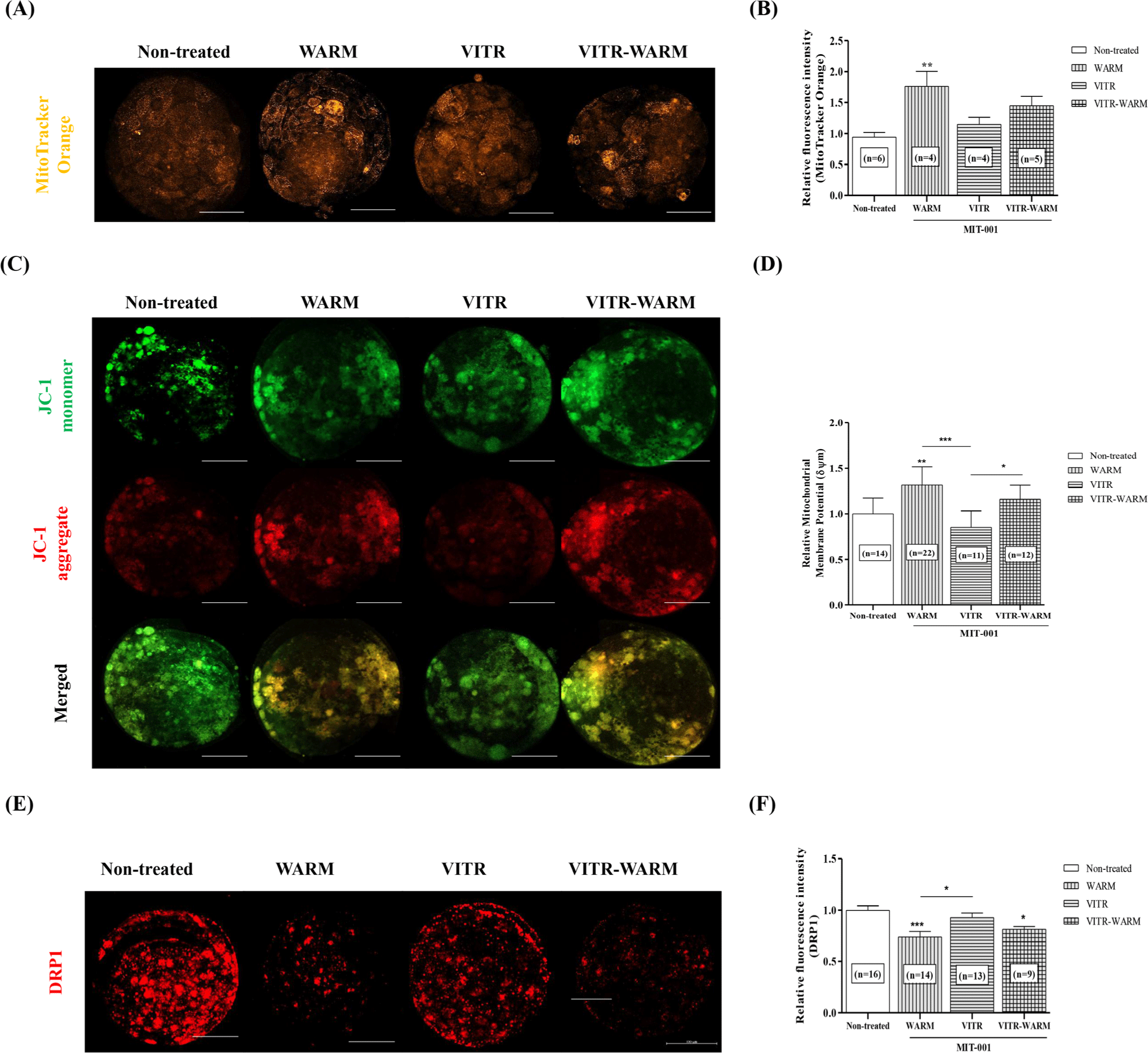
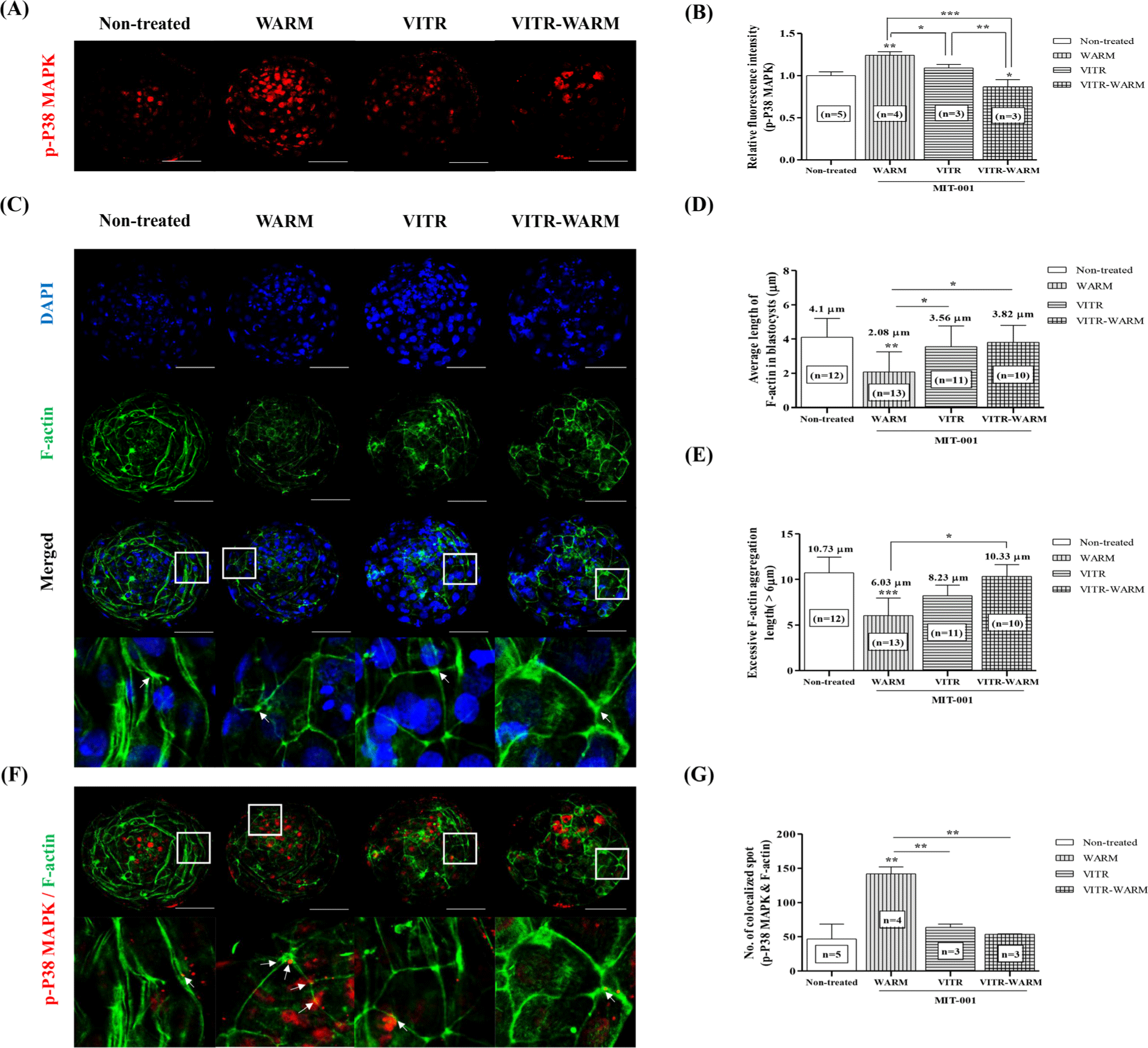
To investigate the effects of MIT-001 treatment on mitochondrial function in vitrified-warmed bovine blastocysts, we performed immunofluorescent staining for MitoTracker Orange, JC-1, and DRP1 (Fig. 4). The activation of mitochondria using MitoTracker Orange and MMP by JC-1 staining showed significantly higher fluorescence intensity in the WARM group (p < 0.01) compared with the Non-treated group (Figs. 4A, 4B, 4C, and 4D). IF staining with DRP1 as a mitochondrial fission marker was significantly reduced in the WARM group (p < 0.001) compared to the other groups (Figs. 4E and 4F). To evaluate the crucial F-actin aggregation and structure during the re-expansion process of vitrified-warmed bovine blastocysts induced by MIT-001, phalloidin-FITC staining, and IF staining of p-phospho (p)-p38 MAPK, an upregulator of F-actin, were performed (Fig. 5). The fluorescence expression of the (p)-p38 MAPK protein increased only in the WARM group (p < 0.01). In addition, analysis of the average length of total F-actin thickness and aggregated F-actin thickness (> 6 μm) was significantly reduced (p < 0.001) in filament thickness in the WARM group (Figs. 5C, 5D, and 5E; Non-treated: 10.73 μm vs. WARM: 6.03 μm vs. VITR: 8.23 μm vs. VITR-WARM: 10.33 μm). Interestingly, bovine blastocysts cultured with MIT-001 (WARM group) during the warming process after vitrification showed a significant increase (p < 0.01) in the co-localization of F-actin and (p)-p38 MAPK compared to the other groups (Figs. 5F and 5G). These results suggest that the addition of MIT-001 during the warming process mitigates cytoskeletal alterations and mitochondrial dysfunction in vitrified-warmed bovine blastocysts.
DISCUSSION
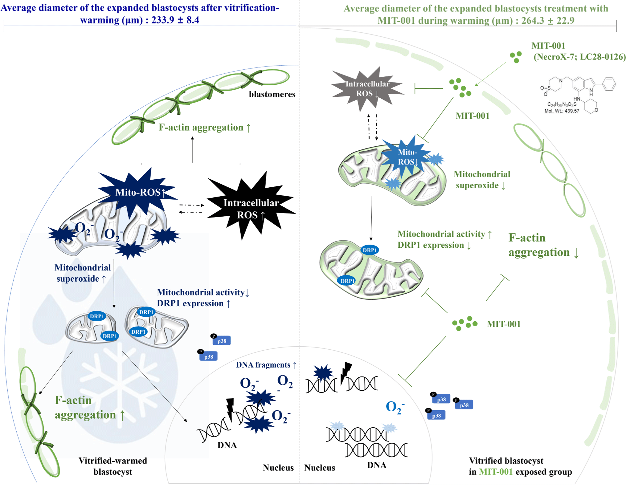
MIT-001 is a novel compound that functions as a mitochondrial-targeted ROS scavenger and has been reported to restore mitochondrial function in various cell types. Here, we demonstrated that MIT-001 treatment during the warming of bovine blastocysts improved mitochondrial function and blastocyst viability by reducing intracellular ROS and mitochondrial-derived superoxide. MIT-001 treatment during the warming period reduced DNA damage, restored cytoskeletal structure, and lowered ROS levels in vitrified-warmed bovine blastocytes. Additionally, MIT-001 decreased DRP1 expression, improved mitochondrial activity, and significantly increased membrane potential and p38 MAPK phosphorylation. The protective effects of MIT-001 on bovine blastocysts were particularly pronounced during re-expansion at specific developmental stages post-vitrification, suggesting that warm-stage-specific treatment with MIT-001 enhanced blastocyst viability (Fig. 6).
Long-term storage of embryos through freezing and vitrification using assisted reproductive technologies is possible; however, research is still needed to improve the quality and survival of blastocysts after warming [5,6]. Various studies have indicated that vitrification significantly disrupts the structures of the cytoskeleton [17], mitochondria [8,18], and cell membranes [19,20], leading to cytological changes in embryos and blastocysts. Recently, it has been established that cellular damage, mitochondrial function, and ROS regulation are crucial factors influencing the re-expansion and survival of cryopreserved bovine blastocysts. A previous study demonstrated that vitrified blastocysts exhibit increased DNA fragmentation and apoptosis compared to in vitro-produced blastocysts [21]. Similarly, reduced survival and re-expansion rates in vitrified porcine blastocysts are closely associated with apoptosis, as evidenced by increased mitochondrial dysfunction and homeostasis [22]. Furthermore, embryos that survive vitrification exhibit defects in cytoskeletal structural stabilization and mitochondrial cytoprotective mechanisms, resulting in increased ROS generation and oxidative stress [23]. Based on this study, the low survival rate of vitrified embryos suggests that cryoinjury impairs embryo quality and developmental competence through oxidative stress, contributing to ROS accumulation. Our study showed that MIT-001 decreased ROS levels and superoxide production after vitrification-warming, but only during the warming period.
Cryopreservation, a key technology in assisted reproduction, induces rapid physicochemical changes in germ cells particularly osmotic stress, pH imbalance, and temperature fluctuations during the freezing and thawing processes [24]. These changes contribute to mitochondrial dysfunction by increasing mPTP opening, ROS accumulation, and intracellular calcium levels, while decreasing mitochondrial membrane potential and ATP production [8]. Such oxidative stress impairs fertilization and embryonic development and is especially known to cause cell damage in bovine embryos, thereby reducing the survival rate of frozen-thawed blastocysts [25]. MIT-001, a mitochondrial-targeted ROS scavenger, appears to be most effective when administered during the thawing phase, where oxidative stress is acutely elevated. While direct evidence specific to cryopreserved embryos is limited, this phase likely provides a critical window for antioxidant intervention, as metabolic activity and ROS generation surge rapidly upon warming. This timing coincides with a rapid surge in oxidative stress, and our results demonstrate that MIT-001 effectively mitigated intracellular stress and improved the survival and quality of bovine blastocysts by counteracting these changes. In contrast, during the freezing phase, metabolic activity in embryos is largely suppressed, resulting in reduced ROS production and mitochondrial activity [26]. Therefore, the scavenging function of MIT-001 under these conditions appears to be limited, which may explain the lack of significant improvement compared to the control group. Moreover, excessive antioxidant exposure in the absence of sufficient ROS may interfere with physiological ROS signaling and cellular homeostasis. Several studies have shown that an excessive reduction of ROS can impair intracellular signaling, mitochondrial functions, and paradoxically decrease cell viability [27–30]. These findings demonstrate that the addition of MIT-001 during the warming process significantly enhanced the survival rate by reducing oxidative stress, leading to improved quality of vitrified-warmed blastocysts (Tables 1 and 2, Figs. 1 and 3). Therefore, our study demonstrates that administration of MIT-001 during the warming phase significantly reduced ROS levels, improved mitochondrial function, and stabilized cytoskeletal structure. These results suggest that the warming phase represents a key intervention window where MIT-001 can effectively alleviate oxidative stress, while untimely or excessive treatment in the freezing or freeze-thaw phases may fail to support cell survival or even disrupt essential redox signaling pathways. Collectively, these findings emphasize that the efficacy of antioxidant treatment depends critically on both timing and dosage.
Various antioxidants, such as melatonin [31] and resveratrol [18], have been utilized as cryoprotectants for the cryopreservation of mammalian blastocysts or spermatozoa to improve early embryo developmental competence and viability [26]. Moreover, supplementation with antioxidants during IVC of bovine blastocysts may contribute to successful blastocyst production [32]. A previous study demonstrated that MIT-001 exerted anti-inflammatory and antioxidant effects on the mitochondria of mouse ovaries by inhibiting mitochondrial peroxide generation [33]. However, few studies have investigated the effects of MIT-001, a mitochondrial-derived peroxide scavenger, on the developmental competence and cryosurvival of bovine blastocysts. Therefore, this study aimed to standardize the survival of blastocysts at the developmental stages prior to vitrification by categorizing them based on their diameter and evaluating the cryoprotective effects of MIT-001 when added to the warming medium. By assessing the potential to mitigate cryo-induced damage and enhance post-warming blastocyst viability using MIT-001, our findings may provide insights into the optimization of embryo cryopreservation strategies for improved reproductive outcomes.
Mitochondrial function is not only associated with ATP supply and ROS regulation during early embryo development [26], but is also known to play a crucial role in cytoskeletal stabilization [15,34], mitochondrial activation, and dynamic responses [24] during the survival of vitrified-warmed blastocysts. DRP1, a key mitochondrial fission factor, is distributed around mitochondria and designated for division [11]. Excessive mitochondrial fragmentation caused by DRP1 hyperactivation has been reported to induce apoptosis during early embryo development, adversely affecting developmental efficiency [11,12]. Yu et al., [1] demonstrated that MIT-001 restores vitrified-warmed bovine blastocysts by enhancing mitochondrial function and cytoskeletal organization [1]. Similarly, we demonstrated that MIT-001 promotes mitochondrial function and maintains MMP in bovine vitrification-warmed blastocysts. Previous studies have indicated that appropriate phosphorylation of p38 MAPK contributes to the stability of F-actin and is associated with the developmental competence of preimplantation embryos, thereby enhancing embryo viability [15]. F-actin cytoskeleton has been reported to be involved in the activation of p38 MAPK [13,14]. In a previous study, we found that F-actin reinforcement improved blastocyst development during early embryonic development in pigs [15]. Additionally, previous results indicate that p38 MAPK activity is required for successful preimplantation development, as its inhibition leads to the reversible arrest of preimplantation development [13–15]. Our results suggest that the increase in p38 MAPK phosphorylation and reduction in F-actin polymerization in the MIT-001 group indicate that MIT-001 enhances cryo-survival by stabilizing F-actin (Fig. 5). These results indicate that MIT-001 enhanced the viability of vitrified bovine blastocysts by promoting the phosphorylation of p38 MAPK, thereby stabilizing F-actin, mitigating mitochondrial superoxide production, and safeguarding mitochondrial integrity (Fig. 6).
CONCLUSION
This study demonstrates that cryoinjury-induced oxidative stress and cytoskeletal disruption significantly compromise the survival and developmental competence of vitrified bovine blastocysts. Notably, supplementation with MIT-001 exclusively during the warming stage significantly improved blastocyst viability by reducing apoptosis and enhancing mitochondrial and cytoskeletal integrity, whereas treatment during vitrification alone or throughout both stages showed no such benefit. These results indicate that the timing of antioxidant intervention is critical, with the warming stage representing a key window of vulnerability to oxidative stress. Our findings suggest that MIT-001 is a promising warming-specific antioxidant strategy for improving cryopreservation outcomes.


