

INTRODUCTION
The corpus luteum (CL), a transient endocrine gland formed following ovulation, plays a crucial role in regulating female reproductive functions by secreting progesterone (P4) [1]. The action of prostaglandin F2 alpha (PGF2α) secreted from the endometrium under estrogen stimulation causes the blood vessels distributed in the CL to constrict, if pregnancy does not occur. The activity of the cholesterol synthesis enzymes is suppressed, resulting in their degeneration [2]. In mammals, the CL is one of the few adult tissues that undergo rapid formation, functional maturation, and structural regression during each estrous cycle [3]. Dynamic remodeling involves extensive alterations in angiogenesis, cell proliferation, and apoptosis [4].
During the initial CL formation stage, ovulation triggers extensive vascular invasion, whereby blood vessels originating from the theca layer rapidly penetrate the ruptured follicles’ granulosa compartment [5]. This newly established vascular network facilitates the efficient delivery of cholesterol and endocrine signals necessary for steroidogenesis [6]. Luteal cells differentiated from granulosa and theca cells synthesize P4 using cholesterol stored in the cytoplasmic lipid droplets, thereby supporting luteal endocrine function [7]. PGF2α secreted from the uterus disrupts luteal blood flow and steroidogenic activity in the absence of pregnancy, thereby initiating functional and structural luteal regression [8]. Consequently, the luteal tissue undergoes atrophy accompanied by lipid degeneration of luteal cells, ultimately forming the corpus albicans [9]. The integrative mechanisms coordinating angiogenesis, steroidogenesis, cell proliferation, and apoptosis during the estrous cycle in pigs remain elusive, although these regulatory components have been individually described.
Cell cycle progression and proliferation are fundamental processes that support rapid growth and functional establishment of the CL during the estrous cycle [10]. After ovulation, luteal cells actively proliferate to expand luteal tissue mass and acquire full steroidogenic capacity, a process tightly regulated by key cell-cycle–associated factors, including cyclin B1 (CCNB1), cyclin-dependent kinase 1 (CDK1), and the mitogen-activated protein kinases/extracellular signal-regulated kinase (MAPK/ERK) signaling components ERK1 and MAPK1 [11]. The activation of the MAPK/ERK pathway facilitates proliferation and differentiation of the luteal cell, thereby promoting luteal maturation and P4 production [11]. Contrastingly, dysregulation of this pathway disrupts folliculogenesis and luteinization [12]. As the estrous cycle advances, proliferative activity progressively declines and apoptotic signaling becomes dominant, driving structural and functional regression of the luteal cell [13]. The balance between pro-apoptotic factors (tumor necrosis factor receptor 1; TNFR1, Bax, and caspase 3; Casp3) and anti-apoptotic proteins, such as Bcl2, ultimately determines luteal cell survival and CL lifespan [14].
Ras and Ras guanosine triphosphatases (GTPases) are small GTP-binding proteins that act as molecular switches to control intracellular signaling associated with proliferation, differentiation, and survival [15]. Ras guanine nucleotide exchange factors (Ras GEFs), such as the son of sevenless homolog 1 (SOS1), mediate Ras activation whereas Ras GTPase-activating proteins (Ras GAPs), including neurofibromatosis type 1 (NF1) and Ras p21 protein activator 1 (RASA1), govern inactivation [16]. These regulatory proteins converge on major signaling cascades, such as the MAPK/ERK and PI3K/AKT-pathways known to affect ovarian follicular growth, steroid hormone production, angiogenesis, and cell-cycle regulation [17]. Although Ras-mediated signaling has been assessed in several reproductive tissues, studies particularly addressing Ras’s role and its GTPases in porcine CL physiology remain limited. Considering the rapid structural alterations and high metabolic demands of the CL, Ras signaling may be a key integrative regulator linking angiogenesis, steroidogenesis, and cellular turnover [18].
However, Ras and its GTPases during the estrous cycle in porcine CL have not been clearly defined. No comprehensive investigation has been conducted to compare the expression of Ras family members (H-, K-, N-, and R-Ras) and their regulatory proteins across distinct estrous cycles. Additionally, the potential interactions among Ras GTPases, steroid hormone receptors, and angiogenic factors have not been examined in pigs. Understanding these molecular relationships is critical because Ras signaling pathways may affect the transition from proliferation and steroidogenesis in the early phase (EP) and middle phase (MP) to apoptosis and regression in the late phase (LP) [19].
Pigs and humans share key reproductive characteristics, including ovarian follicular dynamics, steroid hormone profiles, angiogenic mechanisms, and the balance between proliferative and apoptotic signals during the development and regression of CL [20,21]. These similarities improve the translational value of porcine studies for understanding human ovarian physiology, estrous cycle deficiency, and early pregnancy loss. From a practical perspective, elucidating the CL function in pigs contributes to improved reproductive management in the swine industry, supporting enhancements in estrous synchronization, fertility monitoring, and pregnancy diagnosis [22]. Additionally, the characterization of steroidogenesis, angiogenesis, and cell turnover in the porcine CL provides fundamental insights into the biology of a rapidly remodeling endocrine gland [23,24]. Thus, porcine CL research has agricultural and biomedical significance, strengthening its role as a model for mammalian reproductive physiology.
The angiogenic [25], steroidogenic [26], cell proliferation [27], and apoptotic [28] pathways are dynamically regulated during CL development and regression in pigs. Ras and its GTPases function as the primary molecular switches that control intracellular signaling associated with cell proliferation [29], differentiation [30], angiogenesis [31], and apoptosis [32,33] in different reproductive tissues. Considering these observations, together with the rapid structural remodeling and high metabolic demands of the CL, we hypothesized that Ras-related signaling pathways play an integrated role in coordinating luteal development and regression in pigs. We investigated the phase-dependent expression patterns of the Ras family members (H-, K-, N-, and R-Ras) and their regulatory proteins (NF1, RASA1, and SOS1) in the porcine CL during the EP, MP, and LP. Furthermore, we simultaneously examined the mRNA and protein expression levels of hormone receptors, angiogenic markers, cell cycle regulators, and apoptotic factors. Pathway-level protein–protein interaction (PPI) analysis was performed using the Search Tool for the Retrieval of Interacting Genes (STRING) database to further elucidate the molecular relationships underlying these regulatory processes. Additionally, we aimed to clarify the potential involvement of Ras-related signaling in integrating angiogenesis, steroidogenesis, cell proliferation, and apoptosis during the porcine estrous cycle.
MATERIALS AND METHODS
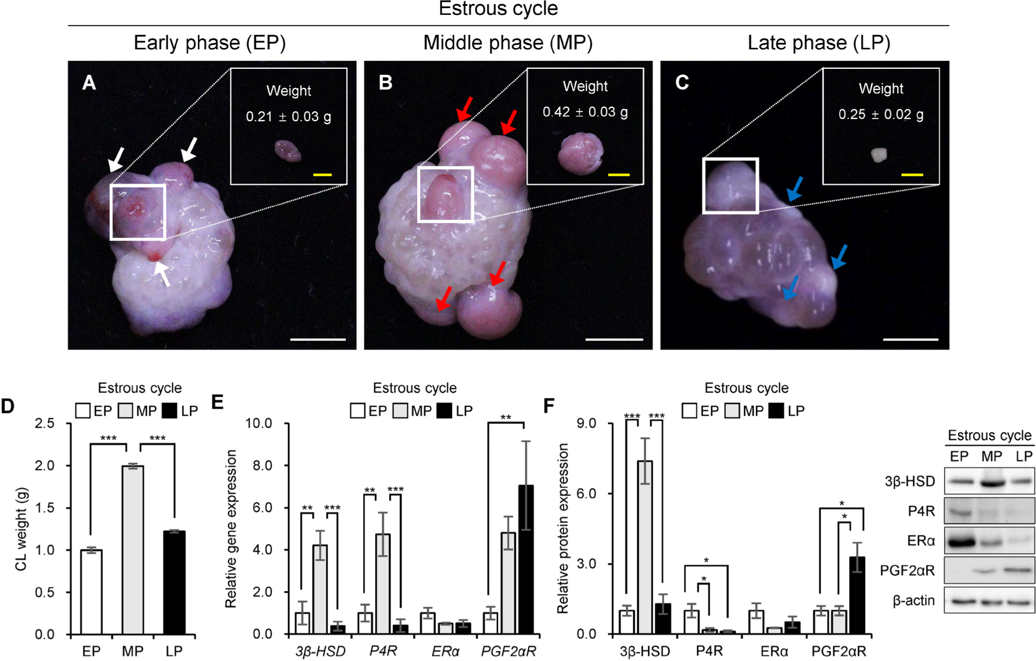
The Institutional Animal Care and Use Committee of the Kangwon National University approved all animal procedures (KW-250716-2). Porcine CL were collected at a local slaughterhouse (Pocheon farm) and transported to the laboratory at 4°C within 2 h following slaughter. The estrous cycle phase was assigned primarily based on ovarian and CL morphology in the laboratory (Figs. 1A, 1B, and 1C). The CL of EP were defined as samples showing visible red blood within the CL, having a relatively smaller overall size than MP CL, and showing blood inside the tissue after isolation and dissection (Fig. 1A). The CL of MP were defined as samples in which blood was not observed and which showed a pinkish luteal coloration (Fig. 1B). The CL of LP were defined as samples with a smaller tissue size than MP CL and ovaries containing developing follicles (Fig. 1C). The expression patterns of 3β-hydroxysteroid dehydrogenase (3β-HSD) and prostaglandin F2 alpha receptor (PGF2αR) were examined only as supportive biological indicators of luteal functional status and were not used for marker-based reclassification of the samples (Figs. 1E and 1F). Subsequently, all isolated CL tissues were weighed and stored at −80°C until further use.
Total RNA was extracted using RNAiso Plus (Takara Bio), according to the manufacturer’s protocol. RNA quality and concentration were measured using an EzDrop 1000C spectrophotometer (Blue-Ray Biotech). Next, 5.0 μg RNA was utilized to synthesize cDNA using a SuPrimeScript cDNA Synthesis Kit (Genetbio) according to the manufacturer’s instructions. Furthermore, cDNA was stored at −18°C until reverse transcriptase polymerase chain reaction (RT-PCR).
Using a qPCR system (Quant3 Studio, Applied Biosystems, Thermo Fisher Scientific), cDNA, along with qPCR mix, primers, and ddH2O, was amplified. Amplification conditions included incubation at 50°C for 2 min, initial denaturation at 95°C for 10 min, followed by 40 cycles of denaturation at 95°C for 15 s and annealing/extension at 60°C for 1 min. The mRNA expression levels were normalized to that of β-actin mRNA. All reactions were performed in triplicate. Primer specificity was confirmed by melt curve analysis. Table 1 lists the primer sequences used in the experiments. We computed the relative mRNA expression levels using the comparative cycle threshold (2–ΔΔCt) method [34].
3β-HSD, 3β-hydroxysteroid dehydrogenase; Ang1, angiopoietin 1; Bax, B-cell lymphoma 2-associated X protein; Bcl2, B-cell lymphoma 2; CCNB1, cyclin B1; CDK1, cyclin-dependent kinase 1; Casp3, caspase 3; ERK1, extracellular signal-regulated kinase 1; ERα, estrogen receptor alpha; H-Ras, Harvey rat sarcoma viral oncogene homolog; K-Ras, Kirsten rat sarcoma viral oncogene homolog; MAPK1, mitogen-activated protein kinase 1; N-Ras, Neuroblastoma rat sarcoma viral oncogene homolog; NF1, neurofibromin 1; P4R, progesterone receptor; PGF2αR, prostaglandin F2 alpha receptor; RASA1, RAS p21 protein activator 1; R-Ras, related RAS viral oncogene homolog; SOS1, son of sevenless homolog 1; Tie2, tyrosine kinase with immunoglobulin-like and EGF-like domains 2; TNFR1, tumor necrosis factor receptor 1; VEGFA, vascular endothelial growth factor A; VEGFR2, vascular endothelial growth factor receptor 2; β-actin, beta-actin.
Luteal tissues were lysed in radio-immunoprecipitation assay cell lysis buffer (GenDEPOT) containing inhibitors (Xpert protease inhibitor cocktail solution and Xpert phosphatase inhibitor cocktail solution; GenDEPOT). The resulting homogenate was centrifuged at 12,000 rpm (13,523×g) and 4°C for 20 min and the supernatants were collected in a fresh tube. Total protein concentration was measured using the Pierce Bicinchonic Acid Protein Assay Kit-Reducing Agent Compatible, according to the manufacturer’s protocol (23250; Thermo Fisher Scientific). Proteins were separated using sodium dodecyl sulfate-polyacrylamide gel electrophoresis and transferred onto polyvinylidene difluoride membranes (Merck Millipore) using a Trans-Blot turbo rapid transfer system (1704150, Bio-Rad), according to the manufacturer’s protocol. The membranes were blocked in blocking solution (5% skim milk in Tris-buffered saline/0.5% Tween-20; TBS-T) at room temperature for 1 h. After blocking membranes, the membranes incubated overnight at 4°C with the appropriate primary antibodies (Table 2). Images were acquired using a chemiluminescence substrate (W3651-012; GenDEPOT) and quantified using a chemiluminescence imaging system (UVITEC Alliance MINI HD9 system, UVITEC). Band intensities were quantified by densitometric analysis and normalized to β-actin as a loading control.
3β-HSD, 3β-hydroxysteroid dehydrogenase; Ang-4, angiopoietin 4; Bax, B-cell lymphoma 2-associated X protein; Bcl2, B-cell lymphoma 2; CCNB1, cyclin B1; Casp3, caspase 3; ERK1/2, extracellular signal-regulated kinase 1/2; ERα, estrogen receptor alpha; H-Ras, Harvey rat sarcoma viral oncogene homolog; HRP, horseradish peroxidase; IgG, immunoglobulin G; K-Ras, Kirsten rat sarcoma viral oncogene homolog; NF1, neurofibromin 1; P4R, progesterone receptor; PGF2αR, prostaglandin F2 alpha receptor; p-ERK1/2, phosphorylated extracellular signal-regulated kinase 1/2; RASA1, RAS p21 protein activator 1; R-Ras, related RAS viral oncogene homolog; SOS1, son of sevenless homolog 1; Tie-2, tyrosine kinase with immunoglobulin-like and EGF-like domains 2; TNFR1, tumor necrosis factor receptor 1; VEGF-D, vascular endothelial growth factor D.
Protein–protein interaction and functional association networks were constructed using the STRING [35] (v.10.0; http://string.embl.de) database, with Sus scrofa selected as the target species. Network edges were generated using a medium confidence score cutoff of 0.400. The molecular action view was used to visualize predicted interaction modes, including positive and negative interactions, activation, and inhibition. STRING-derived interactions were interpreted as predicted functional associations rather than experimentally validated causal relationships. Subsequently, the molecular action of the Ras GTPases (NF1, RASA1, and SOS1), hormone receptors (progesterone receptor; P4R, prostaglandin F2 alpha receptor; PGF2αR, and estrogen receptor alpha; ERα), angiogenesis factors (vascular endothelial growth factor A; VEGFA, VEGF receptor 2; VEGFR2, angiopoietin 1; Ang1, and Tie2), apoptosis factors (TNFR1, Bax, and Casp3), and Ras proteins (H-Ras, K-Ras, N-Ras, and R-Ras) were examined using the STRING database.
Each experiment was conducted with at least three replications. Data was analyzed using SAS ver. 9.4 (SAS Institute). Data was presented as the mean ± SEM and differences between each group (EP vs MP, MP vs LP, and EP vs LP) were calculated using Student’s t-test. Statistical significance was set at p < 0.05.
RESULTS
Fig. 1 shows the morphological characteristics of the porcine ovaries at the EP (Fig. 1A), MP (Fig. 1B), and LP (Fig. 1C) stages of the estrous cycle. The CL showed a substantial increase in size during the MP compared with that during the EP and LP. Consistent with these morphological observations, the tissue weight of the CL (Fig. 1D) was significantly higher in the MP than in the EP and LP (p < 0.001).
Figs. 1E and 1F show the relative expression levels of the steroid hormone receptors. At the mRNA level (Fig. 1E), the 3β-HSD and P4R expressions were significantly increased at the MP compared with the EP and LP, whereas ERα mRNA expression did not significantly differ during the estrous cycle. Contrastingly, PGF2αR mRNA expression was significantly higher at the LP than at the EP and MP (p < 0.05). At the protein level (Fig. 1F), 3β-HSD protein expression was the highest at the MP, whereas P4R protein expression was elevated at the EP (p < 0.001). Similar to the mRNA results, ERα protein expression did not reveal significant differences among the stages, whereas PGF2αR protein expression was significantly increased at the LP (p < 0.05).
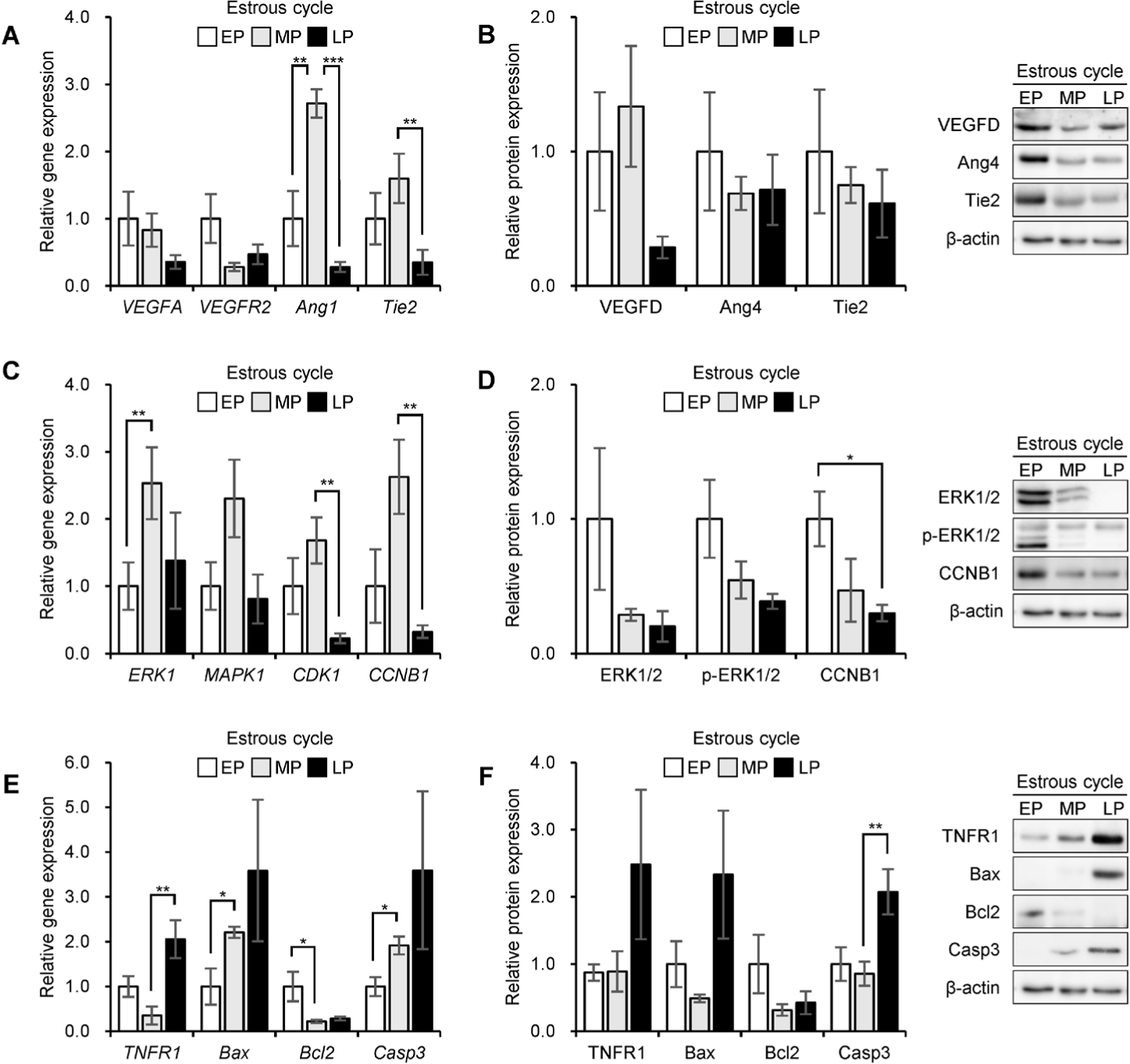
Figs. 2A, 2B, 2C, and 2D show the expression of the angiogenesis-and cell cycle-related factors in the porcine CL during the estrous cycle. The VEGFA and VEGF receptor 2 (VEGFR2) mRNA expression levels (Fig. 2A) were not markedly different among the EP, MP, and LP groups. Contrastingly, the mRNA expression levels of angiopoietin 1 (Ang1) and Tie2 were significantly higher in the MP than in the EP and LP (p < 0.05). At the protein level (Fig. 2B), vascular endothelial growth factor D (VEGFD) protein expression was significantly higher at the MP than at the EP and LP (p < 0.05), whereas angiopoietin 4 (Ang4) protein expression did not significantly differ during the estrous cycle. Tie2 protein expression did not significantly differ during the estrous cycle. The mRNA expression levels of ERK1, CDK1, and CCNB1 (Fig. 2C) were significantly higher in the MP group than in the EP and LP groups (p < 0.05), whereas MAPK1 mRNA expression did not significantly differ during the estrous cycle. At the protein level (Fig. 2D), the expression levels of ERK1/2 and phosphorylated ERK1/2 (p-ERK1/2) were not significantly different among EP, MP, and LP, whereas CCNB1 protein expression was significantly higher in the EP than in the MP and LP (p < 0.05). Fig. 2E and 2F illustrate the expression patterns of the apoptosis-related factors. TNFR1 mRNA expression levels (Fig. 2E) were significantly higher in the LP group than in the EP and MP groups (p < 0.05). Contrastingly, the mRNA expression levels of Bax and Casp3 were significantly increased in MP, whereas Bcl2 mRNA expression was significantly higher in EP than in MP and LP (p < 0.05). At the protein level (Fig. 2F), the expression levels of TNFR1, Bax, and Bcl2 were not significantly different during the estrous cycle, whereas Casp3 protein expression was significantly higher in the LP than in the EP and MP (p < 0.05).
The expression of Ras family members and GTPases identified in the porcine CL was re-validated using mRNA and protein analyses (Fig. 3). The mRNA levels of H-Ras, K-Ras, N-Ras, and R-Ras were significantly changed during the estrous cycle (Fig. 3A). Compared to EP, the expression of Ras family mRNAs revealed significant alterations in MP and was significantly decreased in LP (p < 0.05). Similarly, the protein expression levels of H-Ras, K-Ras, and R-Ras were significantly altered during the estrous cycle, with higher expression noted in the MP and decreased expression in the LP than in the EP (p < 0.05; Fig. 3B). The mRNA expression levels of NF1, RASA1, and SOS1 were not significantly different between the EP and MP, but were significantly changed in the LP (p < 0.05; Fig. 3C). At the protein level, the expression of NF1, RASA1, and SOS1 revealed significant stage-dependent alterations, with changed expression noted in the MP and LP compared to the EP (p < 0.05; Fig. 3D).
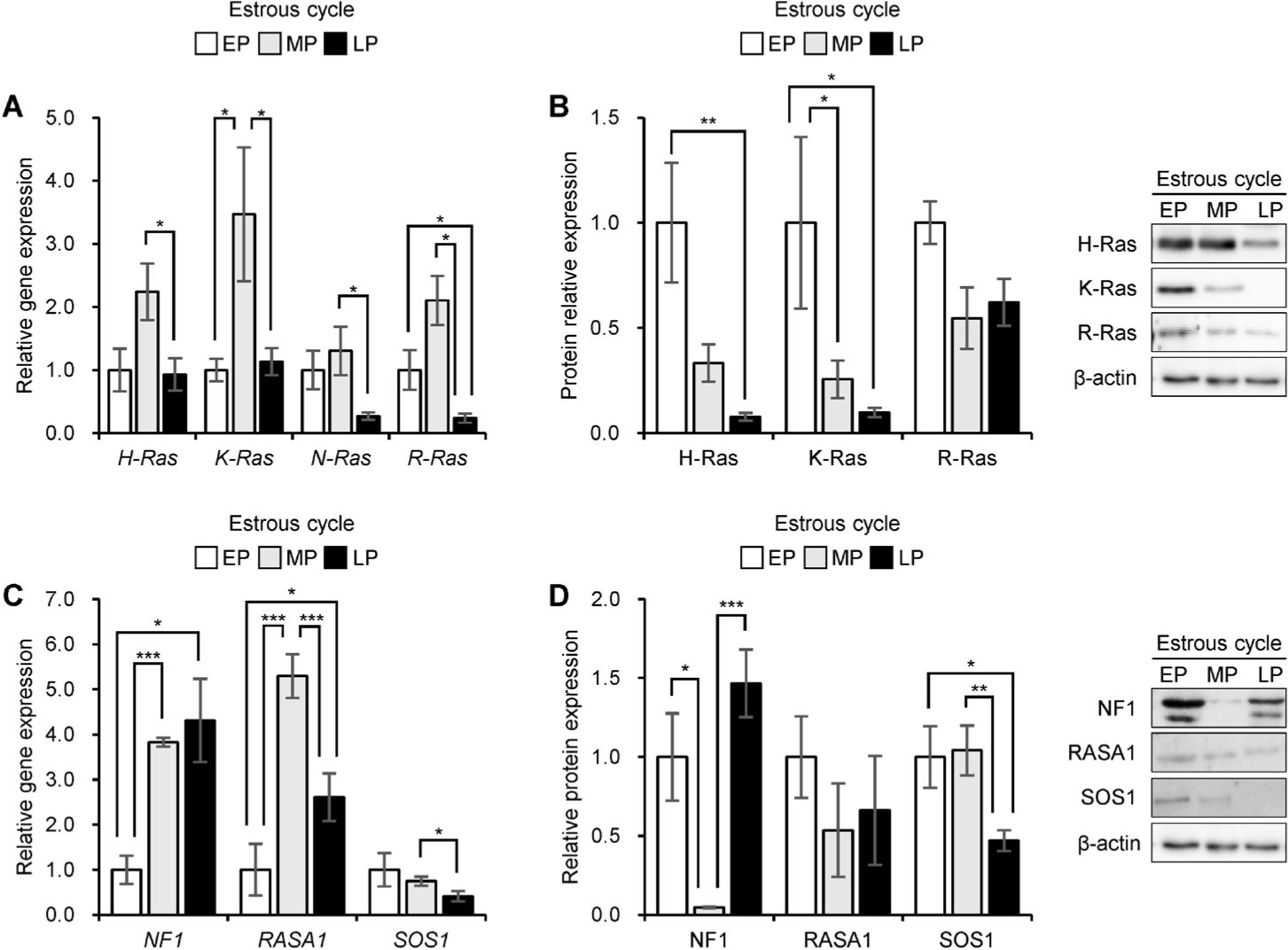
Fig. 4 shows the molecular actions of hormone receptors, angiogenic factors, cell cycle-related factors, apoptotic factors, Ras GTPases, and Ras GTPase proteins. STRING-based molecular action networks were constructed using the same set of proteins arranged in the same positions in Homo sapiens (Fig. 4A), Mus musculus (Fig. 4B), Bos taurus (Fig. 4C), and Sus scrofa (Fig. 4D) to compare conserved interaction patterns across major mammalian models. In the network, ERα was predicted to have a potential activating relationship with P4R (Fig. 4, green arrows), and ERα was also connected to the angiogenic module via VEGFA and its receptor VEGFR2. Additionally, the angiogenic receptors VEGFR2 and Tie2 were associated with Ras-related signaling through SOS1 (Fig. 4, green arrows), suggesting that these molecules may represent candidate links between angiogenic signaling and Ras-related pathways in porcine CL.
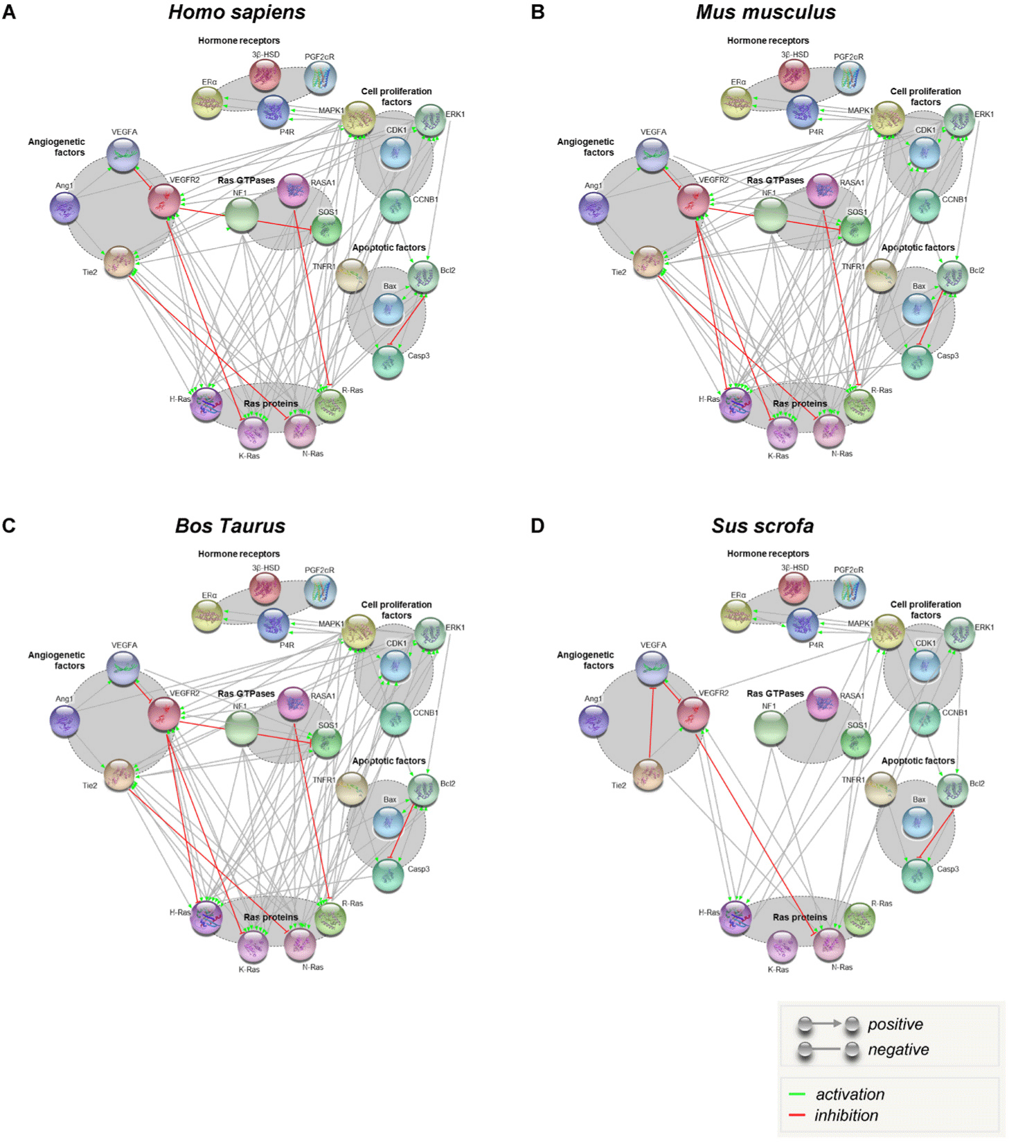
Moreover, the STRING molecular action map indicated that H-Ras and R-Ras were functionally connected to angiogenic receptors and SOS1, suggesting a potential network-level association between angiogenic signaling and Ras GTPase-related pathways (Fig. 4, green arrows). However, Ras GAP (NF1 and RASA1) were predicted to have inhibitory relationships with Ras signaling components (Fig. 4, red lines). Furthermore, Ras-related nodes were located at the interface between upstream hormone receptor/angiogenesis modules and downstream cell cycle (cell proliferation) factors (MAPK1, ERK1, CDK1, and CCNB1) via multiple interaction links (Fig. 4, gray lines). Contrastingly, apoptotic factors (TNFR1, Bax, Bcl2, and Casp3) were primarily connected within the apoptosis module and revealed relatively limited positive molecular interactions with hormone receptors, angiogenic factors, and Ras-related factors in the network (Fig. 4). Thus, although the overall architecture of these interaction modules is broadly conserved across four species, experimental evidence validating how these conserved connections operate in porcine CL is limited. Therefore, combining the species comparative network with stage-dependent expression profiles supports the requirement of porcine-specific studies to clarify the Ras-centered regulatory mechanisms coordinating angiogenesis, cell cycle transition, and luteal regression.
DISCUSSION
The CL is a transient endocrine tissue that undergoes rapid formation, functional maintenance, and regression during the estrous cycle, which requires the coordinated regulation of steroidogenesis, vascular remodeling, cell cycle transition, and apoptotic signaling [36,37]. Thus, we assessed stage-dependent variations in gross morphology and tissue weight, along with the expression profiles of steroid hormone receptor-related factors, angiogenesis- and cell cycle-related factors, apoptosis-related markers, and Ras-related components in the porcine CL, and further integrated these data using a STRING-based molecular action network.
Morphological features and tissue weight data indicated that the porcine CL reached its maximal growth during MP [38]. This observation was supported by 3β-HSD’s expression pattern, which revealed the highest expression at the MP at the mRNA and protein levels [39]. This suggests that the MP corresponds to the period of improved steroidogenic capacity [40]. Additionally, P4R mRNA increased in the MP, whereas P4R protein increased in the EP. Although mRNA and protein levels do not always exhibit identical patterns, this discrepancy suggests that P4 signaling in porcine CL may be regulated at multiple levels, especially during early luteal development. Contrastingly, ERα did not exhibit considerable differences across estrous cycle at either the mRNA or protein level in the present dataset. This indicates that ERα expression itself may remain relatively stable during the estrous cycle. Notably, PGF2αR was the highest at the LP at the mRNA and protein levels, which is consistent with an elevated luteolytic responsiveness as the CL transitions toward regression [41]. Nevertheless, although the expression patterns of 3β-HSD and PGF2αR were consistent with the expected biological characteristics of MP and LP, respectively, 3β-HSD expression alone cannot be regarded as a direct measure of P4 production. Therefore, the interpretation of steroidogenic capacity based on 3β-HSD expression should be considered with caution. In future studies, particularly IHC-based analyses of Ras protein distribution in porcine CL tissues across the estrous cycle, P4 production in each sample will be quantified by ELISA to more accurately confirm the physiological status of the samples prior to further analysis.
Angiogenesis is necessary for CL formation and function, because the developing luteal tissue needs rapid vascularization and subsequent stabilization [17]. In the present study, the angiogenic factors exhibited marker- and level-dependent patterns. VEGFA, VEGFR2, and Ang1 were analyzed at the mRNA level, whereas VEGFD and Ang4 was evaluated at the protein level, primarily due to practical considerations such as assay availability and antibody specificity. At the mRNA level, VEGFA and VEGFR2 did not reveal considerable variations among the stages, whereas Ang1 and Tie2 were elevated in the MP. At the protein level, VEGFD was increased in the MP. Additionally, Ang4 and Tie2 protein did not reveal significant variations among the stages. Thus, luteal vascular remodeling in pigs may be supported by distinct angiogenic mediators depending on the estrous cycle and molecular level. Additionally, the Ang-Tie axis and VEGF family members may contribute in a complementary but not identical way across development and regression.
With respect to cell cycle regulation, the mRNA expression of ERK1, CDK1, and CCNB1 increased in the MP, whereas MAPK1 mRNA did not significantly vary among the stages. However, at the protein level, ERK1/2 and p-ERK1/2 were not significantly different, whereas CCNB1 protein was increased in the EP. Although these patterns appear to be partially inconsistent at the molecular level, such variations can occur because complex regulatory steps, including translation efficiency, phosphorylation dynamics, and protein turnover, control cell cycle progression [42]. Therefore, the present findings indicate that the transcriptional activation of cell cycle regulators is prominent during MP, whereas protein level alterations may reflect earlier proliferative cues or differences in stability/processing during early luteal development.
Regarding apoptotic regulation, stage-dependent changes were noted that differed between transcripts and proteins. At the mRNA level, TNFR1 increased in the LP, Bax and Casp3 increased in the MP, and Bcl2 increased in the EP. At the protein level, Casp3 was increased in the LP, whereas TNFR1, Bax, and Bcl2 did not reveal substantial differences during the estrous cycle. Thus, the transcriptional priming of apoptosis-related factors can be observed earlier; however, the execution marker (Casp3) becomes more evident at the protein level during LP, which is consistent with the structural regression occurring during luteolysis. Although not all apoptotic markers revealed substantial protein level alterations, the combined expression profiles support the concept that luteal regression involves a shift toward progressive signaling as the cell cycle progresses.
Ras-related signaling regulates cell proliferation, differentiation, and survival through pathways such as the MAPK/ERK pathway [43]. It reported that Ras and its GTPases genes dynamic are changed during estrous cycle in porcine cumulus cell [44], but estrous cycle regulation in porcine CL has not been clearly shown. Herein, Ras family members (H-, K-, N-, and R-Ras), Ras GEF (SOS1), and Ras GAP (NF1 and RASA1) revealed differential expression across the estrous cycle at the mRNA and protein levels. These data provide evidence that Ras-related components are dynamically regulated during the porcine estrous cycle and may be linked to luteal functional transition, especially during periods of rapid growth and functional maintenance. Although expression profiling alone does not confirm the direct activation states, these patterns support the requirement for further mechanistic validation of Ras-centered signaling in the luteal cells. Such mRNA-protein discordances observed across multiple targets in this study may be attributed to several study-specific factors, including post-transcriptional regulation, differences in protein turnover and stability between estrous phases, and tissue-level heterogeneity inherent to bulk CL samples composed of mixed luteal, vascular, and immune cell populations. In addition, sampling difference among individual CL and potential sensitivity limitation of estrous phase classification based on ovarian morphology may have contributed to the observed discrepancies. It should also be noted that the present study measured total receptor expression levels at the mRNA and protein level, which do not directly reflect receptor activation or transcriptional activity. Localization analyses such as immunohistochemistry (IHC) or immunofluorescence would be necessary to confirm cell-type-specific expression pattern and functional receptor engagement and are a priority for future studies.
Additionally, the STRING-based molecular action network provided an integrated framework for visualizing predicted functional associations among hormone receptor signaling, angiogenesis-related factors, cell-cycle modules, apoptosis-related nodes, and Ras-related factors. The same protein set was visualized in the same arrangement across Homo sapiens, Mus musculus, Bos taurus, and Sus scrofa. The network predicted potential associations among ERα-P4R, angiogenic receptors (VEGFR2 and Tie2), the Ras activator SOS1, and Ras family proteins, whereas inhibitory relationships were predicted through NF1 and RASA1. Additionally, Ras-related nodes appeared to occupy intermediate network positions connecting the upstream hormone/angiogenesis modules with downstream cell cycle factors (ERK/MAPK-related nodes, CDK1, and CCNB1). Contrastingly, apoptosis-related factors showed revealed relatively limited positive molecular actions with the hormone receptor angiogenesis-Ras modules in the network. Thus, experimental validation in porcine luteal tissue is still limited, although the general architecture of these interactions is conserved across the four species. Additionally, porcine-specific studies are necessary to determine whether these Ras-centered regulatory mechanisms coordinate angiogenesis, cell cycle transition, and luteal regression in the CL.
Although the porcine CL used in this study were classified according to estrous phase, not all genes and proteins examined showed fully consistent phase-dependent patterns. This may partly reflect the biological heterogeneity of CL in polyovulatory pigs and the independent nature of samples collected from slaughtered animals. In future studies, including IHC-based analyses of Ras protein distribution in porcine CL tissues across the estrous cycle, P4 production in each sample will be measured by ELISA to more accurately confirm the physiological status of the samples before further analysis.
In summary, this study demonstrated that porcine luteal development and regression are accompanied by coordinated changes in morphology and tissue weight, stage-dependent expression patterns of steroid hormone receptors, and distinct profiles of angiogenesis, cell cycle, and apoptosis-related markers. Moreover, Ras family members change together with comparative network-based molecular actions. This suggests that Ras signaling may function as an integrative module that links endocrine and vascular cues to intracellular growth and survival pathways during the porcine estrous cycle. These results provide fundamental information for future studies aimed at validating Ras-related mechanisms in luteal cells and enhancing our understanding of luteal physiology in pigs.
CONCLUSIONS
This study showed clear phase-dependent alterations in the porcine CL during the estrous cycle, as evidenced by the morphology, tissue weight, and coordinated expression profiles of the key regulatory factors. CL demonstrated maximal growth at the MP, accompanied by increased 3β-HSD expression, supporting improved steroidogenic capacity during functional maintenance. Contrastingly, PGF2αR expression was the highest at the LP, indicating elevated luteolytic responsiveness during regression. Overall, these results provide fundamental evidence that Ras-related signaling components are dynamically regulated in the porcine CL. Additionally, the findings support the requirement for porcine-specific mechanistic studies to clarify how endocrine and vascular cues coordinate luteal development and regression.


