

INTRODUCTION
Mycotoxin contamination of animal feed is a major problem affecting global livestock production. Mycotoxins are toxic secondary metabolites produced by fungal species such as Fusarium, Aspergillus, and Penicillium, and they remain in the feed even after processing and storage methods. Pigs have a poor ability to detoxify toxins in their bodies, which makes them particularly vulnerable to mycotoxins [1,2]. Deoxynivalenol (DON) and zearalenone (ZEN) are toxins derived from Fusarium, that impair feed intake, growth performance, and reproductive efficiency. Even low concentrations of DON and ZEN, these mycotoxins reduce productivity and cause reproductive disorders, resulting in economic losses [1–3]. In particular, DON is one of the most prevalent Fusarium-derived trichothecene mycotoxins found in cereals, such as wheat, barley, and maize. Upon ingestion, DON exerts various toxic effects, including vomiting, diarrhea, growth retardation, immunodeficiency, and intestinal damage [4–6]. At the cellular level, DON exerts its toxicity primarily by binding to the ribosomal peptidyl transferase center, thereby inhibiting protein synthesis and activating mitogen-activated protein kinase (MAPK) pathways, which lead to ribotoxic stress, oxidative damage, and immune dysfunction [7,8]. These mechanisms cause not only gastrointestinal and immune dysfunctions, but also substantial reproductive disorders in pigs, the species most sensitive to DON exposure [4]. Previous studies have shown that DON impairs ovarian function, damages follicular development, and compromises oocyte quality, thereby reducing fertilization and embryo viability in humans and pigs [9–11]. Deepoxy-deoxynivalenol (DOM-1) is the primary microbial metabolite of DON, generated through the de–epoxidation of the epoxide group at the C12–C13 position by the intestinal microbiota. Since the interaction between the epoxide group and the ribosome causes inhibition of protein synthesis, these structural changes reduce the toxicity of DON [12,13]. Consequently, DOM-1 is considered to be much less toxic than the original compound [12,14,15]. Ruminants are highly protected from DON toxicity because of their diverse gastrointestinal microbiomes that efficiently convert DON to DOM-1 in the rumen [16]. By contrast, pigs have a limited capacity to transform DON into DOM-1 and are more sensitive to DON [17]. DOM-1 has been detected in biological fluids, including plasma, urine, and follicular fluid, indicating its systemic circulation and potential interaction with reproductive tissues [18,19]. Although DOM-1 is considered a lower-toxicity metabolic, its effects on oocyte quality and embryonic development have not been precisely determined [12,14,15].
Conflicting results have been reported regarding the effects of these mycotoxins on reproductive capacity. In bovine studies, DOM-1 induced endoplasmic reticulum (ER) stress in follicular membrane cells, leading to apoptosis [18,20]. By contrast, other studies have reported that DOM-1 is less toxic than DON [12,14,15], and in some cases, no significant differences were observed between DOM-1 treated samples and controls [21]. These conflicting results may be because of differences in experimental models, concentrations, exposure duration, and assessment parameters, suggesting uncertainty regarding the effects of DOM-1. Therefore, understanding the role of DOM-1 in the reproductive context is essential to assess the true risk of DON contamination in swine production and its broader impact on animal and human health.
Given the adverse effects of DON and its metabolite DOM-1 on oocyte quality and embryonic development, it is important to develop effective protective strategies. Resveratrol (Res) is a natural polyphenolic compound found in berries is known to have antioxidant and anti-inflammatory properties. Res has been reported to mitigate DON-induced toxicity, including intestinal inflammation and oxidative stress [22–24]. In addition to its role in gut health, Res has received attention in the field of reproductive physiology. Supplementation with Res improves the quantity and quality of mitochondria, enhances oocyte competence and embryo development rates, reduces ROS levels, and suppresses the expression of apoptosis related genes in bovine and porcine oocytes [24,25–27]. Based on these findings, the present study was designed to evaluate the impact of DON and DOM-1 on porcine oocyte maturation and embryonic development, and to determine whether Res supplementation can counteract their negative effects, thereby improving oocyte quality and developmental potential.
MATERIALS AND METHODS
This study adhered to ethical research protocols for handling porcine ovaries and semen, with all methods officially approved by the Institutional Animal Care and Use Committee (IACUC) of Chungnam National University (Approval No.: 202103A-CNU-002). All chemicals and reagents were purchased from Sigma Chemical, unless otherwise stated. DON and DOM-1 were purchased from Romer Labs.
Porcine ovaries were collected from a local slaughterhouse within 3 h of slaughter. These samples were preserved in 0.9% sterile saline at 30°C and transported to the laboratory in a saline solution supplemented with 75 mg/mL penicillin G and 50 mg/mL streptomycin sulfate. Cumulus-oocyte complexes (COCs) were harvested from 3 to 6 mm antral follicles using a 10-mL syringe. Following the selection of COCs with at least three layers of cumulus cells, incubation was carried out at 38.5°C in a humidified, 5% CO2 incubator. The culture medium consisted of TCM-199 (500 µL), supplemented with 2.5 mM fructose, 0.4 mM L-cysteine, 1 mM sodium pyruvate, 0.13 mM kanamycin, 10% (v/v) pFF, 10 ng/mL EGF, 10 IU/mL PMSG, and 10 IU/mL hCG (MSD Animal Health). After an initial 22 h maturation period, the COCs were rinsed three times in hormone-free in vitro maturation (IVM) medium and further cultured for 20–22 h. Oocyte maturation was confirmed by identifying the extrusion of the first polar body in the perivitelline space after denuding.
After 44 h of IVM, cumulus cells were removed from COCs using 1 mg/mL hyaluronidase in Tyrode’s lactate HEPES buffered medium (114 mM NaCl, 3.2 mM KCl, 2 mM NaHCO3, 0.4 mM NaH2PO4, 10 mM Na-Lactate, 0.5 mM MgCl2·6H2O, 10 mM HEPES, 2 mM CaCl2·2H2O, 0.01% PVA, 12 mM sorbitol, and 0.25 mM Na-pyruvate, with pH 7.2–7.4 and osmotic pressure at 295–310 mOsm; NCSU-W). Maturity was determined by presence of the 1st polar body in the perivitelline space, and only mature oocytes (Stage metaphase II [MII]) were used in the experiments. The selected oocytes were equilibrated in an electrical activation medium containing 0.3 M mannitol, 0.05% BSA, 0.1 mM MgSO4, and 0.1 mM CaCl2. The oocytes were then placed in a Fusion Chamber (BTX, 45-0104) loaded with electrical activation medium; and a direct-current (DC) pulse of 1.8 kV/cm for 30 µs was applied as a single pulse, using a BTX Electro-Cell Manipulator (LF101). After the electrical pulse, the oocytes were placed in porcine zygote medium 5 (PZM-5) with 1.9 mM N-6-dimethylaminopurine (6-DMAP) for 3 h, and cultured in PZM-5 for 6 d at 38.5°C in 5% CO2. The cleavage rate was assessed on day 2, and embryo development was assessed on day 6.
After 44 h of maturation, the degree of cumulus cell expansion was evaluated using the method specified in previous studies [27,28]. Degrees of 0, 1, and 2, indicated the degree of expansion of cumulus cells, respectively All experiments were independently repeated at least three times. For each replicate, approximately 15–20 COCs were randomly allocated to each experimental group.
After 44 h of IVM, cumulus cells were removed from COCs using 1 mg/mL hyaluronidase in NCSU-W. MII-stage oocytes denuded of cumulus cells were placed in 60-µL droplets of modified Tris-buffered medium (mTBM) containing 113.1 mM NaCl, 3.0 mM KCl, 7.5 mM CaCl2·2H2O, 20.0 mM Tris base, 11.0 mM glucose, 5.0 mM Na-pyruvate, 10 µg/mL gentamicin, 1.0 mM caffeine, and 0.2% BSA, with a pH of 7.4 and an osmotic pressure of 290 to 300 mOsm. The mTBM drops were then covered with mineral oil. For insemination, sperm were separated from the diluent and buffer via centrifugation. Centrifugation was performed twice: the first at 500×g for 5 min, and then at 300×g for 2 min. Subsequently, sperm quality was assessed using the CASA system (MEDISCIENCE PLANNING). To achieve the optimal sperm density, 1 mL of mTBM was added to resuspend the sperm pellets. Thereafter, 20 µL of 2×10⁶ sperm/mL was added to the oocytes in 60 µL mTBM droplets and incubated for 6 h in 5% CO2 in air at 38.5°C. Fertilized oocytes were rinsed three times and cultured in 25 µL of PZM-5 at 38.5°C under a 5% CO2 atmosphere for 6 days.
Cleavage and blastocyst rates were evaluated on day 2 and day 6 after in vitro fertilization (IVF). Cleavage and the blastocyst rates were calculated as the percentage of embryos that underwent cleavage and formed blastocysts, respectively, relative to the total number of MII oocytes following fertilization. Depending on the experiment, total blastocysts were counted, and blastocysts were visualized through immunofluorescence staining. The total blastocyst yield was determined on day 6. To evaluate blastocyst quality through immunofluorescence, blastocysts were initially washed in NCSU-W (3 times) and then subjected to Hoechst-33342 staining (5 µg/mL) for 15 min. After the staining, each blastocyst was mounted on a slide with glycerol and a coverslip. Nuclear morphology and total cell counts were analyzed using a fluorescence microscope (Nikon).
Following IVM, cumulus cells were mechanically removed to obtain denuded oocytes for assessment of intracellular ROS and GSH levels. To quantify these levels, oocytes were stained using Invitrogen CellTracker™ Blue (CMF2HC) and the Image-iT™ LIVE Green ROS Detection Kit (Invitrogen), respectively. After being rinsed three times in NCSU-W, oocytes were initially incubated with 25 μM CellTracker™ Blue CMF2HC dye for 15 min at 38.5°C to evaluate GSH. Subsequently, after three times washes in 5% FBS-PBS (v/v), samples were further incubated for 30 min at 38.5°C with 25 μM 5-(and 6)-carboxy-2′,7′-dichlorodihydrofluorescein diacetate for ROS measurement. All oocytes were washed three times after staining, and images were captured using a fluorescence microscope equipped with appropriate filter (GSH: 370 nm; ROS: 460 nm). The average fluorescence intensity was measured by analyzing digital images of the oocytes using ImageJ software. (normalized to the average background intensity).
To assess apoptosis within blastocysts on day 6 of culture, a TUNEL assay was performed using a commercial kit (In Situ Cell Death Detection Kit, Roche Diagnostics). Initially, the embryos underwent a 40 min fixation in 4% paraformaldehyde, followed by three consecutive washes with PBS containing 1% PVA. Subsequently, blastocysts were permeabilized for 40 min with 1.0% Triton X-100. After additional washing steps, samples were treated with the TUNEL reaction mixture (enzyme label solution = 1:9) for 40 min at 38°C under dark conditions. For nuclear visualization, the blastocysts were stained with Hoechst 33342 for 15 min at 38°C, after being washed with 0.05% PBS. Finally, apoptotic signals were visualized and captured using a fluorescence microscope (Nikon Intensilight C-HGFI).
30–50 COCs were collected per group and stored at –80°C until use. Total mRNA was extracted using RNeasy Micro Kit (Qiagen) according to the manufacturer’s instructions. The total RNA concentration was measured using a Biospec-Nano Spectrophotometer (Shimadzu). Complementary DNA (cDNA) was synthesized from mRNA using Maxime™ RT-PCR PreMix (iNtRON). The cDNA was amplified by quantitative real-time PCR (qRT-PCR) using a CFX96 Touch Real-Time PCR System (Bio-Rad Laboratories) and SYBR® Green Master Mix (SmartGENETM). The amplification cycles were as follows: 95°C for 5 min, followed by 39 cycles of 95°C for 10 s and 60°C for 30 s, concluding with a final melting curve from 65°C to 95°C, increasing by 0.5°C every 5 s. Primers were designed from gene sequences from NCBI. The primers used targeted ER stress; Activating Transcription Factor 4 (ATF4), X-box-binding protein-1 (XBP1), and C/EBP Homologous Protein (CHOP) and related to apoptosis; Bcl-2-associated X protein (BAX) and B-cell lymphoma 2 (BCL2), which are listed in Table 1. Relative gene expression levels of each gene were normalized using the expression of Glyceraldehyde-3-phosphate dehydrogenase (GAPDH), using the equation: R = 2−[△Ct sample−△Ct control].
All data were obtained from at least three independent replicates and are presented as mean ± SEM values. Statistical analyses were conducted using GraphPad Prism 10 software, version 10.4.2. Data were analyzed using one-way ANOVA with Tukey tests. p-values < 0.05 were considered to be statistically significant.
Three experiments were conducted to evaluate the effects of DON and its metabolite DOM-1 on porcine oocyte maturation and embryonic development, and to examine the potential protective role of Res.
Preliminary experiments were performed to assess the effects on oocytes, and establish appropriate concentrations of the tested substances. Oocytes were subjected to IVM under the following treatments:
(1) Control (CON); (2) DON 250 ng/mL; (3) DON 500 ng/mL; (4) DON 1,000 ng/mL;
(5) DOM-1 250 ng/mL; (6) DOM-1 500 ng/mL; and (7) DOM-1 1,000 ng/mL.
After 44 h of IVM, maturation rates were assessed and developmental competence was evaluated following parthenogenetic activation by cleavage (day 2) and blastocyst formation (day 6).
To investigate the mechanisms underlying mycotoxin effects, oocytes were matured in vitro under the following treatments (CON, DON 500–1,000 ng/mL, DOM-1 500–1,000 ng/mL). After 44 h of IVM, the following endpoints were assessed. Cumulus expansion was assessed by morphological scoring. ROS and GSH levels were measured in MII oocytes. In addition, the expression of ER stress- and apoptosis-related genes in cumulus–oocyte complexes (COCs) was analyzed using RT-qPCR. Developmental competence following IVF was evaluated by assessing cleavage rates on day 2, blastocyst formation, total cell number, and apoptosis on day 6 using the TUNEL assay.
To test whether Res could mitigate DON-induced damage, oocytes were matured under the following treatments:
The concentration of Res was selected based on previous studies [24,29]. After 44 h of IVM, maturation rates and cumulus expansion were recorded. ROS/GSH levels and ER stress– and apoptosis–related gene expression in COCs were examined. Developmental competence was then evaluated by IVF (cleavage on day 2, blastocyst formation on day 6). The concentration of resveratrol was chosen according to effective doses reported in previous studies.
RESULTS
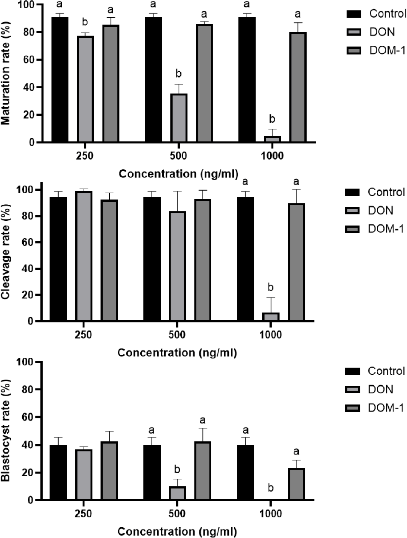
To evaluate the effects of DON and its metabolite DOM-1 on porcine oocyte maturation and subsequent embryonic development, oocytes were subjected to IVM in the presence of increasing concentrations of each mycotoxin, followed by parthenogenetic activation via electrostimulation (Fig. 1). DON exposure resulted in a pronounced, dose-dependent impairment of oocyte maturation and subsequent embryonic development (Table 2). Even at a moderate concentration, DON significantly reduced the oocyte maturation rate (35.00%, p < 0.05) compared with that of control group (91.00%, p < 0.05). In addition, DON markedly compromised blastocyst formation (10.00%, p < 0.05) compared to the control group (39.93%, p < 0.05). At the highest concentration tested, oocyte maturation was almost completely inhibited, and no embryonic cleavage or blastocyst development was observed, indicating severe toxicity of DON toward both nuclear maturation and developmental competence. By contrast, as shown in Table 2, DOM-1 exposure did not significantly affect oocyte maturation or early embryonic cleavage across the tested concentration ranges, and both parameters remained comparable to those of the control group (p < 0.05). However, blastocyst development was adversely affected at the highest concentration of DOM-1 (23.00%), whereas lower concentrations supported blastocyst formation at levels similar to those of the control (39.93%, p < 0.05). These findings suggest that DOM-1 exerts substantially weaker reproductive toxicity than DON, with detrimental effects on embryonic development observed only at elevated concentrations. Based on these results, because a significant difference emerged at concentrations ≥ 500 ng/ml, the treatment range for subsequent experiments was defined as 500–1,000 ng/mL.
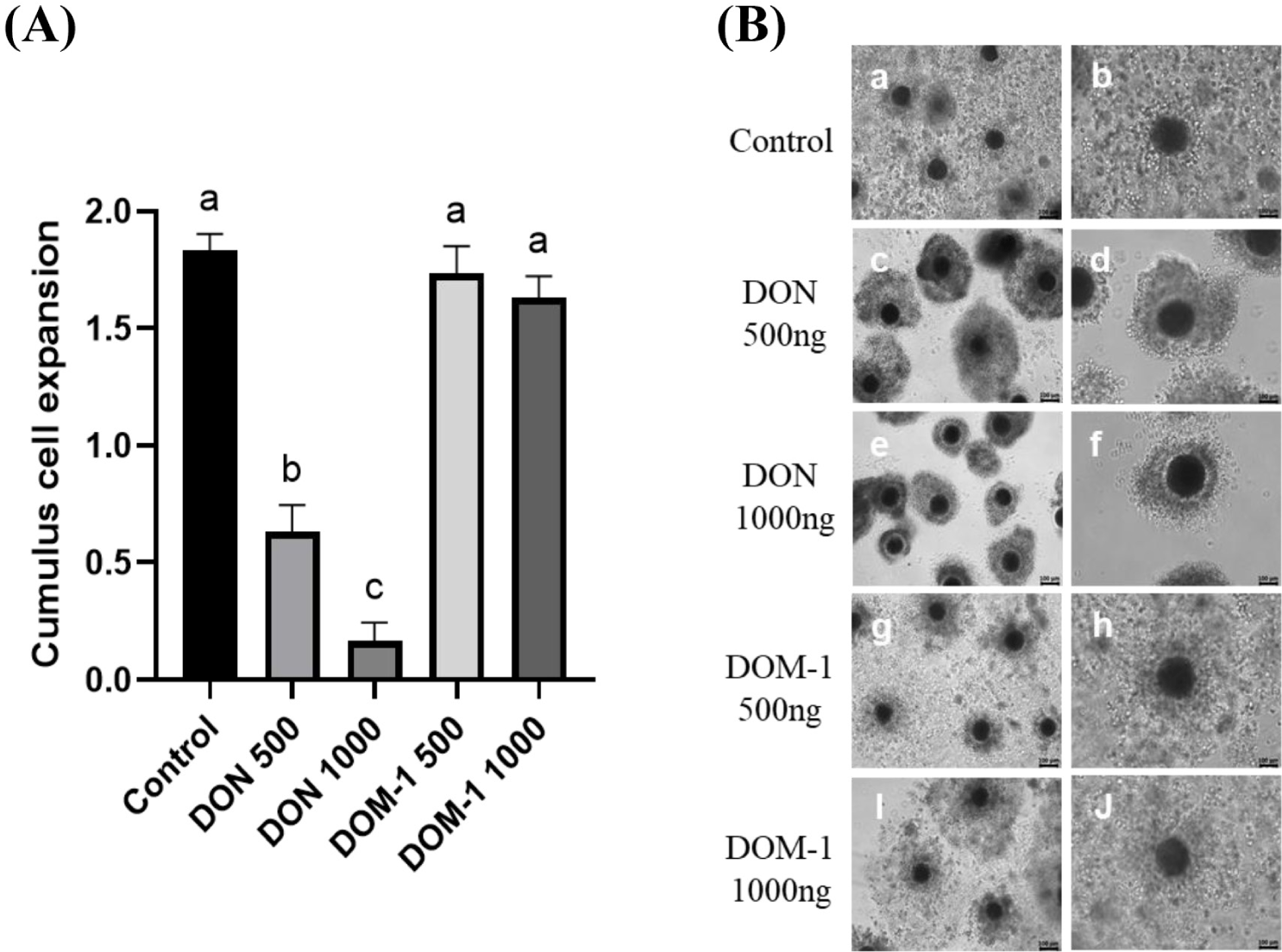
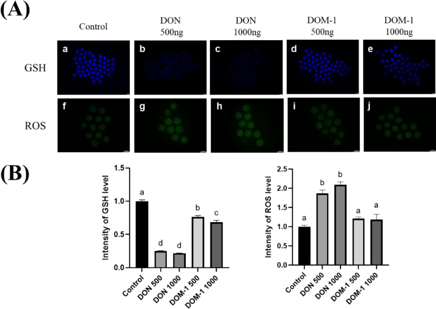
To evaluate the effects of DON and DOM-1 on cumulus expansion, porcine COCs were cultured in a maturation medium supplemented with the respective treatments. After 44 h of IVM, cumulus expansion was observed under a microscope and scored according to established criteria (Fig. 2). The results showed that COCs in both DON-treated groups had significantly reduced cumulus expansion compared with that of the control, with the 1,000 ng/mL DON group displaying a more pronounced reduction than the 500 ng/mL group. By contrast, DOM-1 treatment did not significantly affect cumulus expansion, and the COCs displayed morphology similar to that of the control group. These findings suggest that, unlike DON, DOM-1 has minimal detrimental effects on cumulus cell function during oocyte maturation. To further determine whether these differences in expansion were associated with changes in oxidative stress, intracellular ROS and GSH levels were examined in MII-stage oocytes (Fig. 3). ROS levels were higher in both DON-treated groups than in the control and DOM-1 groups, with the highest levels observed in the 1,000 ng/mL DON group. By contrast, GSH levels were markedly lower in the DON groups than in the control and DOM-1 groups. The DOM-1 groups had slightly lower GSH than the control, but the difference was small. These findings show that DON causes oxidative stress in porcine oocytes by increasing ROS levels and reducing GSH levels, whereas DOM-1 has only a mild effect.
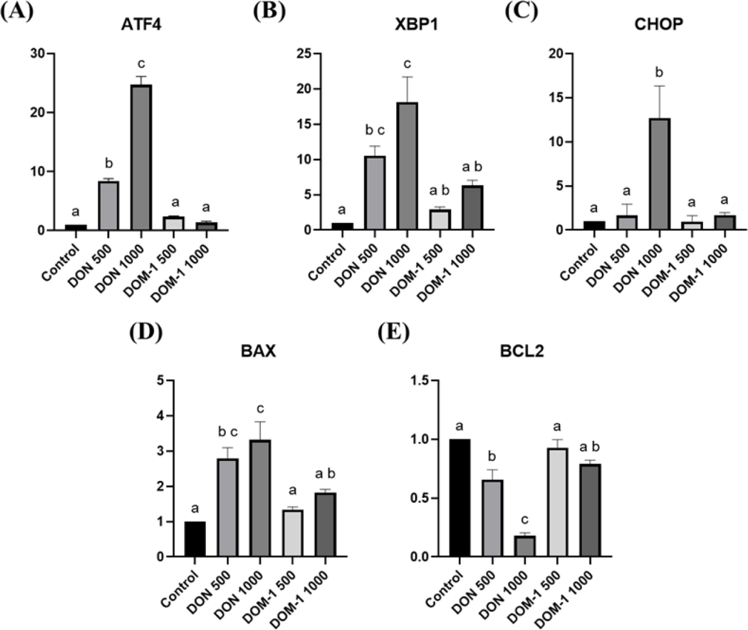
To confirm whether DON and DOM-1 induce apoptosis in porcine COCs during IVM, gene expression analysis was performed using RT-qPCR (Fig. 4). For ER stress-related genes, the expression of activating transcription factor 4 (ATF4) and X-box binding protein 1 (XBP1) was significantly higher in the DON-treated groups, compared with the control, and both genes showed a dose-dependent increase with rising DON concentrations (p < 0.05). The expression of C/EBP homologous protein (CHOP/DDIT3) was significantly elevated only in the 1,000 ng/mL DON group compared with all other groups (p < 0.05). For apoptosis-related genes, the expression of BCL2-associated X protein (BAX) was significantly increased in the DON groups compared with the control, with the 1,000 ng/mL DON group showing the highest expression and significantly exceeding the DOM-1 group (p < 0.05). By contrast, the anti-apoptotic gene B-cell lymphoma 2 (BCL2) was markedly reduced in the DON groups. The 1,000 ng/mL DON group showed the lowest BCL2 expression compared with all other groups (p < 0.05), whereas the 500 ng/mL DON group also showed significantly lower expression than that of both the control and the 500 ng/mL DOM-1 group (p < 0.05).
Because parthenogenetic activation evaluates only the intrinsic developmental competence of oocytes, an additional IVF experiment was conducted to assess fertilization capacity and subsequent embryonic development under conditions closer to physiological reproduction.
Cleavage rates were evaluated 2 d after IVF (Fig. 5A). The cleavage rate in the 500 ng/mL DON group was not significantly different from those in the control and DOM-1 groups (p > 0.05). However, no cleaved embryos were observed in the 1,000 ng/mL DON group. Both DOM-1 groups showed cleavage rates comparable to the control (p > 0.05).
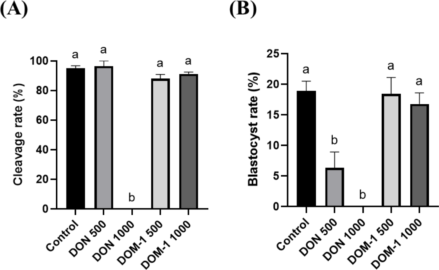
On day 6, blastocyst formation was observed (Fig. 5B). The blastocyst rate was significantly reduced in the DON groups compared with that in control group, and in the 1,000 ng/mL DON group, no embryos developed to the blastocyst stage. By contrast, both DOM-1 groups exhibited blastocyst formation rates similar to those of the control group (p > 0.05). Detailed results are presented in Table 3.
Blastocyst quality was assessed by Hoechst staining (Fig. 6A). Total cell numbers were significantly lower in the 500 ng/mL DON group than in the control and DOM-1 500 ng/mL groups (p < 0.05). No expanded blastocysts were available for staining in the 1,000 ng/mL DON group.
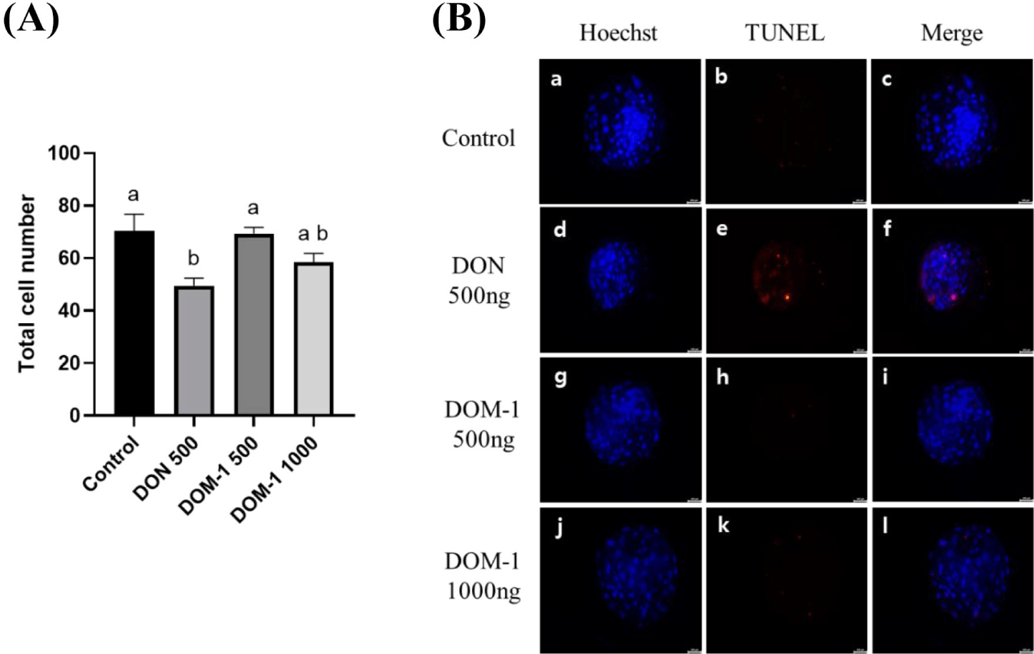
TUNEL staining was performed to determined apoptosis levels (Fig. 6B). Blastocysts derived from the 500 ng/mL DON group contained significantly more TUNEL-positive cells than those from the control and DOM-1 groups (p < 0.05). No significant differences were observed between the control and DOM-1 groups. No expanded blastocysts were obtained from the 1,000 ng/mL DON group for TUNEL analysis.
To investigate whether Res could mitigate the detrimental effects of DON on porcine oocytes, we examined its effects on oocyte maturation, apoptosis-related gene expression, and subsequent embryonic development.
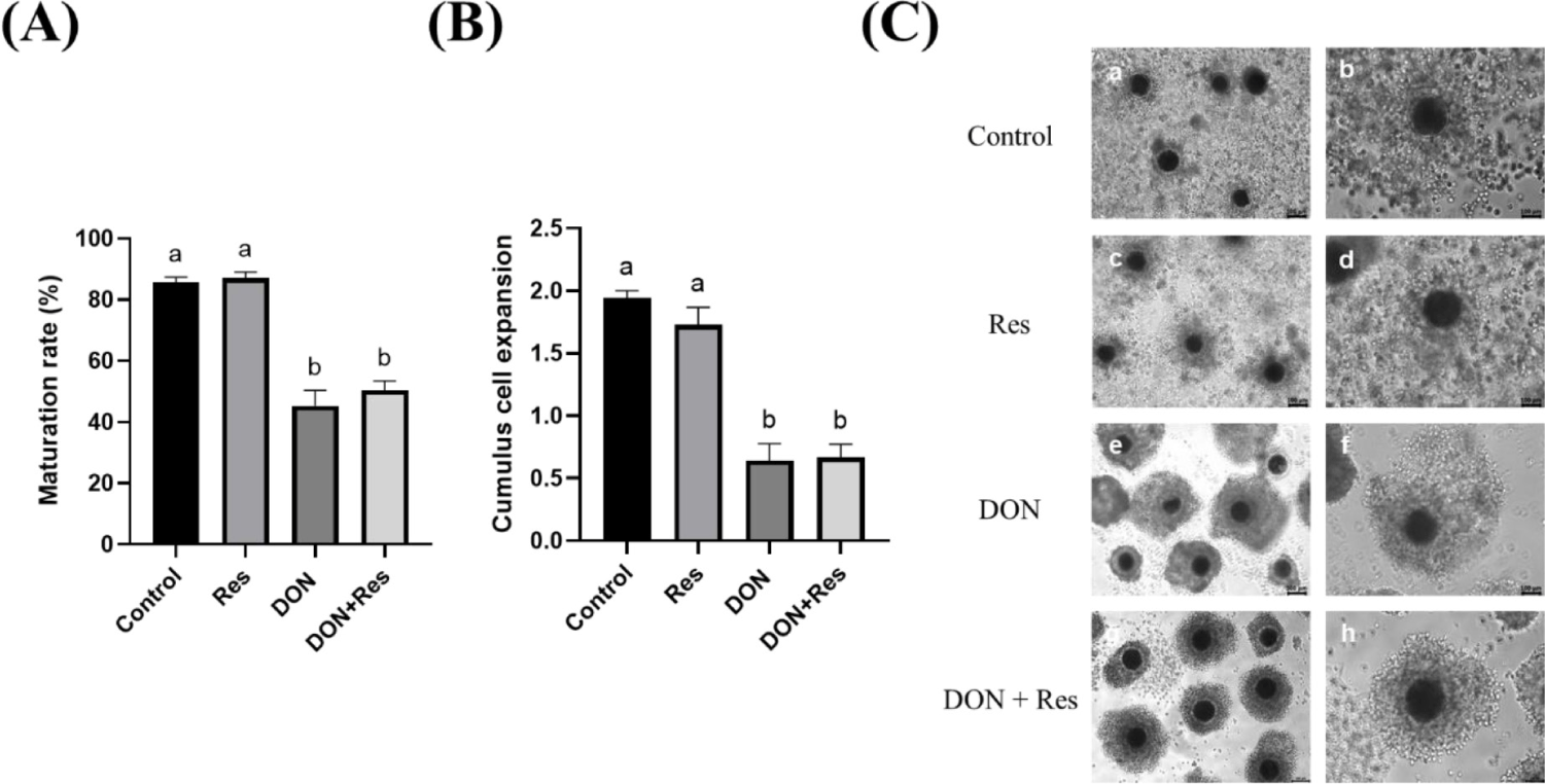
When oocytes were cultured with 2 μM Res, 500 ng/mL DON, or 500 ng/mL DON plus 2 μM Res (DON + Res), no significant differences in maturation rate or cumulus expansion were observed between the control and Res groups. By contrast, both the DON and DON + Res groups showed significantly impaired oocyte maturation and reduced cumulus expansion compared with the control and Res-only groups. Moreover, Res supplementation did not restore the maturation or cumulus expansion of DON-treated COCs (Fig. 7).
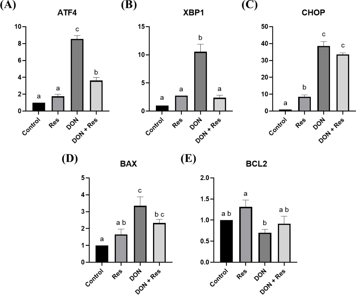
To further assess the protective role of Res, the expression of ER stress– and apoptosis–related genes was examined using RT-qPCR (Fig. 8). As expected, the DON group exhibited significantly elevated expression of ATF4, XBP1, and CHOP compared with the Control and Res groups (p < 0.05). Importantly, Res co-treatment (DON + Res) significantly reduced ATF4 and XBP1 expression relative to DON alone, although CHOP expression remained elevated. For apoptosis-related markers, BAX expression was significantly higher in the DON group than in the control and Res groups, whereas expression in the DON + Res group did not differ significantly from that in the DON group. By contrast, BCL2 expression was significantly reduced in the DON group, whereas its levels in the Res and DON + Res groups were comparable to those in the control group (p > 0.05). These data indicate that Res alleviates DON-induced ER stress but only partially mitigates pro-apoptotic signaling.
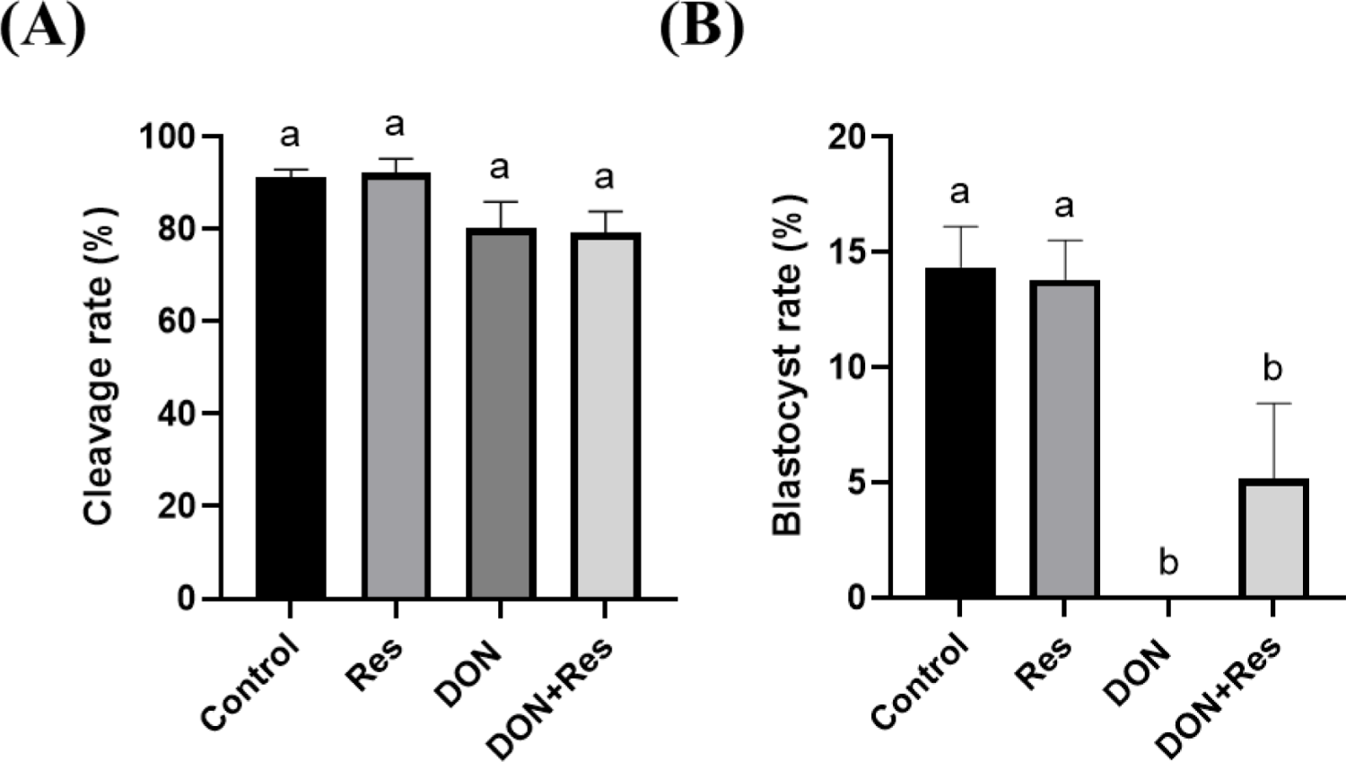
Finally, the effects of Res on the developmental potential of DON-treated oocytes were evaluated using IVF (Fig. 9). On day 2, cleavage rates did not differ significantly among the groups (p > 0.05; Fig. 8A). However, by day 6, the DON group failed to produce blastocysts, whereas the DON + Res group produced embryos that developed to the blastocyst stage. Although the blastocyst rate in the DON + Res group was lower than that in the control and Res groups (p < 0.05), the presence of blastocysts indicates a partial protective effect of Res against DON-induced developmental arrest (Fig. 9B). Detailed results are presented in Table 4.
DISCUSSION
This study demonstrated that DON exerts profound toxicity on porcine oocytes, whereas its metabolite DOM-1 has minimal detrimental effects. DON exposure impairs cumulus expansion, reduces maturation rates, induced oxidative and ER stress, triggers apoptosis, and ultimately compromises developmental competence, by contrast DOM-1 exhibits minimal or no detrimental effects under the same conditions. Furthermore, Res supplementation partially alleviated DON-induced stress responses and increased the proportion of cleaved embryos that developed into blastocysts, highlighting its potential as a protective agent against mycotoxin-induced reproductive toxicity.
Interestingly, our findings suggest that DON-induced developmental arrest may involve two distinct windows of sensitivity in porcine oocytes and embryos. The first window occurs during oocyte maturation, when DON disrupts cumulus expansion, increases oxidative and ER stress, and shiftes apoptotic gene expression profiles, thereby reducing oocyte competence prior to fertilization. The second window occurs during zygotic genome activation (ZGA), which occurs after the 4-cell stage [30]. At this stage, embryonic development becomes highly dependent on de novo transcription and protein synthesis. Given that DON exerts its toxicity by binding to the ribosomal peptidyl transferase center and inhibiting protein synthesis [31,32], it is plausible that DON-exposed zygotes, even if they reach the 4-cell stage, fail to progress further because of impaired transcriptional activation and translational capacity. This dual impact—first at the maturation stage and later during ZGA—provides a mechanistic explanation for the severe reduction in blastocyst formation observed in our study. By contrast, DOM-1, which lacks the epoxide moiety required for ribosomal binding [33,34], did not impair ZGA-dependent development, which is consistent with its minimal cytotoxicity in oocytes. In this study, DON exposure was limited to the oocyte maturation period, therefore, the observed effects were directly attributed to alterations in oocyte quality. Although impaired oocyte maturation may subsequently influence early embryonic development, including stages around the maternal–zygotic transition, this process was not directly examined in the current study. Thus, any potential involvement of DON in the maternal–zygotic transition should be interpreted with caution and warrants further investigation.
During IVM, oocytes rely predominantly on stored maternal transcripts and post-transcriptional control, with minimal de novo transcription; whereas, cumulus cells remain transcriptionally active and exchange metabolites and redox equivalents with the oocyte via gap junctions in transzonal projections. Thus, ROS and GSH depletion detected in MII oocytes likely reflect disrupted cumulus–oocyte metabolic coupling rather than primary transcriptional changes within the oocyte itself [35]. Cumulus cells are key suppliers of cysteine, pyruvate, and NADPH, and help sustain intra-oocyte GSH, which is a principal antioxidant for meiotic competence. Perturbation of cumulus function is therefore expected to reduce oocyte GSH levels and elevate ROS levels, ultimately compromising maturation quality. Our results showing higher ROS levels and lower GSH levels in the DON groups are consistent with this biology and with the literature linking cumulus redox support to oocyte competence [36–38]. To localize the transcriptional response, we assayed COCs and found that DON activated ER-stress–related markers (ATF4, XBP1, and CHOP) and shifted the apoptotic balance toward cell death, with significantly increased BAX expression and markedly decreased BCL2 expression. These changes align with the known actions of DON, which trigger ribotoxic and ER stress pathways, and apoptosis in porcine reproductive cells and embryos [39]. Conversely, DOM-1, a deep-epoxidized metabolite, showed minimal changes in ER stress or apoptosis markers in COCs and had a negligible impact on ROS levels in oocytes, consistent with its markedly reduced cytotoxicity relative to DON. Together, these findings support a model in which DON first impairs cumulus-mediated redox support during maturation (resulting in increased ROS and decreased GSH in the oocyte without changes in oocyte gene-expression), and secondarily drives ER stress–linked apoptosis at the cumulus–oocyte complex level, culminating in poor cleavage and blastocyst outcomes.
The present study examined the potential of Res to counteract the toxic effects of DON on porcine oocytes. Res was selected because of its well-documented antioxidant and anti-apoptotic properties, as previous studies have shown that it reduces intracellular ROS, enhances GSH levels, and improves oocyte competence in mammalian reproduction [25,40]. Previous studies have demonstrated that dietary Res supplementation during gestation and lactation improves the antioxidant status of both sows and piglets, accompanied by modulation of antioxidant-related gene expression and activation of the Kelch-like ECH-associated protein 1 (KEAP1)–NRF2 signaling pathway in the placenta [41]. Considering that measurable levels of DON have been detected in the body fluids of sows and their fetuses that consumed DON-contaminated feed in previous studies [42], we hypothesized that supplementation of the oocyte culture medium with Res would exert antioxidant effects, thereby alleviating DON-induced toxicity and improving cumulus cell function and oocyte maturation. Although Res supplementation did not restore cumulus expansion or nuclear maturation impaired by DON, it exerted beneficial effects at the molecular level within COCs. Specifically, Res can attenuated the DON-induced upregulation of ER stress markers such as ATF4 and XBP1 and stabilized the expression of the anti-apoptotic gene BCL2 [29]. These results suggest that Res partially relieves ER stress in cumulus cells, thereby maintaining a more favorable microenvironment for the enclosed oocytes.
At the developmental level, the protective effects of Res were more apparent. While cleavage rates in the DON + Res group showed only a modest, non-significant trend toward improvement compared with the DON group, blastocyst development was partially restored. Notably, embryos derived from DON + Res oocytes progressed beyond the cleavage stage and reached the blastocyst stage, which was completely blocked in the DON-only group. This finding suggests that although Res cannot fully rescue the maturation process oocytes that succeed in maturation under DON + Res conditions may retain high developmental competence, possibly supported by improved cumulus-mediated signaling or antioxidant defense during maturation.
Collectively, these findings highlight Res as a potential mitigator of DON-induced damage in porcine oocytes, particularly through modulation of ER stress and partial restoration of developmental potential. Nevertheless, the inability of Res to fully restore cumulus expansion, nuclear maturation, and blastocyst rates to control levels underscores the need for additional strategies, possibly involving combined antioxidant or signaling-targeted interventions, to comprehensively overcome the reproductive toxicity of DON.


