

INTRODUCTION
Salmonella is a prominent foodborne pathogen that poses significant challenges to global food safety, particularly in the poultry industry. Salmonella infections are primarily transmitted through the consumption of contaminated poultry products, leading to serious illnesses including gastroenteritis and food poisoning in both humans and animals [1]. Millions of Salmonella infection cases are reported annually worldwide, and these infections impose a substantial economic burden on public health systems and the agricultural sector. In the poultry industry, the economic impact of Salmonella outbreaks is particularly severe, resulting in reduced productivity, increased costs related to recalls and containment efforts, decreased consumer confidence, and barriers to international trade [2]. This global issue emphasizes the need for effective measures to control Salmonella in poultry production.
Traditionally, antibiotics have been the primary method of controlling Salmonella infections in the poultry industry [3]. However, widespread and often indiscriminate use of antibiotics has led to an alarming increase in the number of antibiotic-resistant bacteria, also known as superbugs. The growing prevalence of antibiotic resistance has triggered an urgent need for alternative approaches to manage bacterial infections [4]. A promising alternative to antibiotics is the use of bacteriophages, viruses that naturally target and infect specific bacterial hosts. Bacteriophages offer several advantages over antibiotics, including specificity for pathogenic bacteria without disrupting beneficial microorganisms in the host microbiome [5]. In the context of Salmonella control, bacteriophages have shown potential as highly selective tools for targeting and eliminating pathogenic strains in poultry farming, making them attractive candidates for future intervention. Previous studies have demonstrated the efficacy of bacteriophages in reducing Salmonella levels in poultry, suggesting their potential as viable alternatives to antibiotics [6].
Probiotics have gained increasing recognition for their ability to improve gut health, enhance immune responses, and inhibit the growth of harmful pathogens [7]. Probiotics, particularly those from the Lactobacillus genus, play critical roles in maintaining healthy and balanced intestinal microbiota, promoting overall gut health, and preventing the colonization of harmful bacteria [8]. Several studies have highlighted the importance of Lactobacillus spp. in poultry production, as they contribute to improved feed efficiency, better nutrient absorption, and enhanced disease resistance. The ability of probiotics to modulate gut microbiota and outcompete pathogenic bacteria underscores their value as a preventive strategy for managing Salmonella and other enteric pathogens in poultry [9].
Despite the individual benefits of bacteriophages and probiotics, few studies have explored the synergistic effects of combining these two approaches to enhance pathogen control [10,11]. Although bacteriophages are highly effective at specifically targeting and killing pathogenic bacteria [12], probiotics can simultaneously help restore and maintain healthy gut microbiota by promoting the growth of beneficial bacteria [13]. Therefore, the combined application of bacteriophages and probiotics may offer a more comprehensive solution for pathogen control to reduce the population of harmful bacteria such as Salmonella and promote a balanced gut microbiota that can resist future infections.
In this study, we investigated the synergistic effects of bacteriophages and probiotics in controlling Salmonella infections in the poultry industry. We combined two previously isolated bacteriophages, SLAM_phiST45 and SLAM_phiST56, which were selected for their broad host range and ability to infect Salmonella Typhimurium strains from poultry, swine, and humans. Together, these bacteriophages formed a cocktail that demonstrated superior efficacy in suppressing Salmonella growth compared to single-phage treatments. In addition, we used Lactobacillus reuteri J2M1, a probiotic strain chosen for its robust performance in multiple assays, including acid and bile tolerance, intestinal adhesion, and safety evaluations. L. reuteri J2M1 has previously been shown to have strong probiotic properties and the potential for commercial use in the poultry industry because of its resilience and beneficial effects on gut health.
This study aimed to assess the survival rates of bacteriophages and probiotics as they pass through simulated gastric and intestinal phases, followed by their combined effects on Salmonella suppression and gut microbiota modulation in a cecum fermentation model. Our hypothesis was that the combination of bacteriophages and probiotics would effectively reduce Salmonella populations and improve gut microbiota diversity and stability, contributing to enhanced gut health in poultry. In this study, we aimed to provide new insights into the potential synergistic effects of phages and probiotics as a dual strategy for managing bacterial infections and promoting a healthy microbiome in the poultry industry. This study represents one of the first attempts to combine bacteriophages and probiotics for the dual purposes of pathogen suppression and modulation of the gut microbiota in poultry. The results of this study are expected to provide a foundation for future strategies that can simultaneously address food safety concerns and enhance the overall health and productivity of poultry, thereby contributing to a more sustainable and effective approach for controlling Salmonella and other enteric pathogens in livestock production.
MATERIALS AND METHODS
To isolate a diverse range of lactic acid bacteria (LAB) candidate strains using a culturomics approach, the contents of the cecum, ileum, and jejunum from 5-week-old broilers were used in this study. Briefly, 10 g of each sample was placed in a sample bag (3M) containing 90 mL of 0.1% buffered peptone water (Oxoid) and homogenized using a stomacher for 2 min. The homogenized solution was then serially diluted and plated on Gifu Anaerobic Medium (GAM; Kisan Bio), Bifidobacterium Selective Broth (BS; Kisan Bio), Brain Heart Infusion broth (BHI; BD Difco), Reinforced Clostridial Medium (RCM; BD Difco), and Lactobacilli Man, Rogosa, and Sharpe (MRS) broth (MRS; BD Difco) supplemented with 1.5% Bacto Agar (BD Difco) [14]. The plates were incubated under anaerobic conditions at 37°C for 48 h. Isolated single colonies were harvested and sub-cultured on the same agar plates, followed by identification using 16S rRNA sequencing. Strains with a 16S rRNA gene sequence identity of ≥98% were preserved in 15% glycerol stocks for future experiments. The experimental protocol was approved by the Committee on the Ethics of Animal Experiments of the Chungnam National University. Approval number: CNU-00779.
For metagenomic analysis, 1 mL was removed from each of the three broiler contents that were homogenized in 0.1% buffered peptone water used in the culture procedure. Briefly [15], Genomic DNA (gDNA) was extracted from samples using the DNeasy PowerSoil Pro Kit (Qiagen). Following extraction, the samples were sent to Sanigen, where the V3–V4 region of the 16S rRNA amplicon sequencing was performed using a primer set (forward: 515F, 5’-TCGTCGGCAG CGTCAGATGTGTATAAGAGACAGGTGCCAGCMGCCGCGGTAA-3’; reverse: 806R, 5’-GTCTCGTGGGCTCGGAGATGTGTATAAGAGACAGGGACTACHVGGGTWTC TAAT-3’) on the Illumina Nextseq platform (2 × 300; Order No. O-20231214-16S-396).
Among the bacteria isolated through culturomics and those identified via metagenomics, we selected and organized species with characteristics similar to LAB, such as Lactobacillus, Bifidobacterium, and certain Enterococcus spp. [14]. Subsequently, a Venn diagram was created based on the metagenome and culturome of the selected probiotic candidates and further experiments were conducted using species from the intersection.
To evaluate acid and bile tolerance, we performed a method modified from a previous study [16]. First, to prepare the acidic medium, MRS broth was adjusted to pH 2.5 with 6 N HCl (Sigma-Aldrich). The broth was autoclaved and then supplemented with pepsin from porcine gastric mucosa (Sigma-Aldrich) at 1,000 units/mL using a 0.45 μm syringe filter (Sartorius Korea Biotech). For the acid tolerance test, 100 μL of overnight-cultured bacteria were inoculated into 10 mL of the acidic broth and incubated at 37°C for 3 h, after which colony-forming units (CFU) were counted on MRS agar. Artificial bile broths were prepared by mixing Oxgall (Acumedia Manufacturers) with MRS broth at final concentrations of 0.3% and 1%. Next, 100 μL of overnight-cultured bacteria were inoculated into 10 mL of the artificial bile broths at different concentrations and incubated at 37°C for 24 h. The survival rate was calculated by comparing the final CFU/mL to the initial CFU/mL.
For the cell adhesion experiment, primary intestinal cells from 10-week-old broilers provided by the Daejeon Poultry Research Unit of Chungnam National University (Daejeon, Korea) were seeded in a 24-well plate at a concentration of 2.4 × 10⁵ cells/cm2. The bacterial culture was washed five times with filter-sterilized phosphate-buffered saline (PBS; LPS Solution) and then resuspended in high-glucose Dulbecco’s modified Eagle medium (DMEM)/F12 (Gibco) to a final concentration of 1.5 × 10⁷/mL. The cells were then incubated in a humidified environment at 37°C with 5% CO2 for 3 h. To harvest the bacteria-adhered cells, the wells were washed five times with PBS buffer, and then 200 µL of trypsin-ethylenediaminetetraacetic acid (EDTA) solution (Sigma-Aldrich) was added. The cells were incubated in a humidified environment at 37°C with 5% CO2 for 10 min. After incubation, the cells were harvested, serially diluted with 0.85% NaCl, and plated onto MRS agar plates for CFU counting [17].
In this study, the antibacterial activity of the selected probiotics was assessed using three strains of two major poultry pathogens, Salmonella Typhimurium and Campylobacter jejuni [18]. Most pathogens were directly isolated from diseased livestock and obtained from the Korea Veterinary Culture Collection (KVCC). The details of all pathogens used in this experiment, including the culture methods, are summarized in Supplementary Table S1. First, cultures of pathogens grown overnight for 24 h were spread on agar plates, and then 5 μL of overnight-cultured probiotics were dotted on the plates and incubated at 37°C for 24 h. Finally, the inhibition zone was calculated by comparison to the lysis zone of the positive control, Lactobacillus rhamnosus GG (LGG) [19].
To investigate the antibiotic susceptibility, the disc diffusion method, following the standard Kirby-Bauer method [20] was used. First, 100 μL of overnight-cultured bacteria were plated on MRS agar and incubated at 37°C for 48 h. Then, antibiotic discs were placed on the surface of each plate, followed by an additional incubation at 37°C for 24 h. The antibiotic discs used in this experiment (Flinn Scientific) were ampicillin (10 μg), chloramphenicol (30 μg), kanamycin (30 μg), penicillin (10 μg), tetracycline (30 μg), and vancomycin (30 μg). Antibiotic susceptibility of the probiotics was determined according to the guidelines provided in the product manual (Supplementary Table S2).
Caenorhabditis elegans fer-15(b26)II; fem-1(hc17)IV was obtained from the Caenorhabditis Genetic Center (Minnesota, USA) and maintained on Nematode Growth Medium (NGM; 3.5 g Bacto Peptone [BD Difco], 3 g NaCl [Sigma-Aldrich], and 20 g [Bacto Agar]) plates at 15°C. The standard feed for C. elegans, Escherichia coli OP50 (OP50), was cultured in Luria-Bertani (LB; BD Difco) broth at 37°C with shaking at 160 rpm for 24 h. To prepare live bacterial lawns for C. elegans feeding, the bacterial pellet was collected by centrifugation at 8,000 rpm for 5 min, washed twice with sterile M9 buffer (3 g KH2PO4, 6 g Na2HPO4, and 5 g NaCl dissolved in 1 L distilled water, autoclaved), and 1 mL of 1 M MgSO4 (Sigma-Aldrich) was added. The bacterial pellet was concentrated to a final concentration of 2.5 mg/mL (wet weight) in M9 buffer and suspended in NGM plates for further use [21].
For the lifespan study, eggs were harvested from adult worms by bleaching with a sodium hypochlorite-sodium hydroxide solution (Sigma-Aldrich). The newly hatched larvae were then synchronized at the L1 stage on NGM plates maintained at 25°C. After 3 day, the worms, now at the L4 stage, were transferred to 35-mm NGM plates that had been pre-seeded with OP50, LGG, and other probiotics selected through multi-omics techniques. The experiment was repeated thrice, with the worms moved to fresh bacterial lawns every alternate day until all the worms had perished [14].
The growth conditions and other details of the Salmonella Typhimurium strains used for bacteriophage isolation are provided in Supplementary Table S1. Briefly, a total of 61 phages were isolated using 11 Salmonella Typhimurium strains derived from poultry, swine, and humans, with the information summarized in Supplementary Table S3.
Bacteriophage isolation was performed using sludge samples obtained from the Nanji, Tancheon, Seonam, and Jungnang sewage treatment plants (Seoul, Korea). Briefly, 20 mL sludge sample, 20 mL of fresh Tryptic Soy Broth (TSB; BD), and 1 mL of overnight Salmonella culture were mixed and incubated at 37°C, 160 rpm for over 16 h. After centrifugation (10,000 rpm, 5 min), 100 μL of the filter-sterilized supernatant was mixed with 300 μL of bacterial culture and 4 mL of TSB soft agar medium (0.4%). The mixture was then poured onto preprepared TSB agar (TSA) plates (1.5% agar) and incubated at 37°C for 6 h. Single plaques were isolated two to three times, and phage stocks were prepared by plate elution and filter sterilization [22].
After adding 300 μL of bacterial culture to 4 mL of TSB soft agar medium, the mixture was poured onto a prepared TSA plate. The plate was left at room temperature for approximately 15 min to allow for solidification. Then, 10 μL of Salmonella Typhimurium-infecting phages diluted to a concentration of 106 plaque-forming units (PFU)/mL were dotted onto the prepared plate and incubated at 37°C for 6 h to observe bacterial lysis [23]. Based on the host range test, the two selected phages were named SLAM_phiST45 and SLAM_phiST56, and were propagated using Salmonella Typhimurium KVCC-BA0000422 (ST422) and KVCC-BA0000008 (ST008) as host bacteria, respectively. Salmonella Typhimurium KVCC-BA0000422 used in this study is a multidrug-resistant bacterium that is resistant to five out of the six tested antibiotics (Supplementary Fig. S1).
The phage lysate stock was concentrated over 100-fold using polyethylene glycol (PEG; Sigma-Aldrich) precipitation and then centrifuged at 33,000 rpm for 6 h with CsCl (Sigma-Aldrich) solutions at densities of 1.3, 1.4, 1.5, 1.6, and 1.7 g/mL [24]. Next, the phage stock was dialyzed overnight in SM buffer (50 mM Tris–HCl, 100 mM NaCl, and 10 mM MgSO4; pH 7.5) and used for TEM analysis. Briefly [25], 10 μL of the phage stock diluted to 1 × 10⁹ PFU/mL was placed on a carbon-coated copper grid and incubated for 5 min. The grid was then negatively stained with 10 μL of 2% uranyl acetate. The phage morphology was observed using EF-TEM (LIBRA 120, Carl Zeiss) at an accelerating voltage of 120 kV and a magnification of 200,000×g.
One-step growth analysis was performed to determine the burst size and latent period of the selected phages. In summary [26], 1% of an overnight culture was added to 20 mL of fresh TSB medium and incubated at 37°C, 160 rpm until the optical density (600 nm) reached 0.2 (approximately 1 × 10⁷ CFU/mL). The phages were then added at a multiplicity of infection (MOI) of 0.01 and allowed to adsorb to the bacterial cells by incubating without shaking for 5 min. Next, 1 mL of the sample was extracted and centrifuged at 13,500 rpm for 5 min. After filter sterilization, the supernatant was diluted for PFU counting. The remaining samples were removed from the incubator every 5 min, and 1 mL was extracted for further PFU counting.
The pH and temperature stability of the two phages were tested under various conditions [27]. For the temperature stability test, 1 mL of the phage stock diluted to 1 × 10⁹ PFU/mL was mixed with 9 mL of SM buffer and incubated at different temperatures for 1 h, followed by PFU counting. For the pH stability test, 1 mL of the phage, diluted to the same concentration, was added to 9 mL of SM buffer at various pH levels. The mixture was then incubated at 37°C for 1 h, after which PFU counts were performed.
Genomic DNA was extracted from the filter-sterilized PEG-precipitated phage stock after treatment with DNase and SDS using a DNeasy Blood & Tissue Kit (Qiagen) [28]. The obtained gDNA was prepared for ligation sequencing and purification according to the Oxford Nanopore Technologies (ONT) protocol (SQK-LSK109). Next, gDNA was sequenced on the R9.4.1 flow cell using the MinION Mk1B device provided by ONT. Reads of at least 40,000 bp were assembled into a single genome using the Flye (v_2.9.1) assembler [28].
Open reading frames (ORFs) were predicted using RAST [29], Phanotate [30], and Prodigal [31], and ORFs identified by at least two of the programs were selected. The ORFs were then compared with a non-redundant database using the Basic Local Analysis Search Tool for Proteins (BLASTP) algorithm and functionally annotated. Based on the completed annotation table, SLAM_phiST45 and SLAM_phiST56 were submitted to National Center for Biotechnology Information (NCBI) BankIt, where they were assigned the accession numbers PP948674 and PP948675, respectively. tRNA-encoding genes were identified using ARAGORN (v1.2.41) [32]. The circular genome map of SLAM_phiST1N3 was prepared using CGView [33] based on the FASTQ file obtained from BankIt.
In the past, bacteriophage classification was primarily based on morphological characteristics observed through TEM analysis. The families Siphoviridae, Podoviridae, and Myoviridae, which belong to the Caudovirales order, are the primary representatives. However, with the recent dissolution of the Caudovirales order, morphological classification is no longer used [34]. In the present study, we initially used genome-based BLAST for nucleotides (N) searches to classify SLAM_phiST45 and SLAM_phiST56 as subfamilies. Next, we performed a genus-level classification by conducting BLASTN analyses on species belonging to these subfamilies, as defined by the International Committee on Taxonomy of Viruses (ICTV) [35]. Finally, species-level classification was performed by calculating the Average Nucleotide Identity (ANI) with closely related species identified from the BLASTN results. ANI analysis was performed using three different methods. ANIb [36] was calculated using BLAST+ and ANIm [37] was based on MUMmer with JSpeciesWS, whereas orthoANI [38] was calculated using the OrthoANIu algorithm available on the EzBioCloud server.
The selected phages were used to inhibit Salmonella in a liquid medium through single-phage and phage cocktail applications. Briefly, Salmonella overnight culture was inoculated at 1% in fresh TSB medium to reach an OD (600 nm) of 0.3, followed by treatment with a single phage or phage cocktail at an MOI of 1. Optical density was then monitored hourly under shaking conditions (160 rpm) at 37°C [39].
To determine the synergistic effect of SLAM_phiST45 and SLAM_phiST56, the frequency of occurrence of BIM was assessed for both individual phages and the phage cocktail. Briefly [40], a 1% overnight culture was inoculated into fresh medium and grown to an OD (600 nm) of 0.2. The culture was then mixed with either individual phages or the cocktail at an MOI of 100 (approximately 1 × 10⁷ CFU/mL bacteria and 1 × 10⁹ PFU/mL phage) and incubated at 37°C for 20 min to allow phage adsorption. Finally, the mixture was added to 4 mL of TSB soft agar and poured onto TSA plates, followed by incubation at 37°C for 24 h. BIM frequency was calculated by determining the ratio of the number of surviving colonies on other plates to the number of surviving colonies after the phage cocktail treatment.
The EOP was assessed for the two phages used in the phage cocktail against 11 different Salmonella Typhimurium strains, including their respective host bacteria. First, 11 overnight Salmonella cultures were mixed with appropriately diluted phages in TSB soft agar, and the mixture was poured onto TSA plates. The plates were allowed to solidify at room temperature for 15 min and then incubated at 37°C for 6 h. The EOP was calculated by dividing the average PFU of the target bacteria by the average PFU on the host bacteria [41].
To simulate the chicken gastrointestinal phase and test the stability of probiotics and phages, a modified version of a previously described method was used [42]. For probiotics, 5 mL of the overnight culture was centrifuged at 8,000 rpm for 5 min, and the supernatant was removed. The pellet was resuspended in 5 mL chicken gizzard digestive juice (1 M NaCl, 10 g/L pepsin from porcine gastric mucosa [Sigma-Aldrich], pH 2.5) or intestinal digestive juice (3.5% bile extract [Sigma-Aldrich] and 0.35% pancreatin from porcine pancreas [Sigma-Aldrich], pH 6.0). The gastric phase was incubated for 1 h, and the intestinal phase for 3 h at 41°C with shaking. For phage stability, 100 µL of phages concentrated to 1 × 10¹⁰ PFU/mL using PEG precipitation was added to 9.9 mL of gizzard digestive juice and intestinal digestive juice and incubated under the same conditions. Loss occurring during the gastric and intestinal phases was measured by comparing the CFU of probiotics and PFU of phages before and after the experiment.
The chicken gastrointestinal phase primarily tested the stability of the bacteria and phages, whereas the cecal fermentation phase focused on assessing Salmonella inhibition and changes in the microbiota caused by the phages or probiotics. Briefly [43], 1 g of cecum was added to 10 mL of modified Gifu anaerobic medium (mGAM, HIMEDIA) along with Salmonella and probiotic cultures at 1 × 10⁷ CFU/mL each. The phage cocktail was added at a concentration of 1 × 10⁷ PFU/mL. The mixture was incubated with shaking at 41°C, similar to the gastrointestinal phase, and Salmonella reduction as well as changes in the gut microbiota were observed after 24 h. First, the relative abundance of Salmonella was analyzed using previously validated primers (Supplementary Table S4) [44] on genomic DNA extracted from the samples before and after fermentation [45]. Reverse transcription-polymerase chain reaction (RT-qPCR) was performed using the miScript SYBR Green PCR Kit and the CFX96 Real-Time System (Bio-Rad). Next, to analyze changes in the gut microbiota, the gDNA from each sample was sent to Sanigen, where 16S rRNA amplicon sequencing of the V3–V4 region was performed using a primer set (Forward: 515F, 5’-TCGTCGGCAGCGTCAGATGTGTATAAGAGACAGG TGCCAGCMGCCGCGGTAA-3’; Reverse: 806R, 5’-GTCTCGTGGGCTCGGAGATGTG TATAAGAGACAGGGACTACHVGGGTWTCTAAT-3’) on the Illumina Nextseq platform (2 × 300; Order No. O-2024523-16S-154).
RESULTS
In the phylogenetic analysis, 394 bacterial strains were isolated by culturing the cecum, ileum, and jejunum of chickens in five different media under anaerobic conditions (Fig. 1A). The most frequently isolated species were L. reuteri (18.3%), L. crispatus (16.2%), and L. salivarius (11.4%; Supplementary Fig. S2A). L. reuteri was the most abundant species in the cecum (19.3%) and ileum (22.7%), whereas L. salivarius was the most commonly detected species in the jejunum (20.5%; Supplementary Figs. S2B, S2C, and S2D). Next, for the metagenomic analysis, DNA was extracted from the contents of the three intestinal sections to analyze the actual gut microbiota (Fig. 1B). The overall abundances were as follows: Ruminococcus (15.4%), Oscillospira (12%), Clostridium (10%), and Lactobacillus (8.4%; Supplementary Fig. S3A). These four genera were the most abundant across all three intestinal sections (Supplementary Figs. S3B, S3C, and S3D). However, significant differences were observed in the alpha and beta diversity analyses of the metagenomic data from the three intestinal sections. In particular, the jejunum showed significant differences compared to the other two sections in both the Chao and Berger–Parker indices (Supplementary Figs. S4A and S4B). Similarly, results from weighted and unweighted UniFrac analyses showed distinct clustering of the jejunum compared to the other two intestinal sections (Supplementary Figs. S4C and S4D).
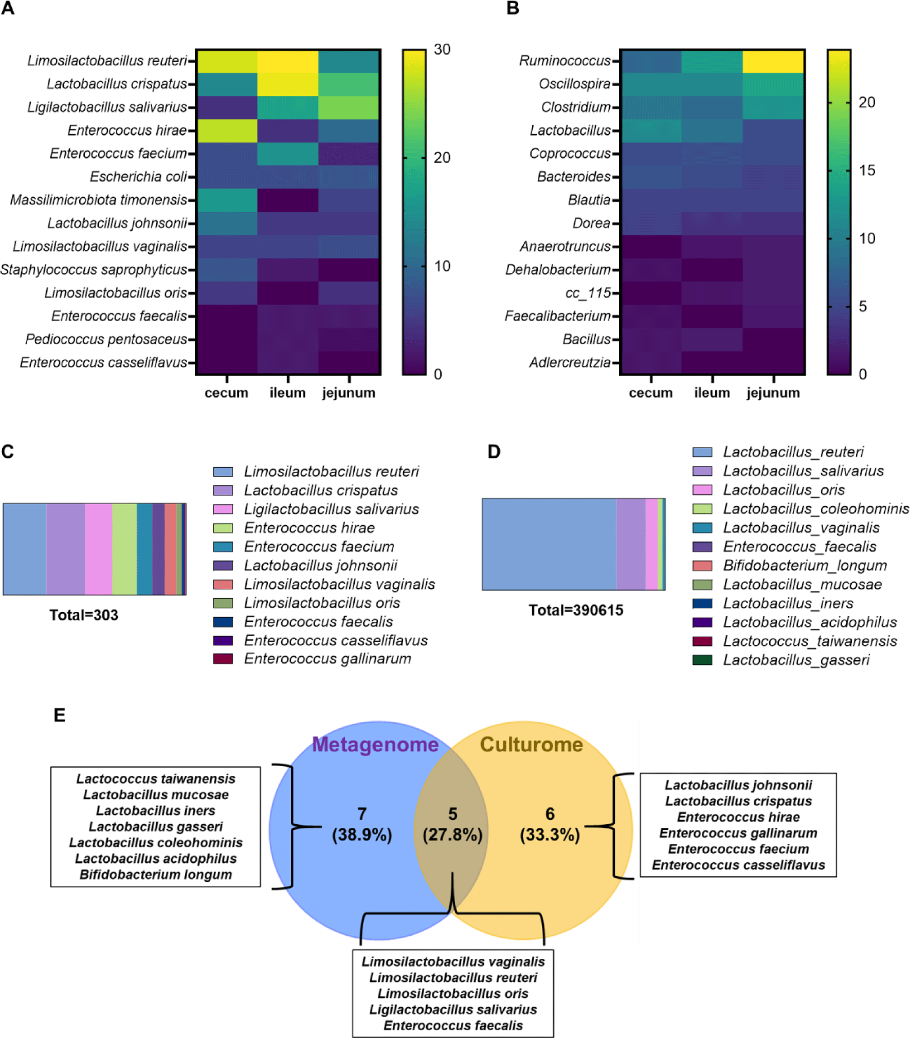
Subsequently, bacterial species from the Lactobacillus, Enterococcus, and Bifidobacterium genera found in both the culturomic and metagenomic analyses were chosen as probiotic candidates. Interestingly, L. reuteri showed the highest abundance in both the culturome (23.8%) and the metagenome (73.3%) results (Figs. 1C and 1D). When the two datasets were presented using a Venn diagram, five species, Lactobacillus vaginalis, L. reuteri, Lactobacillus oris, L. salivarius, and Enterococcus faecalis, were found at the intersection (Fig. 1E). Examination of the probiotic-based multi-omics results for each intestinal section revealed that L. reuteri, L. vaginalis, and L. salivarius were always included at the intersection (Supplementary Figs. S5A, S5B, and S5C). Five species were selected as probiotic candidates using a multi-omics-based approach, and probiotic activity tests were subsequently conducted.
In a previous study, five species were selected through a multi-omics analysis based on culturomic and metagenomic approaches. The number of strains of each species, which were isolated using culturomic analysis and stored as glycerol stock, was as follows: 72 strains of L. reuteri, 45 strains of L. salivarius, 19 strains of L. vaginalis, nine strains of L. oris, and four strains of E. faecalis. However, for industrial applicability and convenience, strains that could not be cultured under aerobic conditions were excluded. Ultimately, an acid tolerance experiment was conducted using 20 strains of L. reuteri, six strains of L. salivarius, 12 strains of L. vaginalis, five strains of L. oris, and one strain of E. faecalis (Fig. 2A). After excluding E. faecalis I2B14, which had a survival rate of 70.5%, the two strains with the highest survival rates among the four Lactobacillus spp. were selected and bile tolerance experiments were conducted.
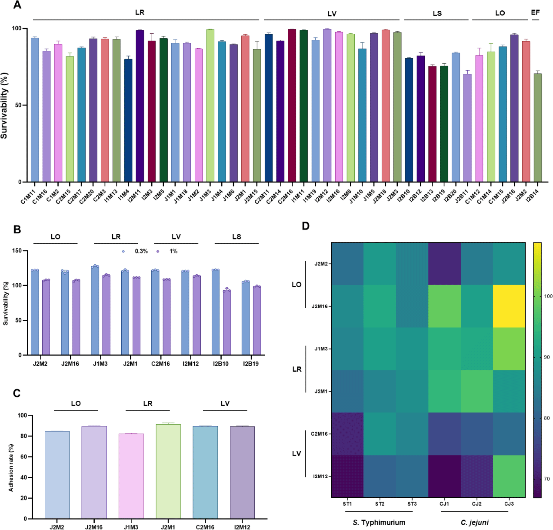
The eight Lactobacillus strains selected for the acid tolerance experiment were exposed to bile salts at concentrations of 0.3% and 1% for 24 h. When exposed to 0.3% bile salts, the CFU of all strains increased compared with the initial count. In contrast, when exposed to 1% bile salts, L. salivarius I2B10 and I2B19 showed a decrease in cell density to 93.65% and 98.74% of the initial count, respectively. However, the other six strains showed an increase in CFU compared to the initial count (Fig. 2B). A lower survival rate of L. salivarius in both acid tolerance and bile tolerance experiments was also observed in a previous study [46]. In addition, the viability of the L. salivarius strains decreased rapidly when the bacteria were stored in a refrigerator. As a result, due to their relatively low acid and bile tolerance and the rapid decline in viability during refrigeration, L. salivarius strains were excluded from subsequent experiments. Next, a cell adhesion experiment was conducted by culturing the selected Lactobacillus strains on primary chicken cells for 3 h and then comparing the bacterial counts to the initial concentration. L. reuteri J2M1 showed the highest adhesion rate of 91.6%, whereas the other strains demonstrated adhesion rates above 80% (Fig. 2C).
To assess the antimicrobial activity of Lactobacillus strains, we used Salmonella Typhimurium and Campylobacter jejuni, which have the highest prevalence in poultry farms. Information regarding the pathogens used is summarized in Supplementary Table S1. Three strains were tested for each species. Antimicrobial activity was measured by comparing the lysis zone with that of the positive control L. rhamnosus GG. On average, the strains with the highest antimicrobial activity were L. oris J2M16 (94.16 %), L. reuteri J1M3 (92.69 %), and L. reuteri J2M1 (88.25 %; Fig. 2D). In addition, to test the stability of Lactobacillus strains, an antibiotic resistance experiment was conducted using six antibiotics commonly used in the livestock industry. L. reuteri J2M1 and L. vaginalis C2M16 were sensitive to all antibiotics, except kanamycin, whereas the other four strains showed resistance to both kanamycin and tetracycline (Table 1).
Antibiotic susceptibility of the three final Lactobacillus spp. was tested against six commonly used antibiotics in the livestock industry.
C. elegans is a well-established model organism widely used as an in vivo alternative for studying metabolic and genetic processes. We selected it as a surrogate model for poultry and conducted experiments to compare its lifespan with that of poultry fed a standard diet. In this study, C. elegans was fed a standard diet of E. coli OP50 from stages L1 to L4. From the L4 stage onwards, they were administered either OP50, LGG, or the Lactobacillus strains selected in the previous experiments, and their lifespan was evaluated. All treatment groups showed a significant increase in lifespan compared with the OP50 group. Notably, L. reuteri J2M1, L. oris J2M2, and L. vaginalis C2M16 exhibited a trend of extending the lifespan even more than the positive control, LGG. On the 6th day of the experiment, all treatment groups had a survival rate of approximately 70 %. However, 2 day later, on the 8th d, the survival rate of OP50-fed C. elegans dropped significantly to 30%. In contrast, L. oris J2M2 showed survival rates of 61%, L. reuteri J2M1 50%, and L. vaginalis C2M16 46.7% on the 8th d. In particular, although all nematodes in the OP50 group died by the 13th day, 20% of the L. reuteri J2M1 group remained alive (Figs. 3A, 3B, and 3C).
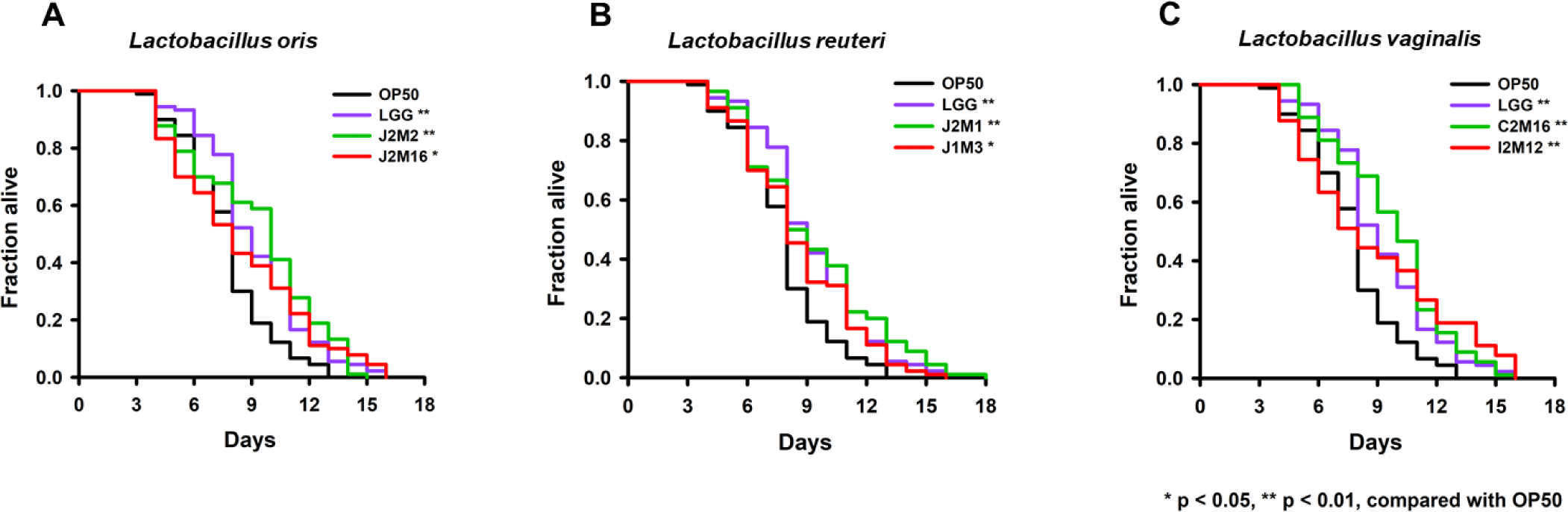
Based on the C. elegans lifespan assay, L. reuteri J2M1, L. oris J2M2, and L. vaginalis C2M16 were selected as probiotic candidates. When comparing the results of the five probiotic activity assays, including the lifespan assay, L. reuteri J2M1 showed superior tendencies in all characteristics except in the acid tolerance test. Consequently, L. reuteri J2M1 was chosen as the probiotic strain for the final experiment to evaluate its phage-probiotic synergistic effects.
Initially, 61 bacteriophages were isolated from the sludge samples of 11 strains of Salmonella Typhimurium derived from poultry, swine, and humans. These phages were subjected to host range testing against 11 Salmonella Typhimurium strains (Supplementary Table S5). Ultimately, phage 45, which infected eight of the 11 Salmonella strains, and phage 56, which uniquely infected the poultry-derived ST008 strain, were selected and named SLAM_phiST45 and SLAM_phiST56, respectively. Taken together, these phages demonstrated a host range that covered 10 of the 11 tested strains.
SLAM_phiST45 exhibited characteristics of the Siphoviridae family, which belonged to the now-defunct family, featuring a non-enveloped head (diameter 75 ± 6 nm, n = 8) and a non-contractile tail (length 182 ± 6 nm, n = 8; Fig. 4A). In contrast, SLAM_phiST56 was classified under the Myoviridae family, which is also part of the defunct family, and displayed a non-enveloped head (diameter 59 ± 7 nm, n = 9) and contractile tail (length 115 ± 10 nm, n = 9; Fig. 4B).
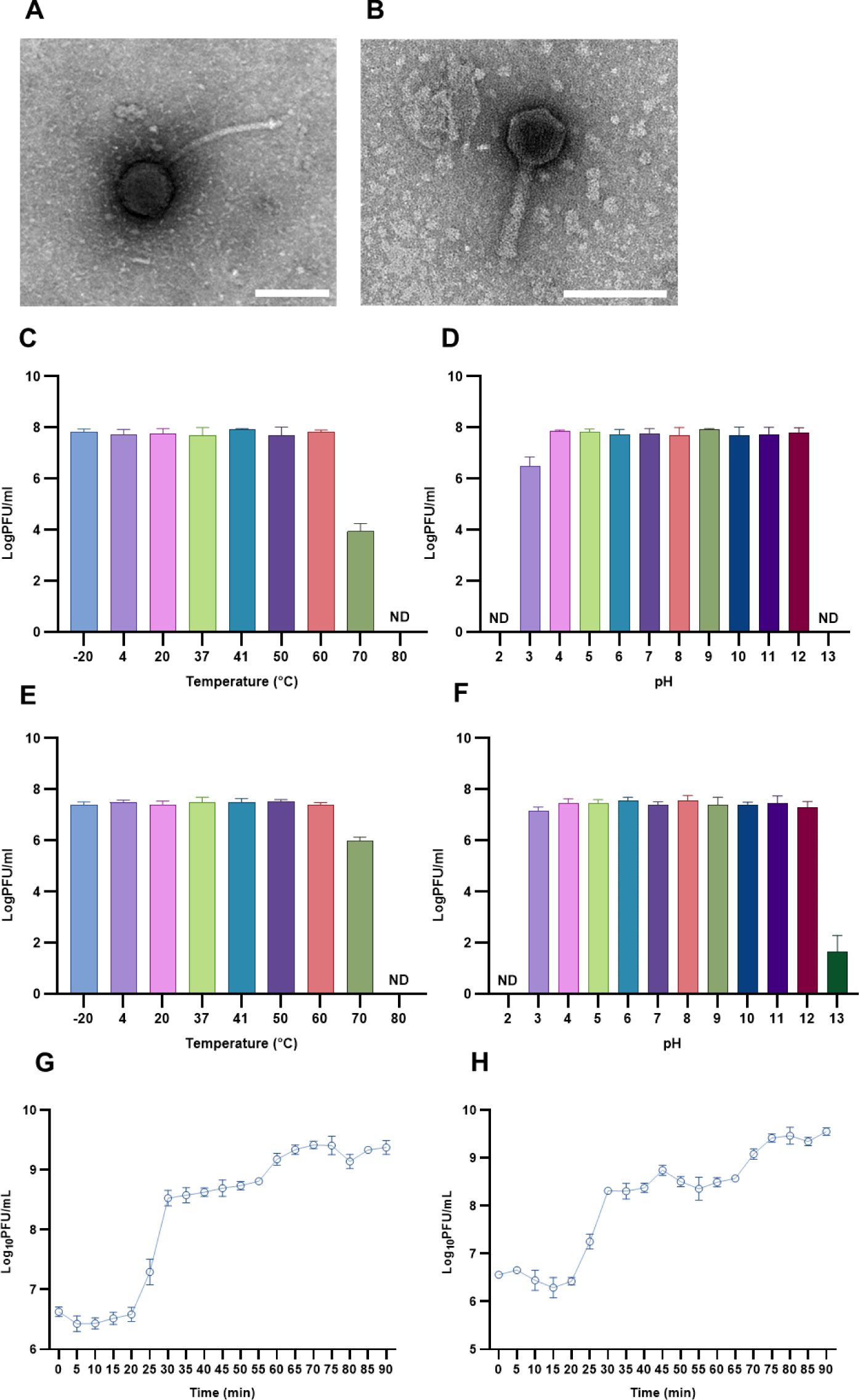
In the thermal stability test, SLAM_phiST45 showed a reduction of around 3 logs at 70°C, whereas SLAM_phiST56 exhibited a decrease of approximately 1 log. In the pH stability test, both phages struggled to survive at pH values of 2 and 13. However, at pH 3, SLAM_phiST45 demonstrated relative stability, with a reduction of approximately 1.5 log, whereas SLAM_phiST56 remained mostly unaffected (Figs. 4C, 4D, 4E, and 4F).
Analysis of the one-step growth curves for SLAM_phiST45 and SLAM_phiST56 showed that, although both phages had a latent period of 20 min, their burst sizes differed (approximately 140 and 98, respectively; Figs. 4G and 4H).
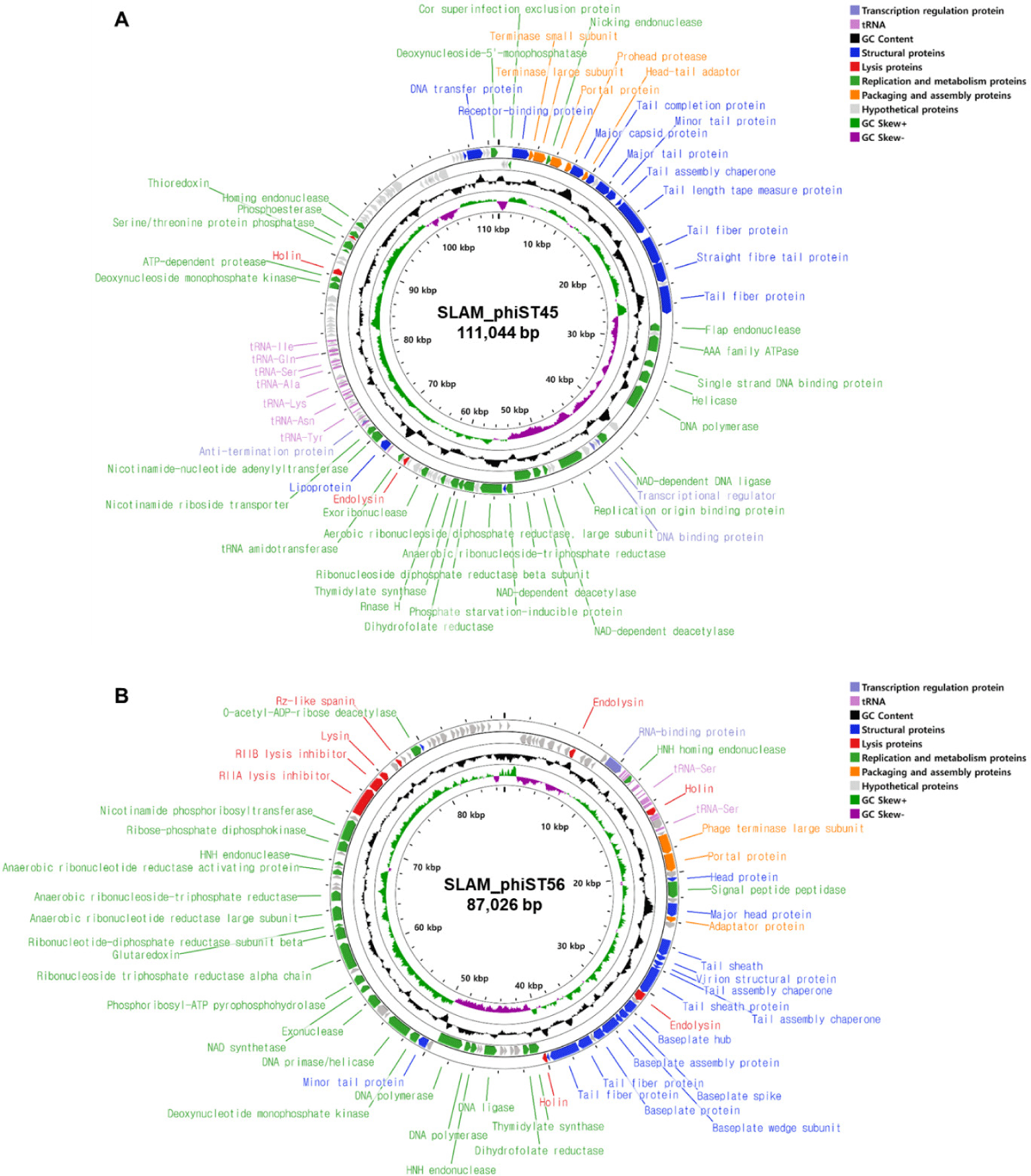
The genome of SLAM_phiST45 was 111,044 bp long, with a G+C content of 40.1%. In total, 130 ORFs were predicted, with 101 ORFs on the forward strand and 29 on the reverse strand, along with 29 tRNA genes (Fig. 5A). Among these, 55 ORFs (41.9%) were functionally annotated using BLASTP. The genome of SLAM_phiST56 was 87,026 bp in length, with a G+C content of 38.8%. It contains 104 ORFs, with 80 on the forward strand and 24 on the reverse strand, along with 26 predicted tRNA genes (Fig. 5B). Similarly, 52 ORFs (50.0%) were functionally annotated. Both phages were classified into six functional groups: structural, lysis, replication and metabolism, packaging and assembly, transcriptional regulatory, and hypothetical proteins. No genes related to integrases or antibiotic resistance were identified in either phage (Supplementary Tables S6 and S7).
To classify the phages based on their genomes, BLASTN searches were conducted using the SLAM_phiST45 and SLAM_phiST56 genome sequences. BLASTN analysis indicated that the most closely related species, SLAM_phiST45, belongs to the Markadamsvirinae subfamily (Supplementary Table S8). Based on this finding, we compared its genome with those of 58 Salmonella-infecting species from the Epseptimavirus genus and 26 species from the Tequintavirus genus, which are part of the Markadamsvirinae subfamily classified by the ICTV in 2023 (78 Epseptimavirus spp. and 70 Tequintavirus spp. in total). The results revealed that SLAM_phiST45 belongs to the Epseptimavirus genus and is most closely related to Salmonella phage BD13 (Fig. 6A). ANI calculations using three different methods showed that SLAM_phiST45 and Salmonella phage BD13 shared ANIu (97.39 %), ANIb (96.71 %), and ANIm (97.70 %), suggesting that they belong to the same species (Table 2).
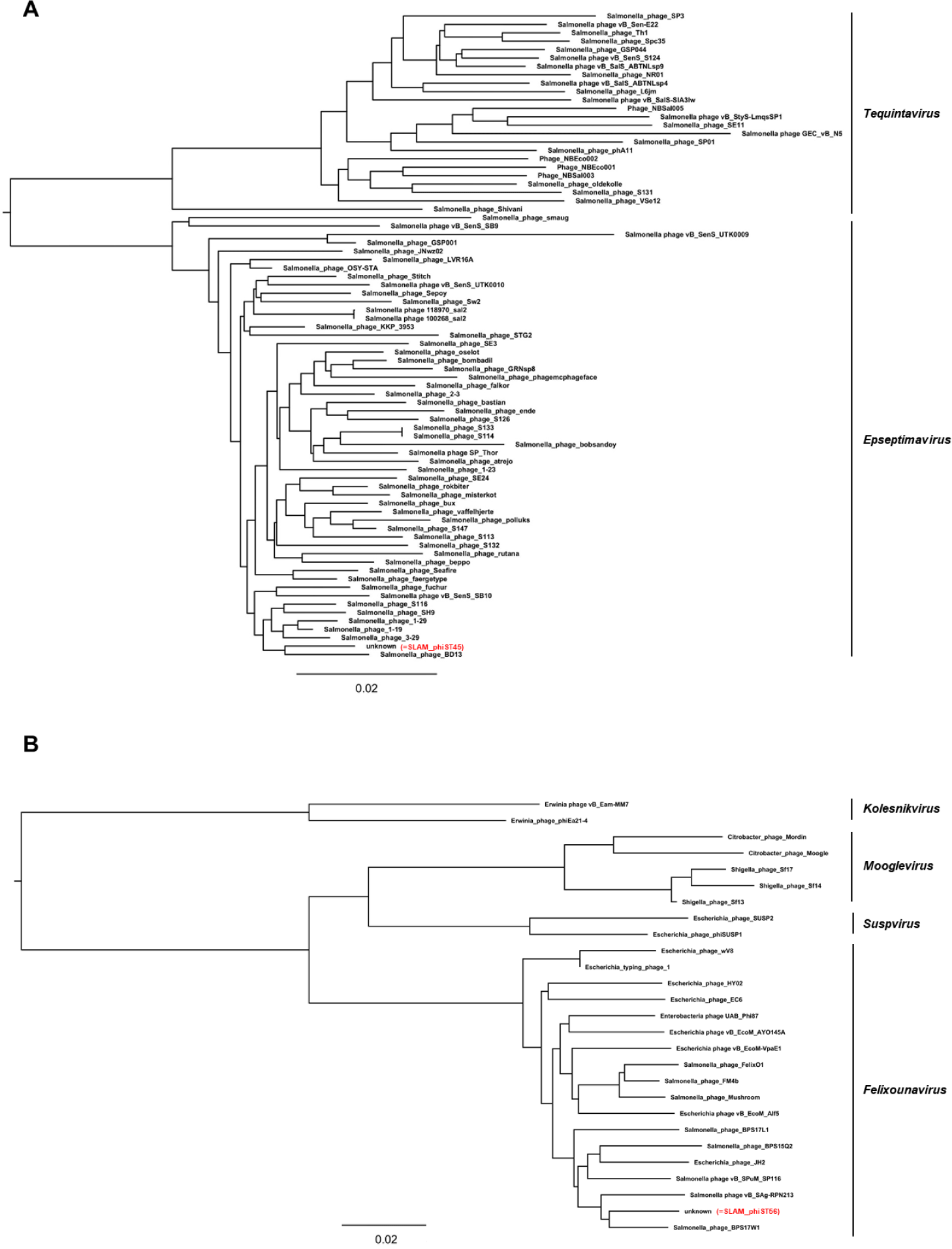
BLASTN analysis revealed that the closest relatives of SLAM_phiST56 predominantly belonged to the Ounavirinae subfamily (Supplementary Table S9). We then compared the genome with that of all viruses in the four genera of the Ounavirinae subfamily classified by the ICTV: Felixounavirus (17 species), Kolesnikvirus (two species), Mooglevirus (five species), and Suspvirus (two species). These results suggest that SLAM_phiST56 belongs to the Felixounavirus genus, with the highest similarity to Salmonella phage BPS17W1 (Fig. 6B). Three ANI analyses between SLAM_phiST56 and Salmonella phage BPS17W1 yielded ANIu, ANIb, and ANIm values of 97.09 %, 96.49%, and 96.94 %, respectively, indicating that they are the same species (Table 2).
The inhibitory effects of SLAM_phiST45 and SLAM_phiST56 on host bacteria ST422 and ST008 were evaluated in liquid culture (Fig. 7A). Briefly, 1% overnight cultures of ST422 and ST008 were inoculated into fresh TSB broth. When the OD (600 nm) reached 0.3, the two phages were added at an MOI of 1. For ST008, although there was a consistent inhibition of bacterial growth compared to the control from the time of phage addition, no clear lysis (OD ≤ 0.05) was observed. In contrast, ST422 showed a rapid decline in bacterial density after phage addition, with clear lysis observed for 2–3 h, followed by bacterial regrowth. In addition, a phage cocktail was used to suppress both bacterial strains, with the phages added simultaneously under the same conditions. Interestingly, the phage cocktail resulted in a more significant inhibition of bacterial growth than the individual phages. For ST008, although clear lysis was not achieved, significant bacterial inhibition was observed after approximately 8 h. Furthermore, in the case of ST422, the phage cocktail maintained clear lysis for approximately 11 h, demonstrating at least 8 h of additional inhibition compared with the use of a single phage.
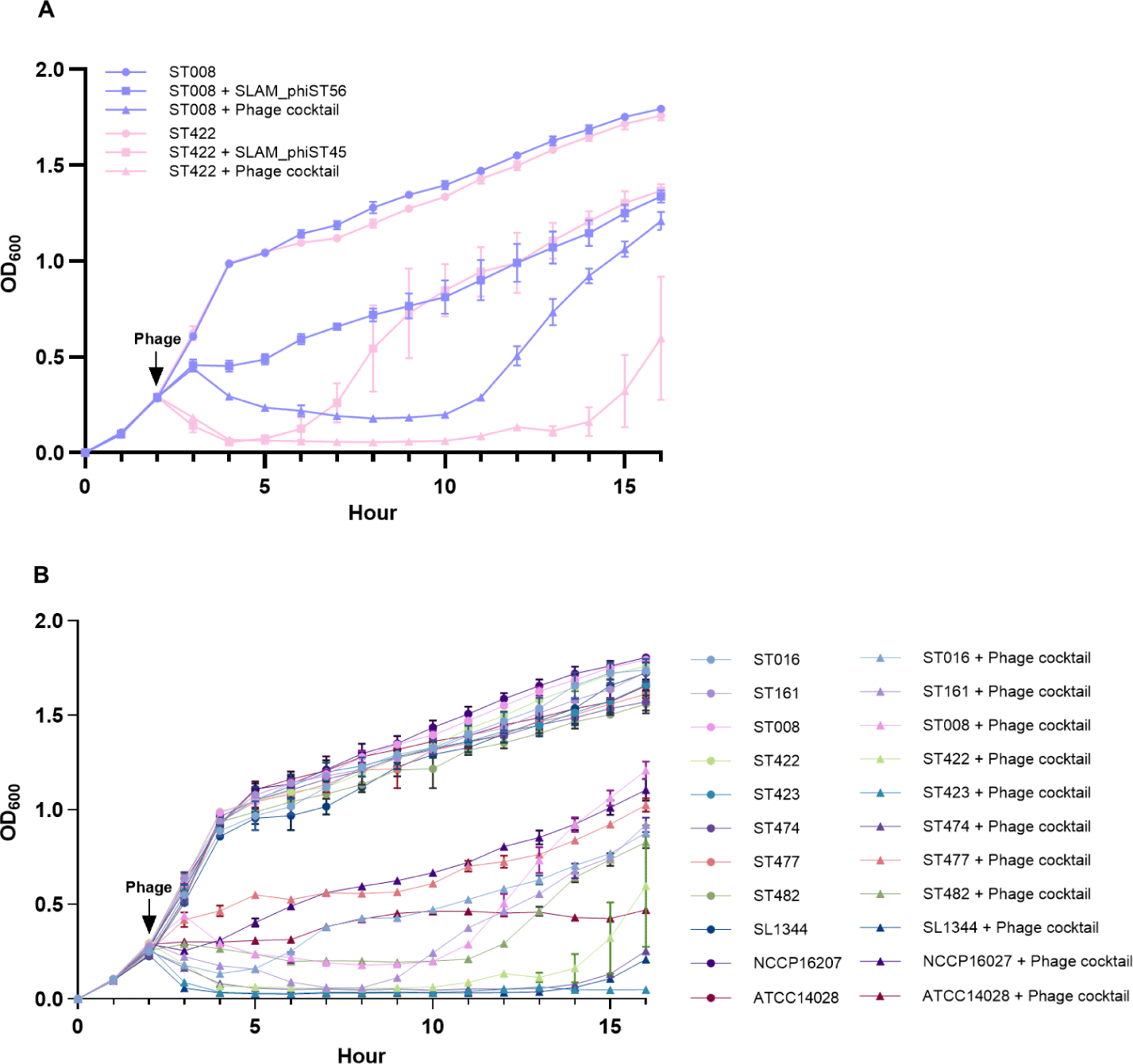
The efficiency of plating (EOP) test is a method for quantifying the infection efficiency of phages across various bacterial strains and evaluating how effectively a particular bacteriophage can propagate in different host cells. In this study, we performed an EOP test to analyze the cause of the significant reduction in bacterial growth observed with SLAM_phiST45 and SLAM_phiST56 in liquid culture inhibition tests (Table 3). Interestingly, when both phages exhibited high EOP values for a specific strain, bacterial inhibition in the liquid culture was also highly effective. Specifically, ST423, ST474, and SL1344 showed more effective inhibition than ST422 (Fig. 7B). These findings suggest a strong potential for selecting more effective phages when using phage cocktails for therapeutic applications.
An EOP test was conducted to demonstrate the synergistic effect of the phage cocktail in the liquid inhibition test.
A BIM test was conducted to identify and study bacterial mutants that were resistant to bacteriophages, serving as a tool for evaluating the effectiveness of phage therapy and providing valuable insights for the development of effective phage cocktails. In this study, we investigated the synergistic effects of SLAM_phiST45 and SLAM_phiST56 by comparing the BIM frequency when using a phage cocktail versus single phages (Table 4). The results showed that the BIM frequency was 2.5 times higher for SLAM_phiST45 and 12.5 times higher for SLAM_phiST56 when treated with single phages than when treated with the phage cocktail. This demonstrates that measuring BIM frequency can be a useful indicator of the efficacy of phages or phage cocktails.
| Phage treatment | BIM frequency avreage | Fold-change |
|---|---|---|
| SLAM_phiST45 | 3.67 × 10⁴ CFU/mL | 2.5 |
| SLAM_phiST56 | 1.83 × 10⁵ CFU/mL | 12.5 |
| Phage cocktail | 1.47 × 10⁴ CFU/mL | 1 |
Finally, we investigated the inhibition of Salmonella and changes in the gut microbiota using a mixture of the previously selected probiotic L. reuteri J2M1 and a phage cocktail in a simulated chicken gut system. First, we evaluated the survival rates of J2M1, SLAM_phiST45, and SLAM_phiST56 during the simulated chicken gastric phase and observed reductions of approximately 1.8, 2.1, and 0.9 log, respectively (Figs. 8A, 8B, and 8C). Next, during the simulated chicken intestinal phase, all treatments showed less than a 1 log reduction, indicating relatively stable survival (Figs. 8D, 8E, and 8F).
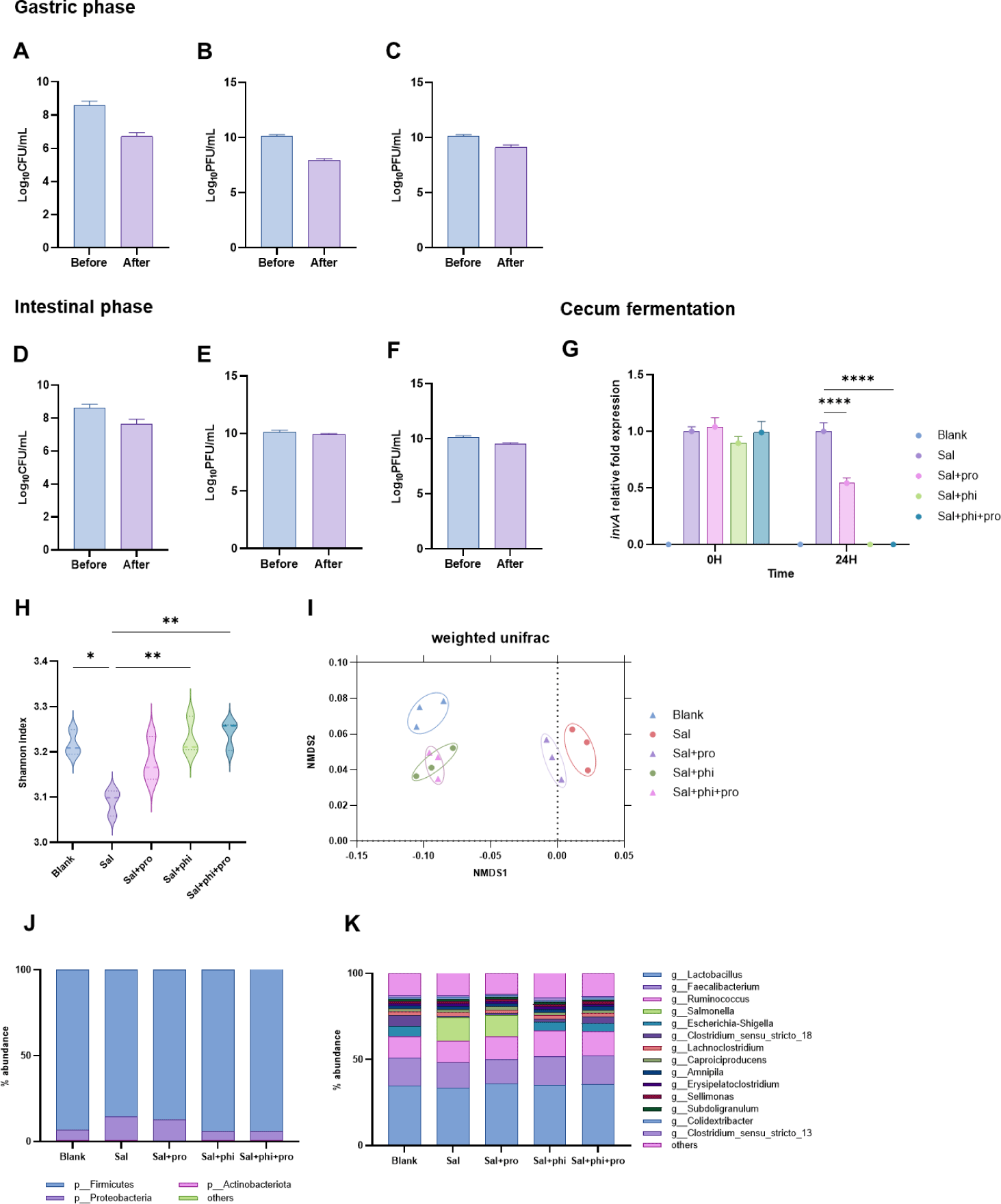
Finally, the cecum fermentation stage was conducted in five experimental groups: a blank group with only cecum contents; a sal group with Salmonella added; a sal+pro group with Salmonella and the probiotic; a sal+phi group with Salmonella and the phage cocktail; and a sal+phi+pro group with Salmonella, the probiotic, and the phage cocktail. The results showed that the most effective groups for reducing Salmonella were the sal+phi and sal+phi+pro groups, which nearly eradicated Salmonella within 24 h. In contrast, the sal+pro group showed a reduction in Salmonella detection compared with the control group (Fig. 8G).
Changes in the microbiota were analyzed using alpha and beta diversity metrics. Alpha diversity showed similar values for all groups except for the sal group (Fig. 8H). However, beta diversity revealed distinct clustering, with the blank and phage-treated groups clustering closely, whereas the Sal and Sal + Pro groups formed another cluster (Fig. 8I). The gut microbiota was analyzed at the phylum and genus levels, and similar patterns were observed in the phage-treated and blank groups, as well as the sal and sal+pro groups, showing nearly identical microbial communities (Figs. 8J and 8K).
For a more precise analysis of the synergistic effect of J2M1 and the phage cocktail, we selected genera that showed significant differences between the sal+phi and sal+phi+pro groups among those with an overall abundance of over 1%. The selected genera were Oscillibacter, Clostridium sensu stricto 18, and Clostridium sensu stricto 13 (Supplementary Figs. S6A, S6B, and S6C). Compared to the sal+phi group, the sal+phi+pro group showed a significant increase in Oscillibacter and Clostridium sensu stricto 18, whereas Clostridium sensu stricto 13 showed a significant decrease. Oscillibacter is more abundant in healthy individuals than in patients with inflammatory bowel disease (IBD), and Clostridium sensu stricto 18 includes species such as Clostridium butyricum, which is used as a probiotic. Conversely, Clostridium sensu stricto 13 includes pathogenic species such as Clostridium perfringens and Clostridium difficile. Although further metagenomic studies at the species level are required, these findings suggest a potential synergistic effect of phages and probiotics.
DISCUSSION
Isolation of LAB using a culture-based approach has been extensively studied in various animals, including humans [47]. Therefore, the isolation of various probiotics, such as Lactobacillus spp., using culturomics in the poultry industry may no longer be novel [48]. In this study, four different Lactobacillus spp. were selected from various intestinal contents of chickens using a multi-omics approach that simultaneously used culturomic and metagenomic techniques. Subsequently, L. reuteri J2M1 was selected through various analyses and lifespan assays using an in vivo alternative nematode model, C. elegans. L. reuteri was found in a probiotic-based multi-omics analysis of the cecum, ileum, and jejunum. This suggests the potential for developing host-specific probiotics through culturomics- and metagenomics-based analysis as well as provides evidence of balanced microbial community dynamics [49]. In particular, L. reuteri J2M1 demonstrated strong performance in acid tolerance, bile tolerance, and intestinal cell adhesion tests, making it suitable for commercial use. In addition, its safety for commercial use was validated using antibiotic resistance tests. Although it showed lower antimicrobial activity against chicken-derived pathogens than the positive control, L. rhamnosus GG (LGG), it tended to extend the lifespan of C. elegans in lifespan assays compared to LGG. Although antimicrobial activity is an important characteristic of probiotics, it was not a significant issue in this study, because bacteriophages, which specialize in antimicrobial activity, were applied simultaneously.
In recent decades, the steady emergence of superbacteria has led to extensive research into phages as alternatives to antibiotics [49]. Although more research is needed to determine whether phages are suitable for human use, previous studies, including those on the application of phages in the livestock industry and clinical trial results in humans, have provided evidence to support their safety [50]. In this study, phages targeting Salmonella Typhimurium, a significant problem in the poultry industry, were isolated. A total of 61 phages were obtained from various Salmonella Typhimurium strains isolated from infected chickens, pigs, and humans. Among these, two phages, SLAM_phiST45 and SLAM_phiST56, were selected for their broad host range, including chicken-derived and Salmonella strains from pigs and humans. Notably, in an experiment to inhibit Salmonella Typhimurium KVCC-BA0000422, which is the host bacterium of SLAM_phiST45 in liquid medium, the phage cocktail showed approximately 8 h of suppression compared to the use of SLAM_phiST45 alone. To provide evidence for the synergistic effect of the two phages, we conducted EOP and BIMs tests. Notably, the results of the EOP test showed a strong correlation with those of the liquid inhibition test using different strains in addition to the host bacteria of the two phages. When both phages exhibited high EOP values against a particular strain, bacterial inhibition in the liquid medium increased significantly. These findings suggested that the EOP test can serve as a simple and useful tool for selecting an effective phage cocktail to target specific bacteria. The EOP and BIMs test results were consistent with those of previous studies using phage cocktails [51]. In addition, the commercial potential of the two phages was confirmed through temperature and pH stability tests, and whole-genome sequencing revealed that they were safe phages, free from integrase or toxic factors. TEM revealed that SLAM_phiST45 and SLAM_phiST56 exhibited morphological characteristics similar to those of the defunct Siphoviridae and Myoviridae families. Furthermore, genome-based classification suggested that the phages belonged to the Epseptimavirus and Felixounavirus genera, respectively, and species-level classification was completed through three distinct ANI analyses. Although the phages were not identified as new species, their identity with closely related species was < 98%, indicating that they were likely not of the same strain.
Various bacteriophage application methods have been used, including the use of single phages, phage cocktails, and phage-antibiotic combination [52,53]. On the other hand, although there have been some previous studies on the synergistic effects of phages and probiotics [11,54], the number of experiments aimed at treating pathogenic infections while simultaneously improving gut microbiota is limited [10,11,55–56]. In this study, we investigated the synergistic effects of phage cocktails and probiotics on L. reuteri J2M1, SLAM_phiST45, and SLAM_phiST56 strains. First, we tested the survival rates of the probiotic and phages during passage through the chicken gastric and intestinal phases and observed a reduction in pathogens and changes in the microbial community in a cecum infected with Salmonella. Similar to previous studies that used a gut simulation system, we confirmed that the application of phages significantly reduced Salmonella [57]. However, there is no evidence of a synergistic effect of phages and probiotics on alpha and beta diversity, including pathogen reduction. By contrast, when comparing the microbial communities of the sal+phi and sal+phi+pro groups at the genus level, we identified three bacterial genera that differed significantly. First, the Oscillibacter genus, which belongs to the Ruminococcaceae family, primarily produces short-chain fatty acids through carbohydrate fermentation [58]. In addition, it is likely to have anti-inflammatory properties, and its association with IBD has been studied [59]. Second, Clostridium sensu stricto 18 and 13 are terms used in the SILVA database to represent specific groups within the Clostridium genus, which likely include species such as Clostridium butyricum and Clostridium perfringens, respectively [60]. In the sal+phi+pro group, Clostridium sensu stricto 18 (including Clostridium butyricum) increased significantly, whereas Clostridium sensu stricto 13 (including Clostridium perfringens) decreased significantly. The combination of phages and probiotics led to changes in the abundance of specific bacterial strains. Interestingly, when L. reuteri J2M1 was added, there was an increase in the Oscillibacter genus, which may have anti-inflammatory properties, and in the Clostridium genus, which includes beneficial species such as Clostridium butyricum. However, when only phages were used, an increase in the Clostridium genus, including Clostridium perfringens, was observed. These findings suggest that, although phage treatment alone may control targeted pathogens, it can lead to a reduction in beneficial bacteria and an increase in other pathogenic species. However, the synergistic effects of phages and probiotics may mitigate these negative effects.
CONCLUSION
This study employed a multi-omics approach to efficiently identify probiotics and bacteriophages while optimizing phage selection and classification. By integrating culturomics and metagenomics, L. reuteri, the most abundant species in the cecum, ileum, and jejunum, was successfully isolated, demonstrating the efficiency of this method in identifying host-specific probiotics. Additionally, in vitro experiments confirmed the reliability of EOP tests for phage selection through strong correlation with liquid inhibition assays. A streamlined phage classification method was also established using whole-genome analysis, with BLASTN and ICTV standards for genus-level classification and ANI analysis for species-level identification. Furthermore, in a simulated poultry gut model, the combination of phages and probiotics improved microbial balance by increasing beneficial genera and reducing pathogenic genera, suggesting probiotics can mitigate potential adverse effects of phage therapy. Although in vivo validation was not performed, these in vitro findings offer valuable insights for optimizing phage applications as a sustainable alternative to antibiotics in agriculture.


