

INTRODUCTION
The global meat industry generates an estimated 150 million tons of slaughter by-products each year, encompassing both edible and inedible parts. The proportions of these by-products vary by animal species and account for more than half of an animal’s body weight. While a certain portion of these materials is utilized for human consumption and various related industries, a substantial amount remains underutilized [1,2]. Efficient use of these by-products not only generates economic value and supports the animal production industry but also helps reduce environmental pollution.
The global pork industry continues to grow sustainably, with pork ranking as the second most consumed meat worldwide, after poultry [3]. Bones constitute approximately 10%–12% of a pig’s total body weight, meaning that a large amount of this by-product is generated from slaughters worldwide. Pork bones have long been used in traditional dishes such as Paigu Luobo Tang (a classic Chinese pork bone soup) and Gamjatang (a Korean pork backbone soup) [4]. These dishes are widely believed to be nutritious and offer numerous health benefits for humans [5,6]. In many Asian countries, pork bones are typically displayed and sold in retail outlets such as butcher shops and supermarkets. Due to their sharp edges and bulky structure, pork bones are unpackaged or minimally overwrapped during retail display, which may compromise their quality.
Several active substances (e.g., collagen, gelatin, peptides, etc.) in bones have been reported [7]. Amongst gelatin is a product obtained by partial hydrolysis of collagen molecules [8]. In vitro and in vivo studies have shown that gelatins derived from bovine and pork by-products exhibit high biological activities such as antioxidant and anti-aging [9,10]. To date, gelatin is commonly extracted from animal sources using traditional methods that combine acids and commercial proteases at temperatures around 70°C [11,12]. In connective tissues, collagen predominantly exists in a cross-linked form, making it difficult to dissolve under normal conditions. Therefore, mild acids (e.g., acetic acid and hydrochloric acid, etc.) are required to swell the tissues and weaken the interchain cross-linkages before collagen extraction [8,13]. Furthermore, when extracted at high temperatures above 65°C–75°C (the denaturation transition temperature of collagen), the resulting product usually has a lower gel strength and emulsion stability compared to that extracted at a lower temperature (45°C–55°C) [14,15]. In this solidified form, it becomes challenging to dissolve the gelatin during analyses of its properties. Additionally, the use of commercial proteases and synthetic chemicals (e.g., inorganic and organic acids) to extract them is costly and raises health concerns [16].
Vinegar contains a considerable amount of acetic acid [17]. It is widely known as an acidic condiment commonly used in many culinary cultures and in the food processing industry [18]. This acidic seasoning may serve as an effective and safe alternative to synthetic acids in the pretreatment step for the collagen extraction. Meanwhile, enzymes from plant origin are highly effective in hydrolyzing connective tissues and proteins [19,20]. In particular, actinidin from kiwifruit and zingibain from ginger are abundant and readily available. Ginger and kiwifruit are cultivated globally in abundant quantities, ensuring sufficient supply to all markets at affordable prices [21]. These plant-derived enzymes have recently been used to extract gelatin from bovine skin with high efficiency [22]. Therefore, these plant-derived proteases can be effective alternatives to costly commercial enzymes for converting meat by-products into bioactive materials.
Given the large volume of pork bones produced by the pork industry, effective utilization of this by-product at both household and industrial scales is essential. However, to our knowledge, no studies have been conducted to assess its shelf-life characteristics or nutritional composition, and develop efficient methods for its utilization. The objective of this study was to assess the shelf-life characteristics and nutritional composition of pork bones and to develop a novel method for extracting their bioactive gelatin. Our findings showed that under the same storage conditions, the shelf-life indices differed between the leg bone and the backbone. The pork bones were identified as a rich source of collagen, essential amino acids, and unsaturated fatty acids. The use of vinegar and plant-derived protease extracts at moderate extraction temperature yielded low-molecular-weight gelatin peptides with strong antioxidant activity.
MATERIALS AND METHODS
In the study, two main bone items (leg and backbone) were used. The bones were collected from commercial growing-finishing pigs (n = 10, at 185-day-old and body weight of about 110 kg) at 24 h after slaughter in a practical plant of the National Institute of Animal Science. Chemicals used for shelf-life, nutritional composition analysis, and antioxidant assays were purchased from Sigma-Aldrich. Chemicals used for sodium dodecyl-sulfate polyacrylamide gel electrophoresis (SDS-PAGE) were purchased from Bio-Rad. Ginger rhizome and kiwifruit were purchased at a local supermarket (Jeonbuk, Korea). Apple vinegar was purchased from Ottogi.
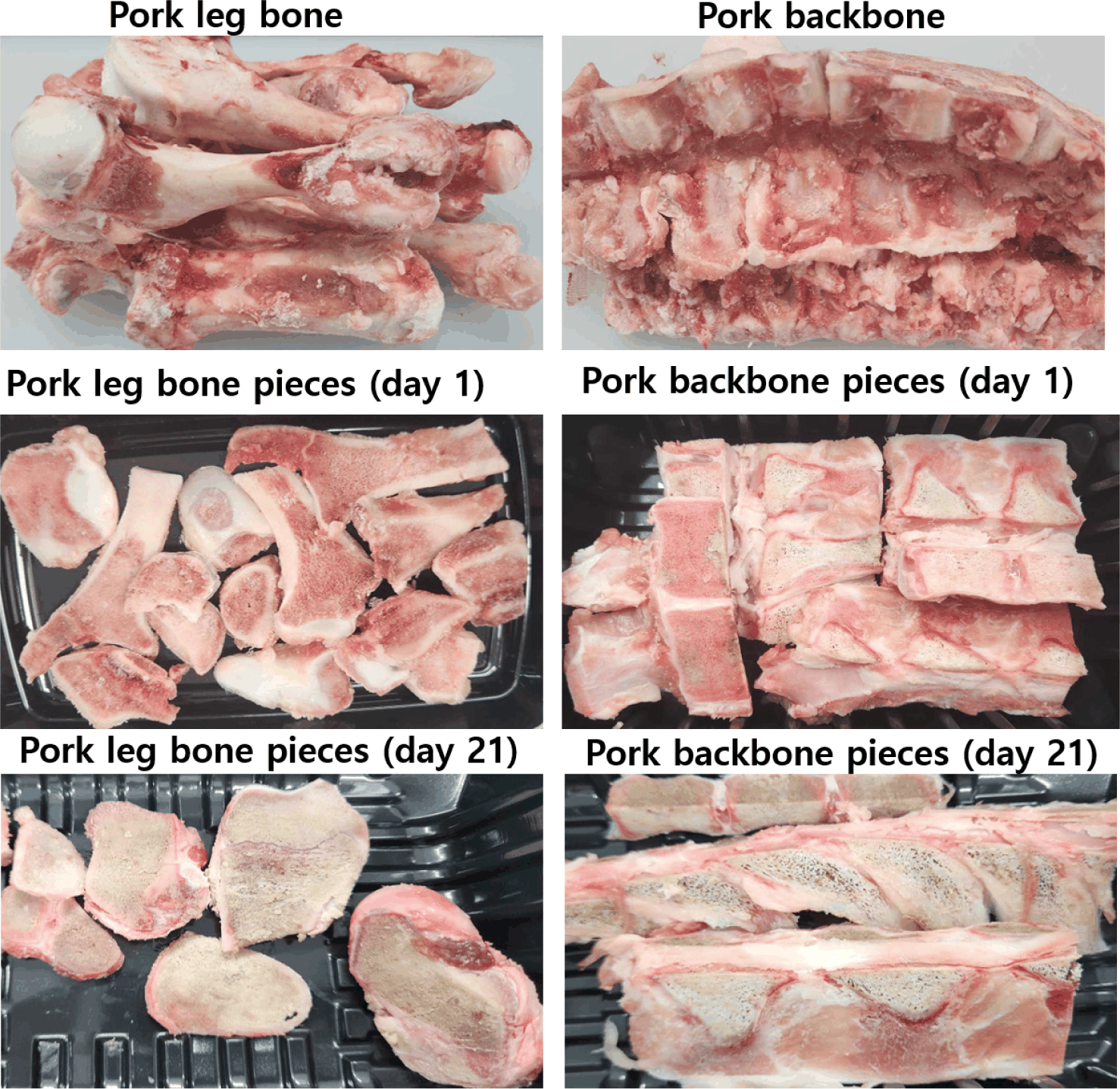
For each type of bone, the samples collected from the 10 carcasses were pooled together (not separated for each animal). The bones were cut into 2.0 cm-thick pieces using an electric saw (JWB 400S, Jiwoo Tech). For shelf-life evaluation, bone pieces from each type of bone were randomly selected, placed on plastic trays (approximately 200 g per tray), and overwrapped with plastic film. All samples were then displayed in refrigerated cabinets at 4°C. At the end of each storage period (1, 7, 14, and 21 days), the samples were analyzed for shelf-life parameters, including microbiological indicators, color, and protein and lipid oxidation. Representative images of the pork bones examined are presented in Fig. 1. For each storage period, six trays per type of bone were randomly taken and analyzed for the specified parameters. After microbial sampling and color measurement, the bones were trimmed of all lean and fat tissues, immersed in liquid nitrogen for 5 min, ground into powder using a specialized bone grinder (Hanil Electric), and sieved through a 40-mesh screen. The powdered bone samples were subsequently used for analysis of other remaining shelf-life parameters. Nutritional composition analysis and gelatin extraction were conducted on the 1-day stored samples.
Microbiological sampling was performed by rubbing a sterile sponge several times over a 25 cm² (5 × 5 cm) surface area of each sample. The sponge was then placed into a sterile vial containing 10 mL of peptone water. After stirring for approximately 1 min, serial dilutions were prepared using diluent vials containing 9 mL of 0.85% saline. The total aerobic plate count (APC) was determined by spreading 1 mL of each diluted sample onto Petrifilm APC plates and incubating for 48 h at 37°C. Results were expressed as the logarithm of colony-forming units per square centimeter (Log10 CFU/cm2).
During storage, changes in surface color were measured using a colorimeter with a D65 illuminant*C and 2° observer (CR-400, Minolta). Before using, the device was standardized with a white tile (Y = 86.3, X = 0.317, and y = 0.324). For each sample tray, color values were recorded at five different air-exposed locations on the upper surface (avoiding marrow areas) of the bone tissue after removing the wrapping film. The measured color parameters included CIE L*, CIE a* (indicating the bright red color of bone tissues upon exposure to oxygen), and CIE b*. Additionally, the a* values obtained at each storage stage were used to assess the discoloration degree of the bone samples over time. Discoloration was calculated as the percentage loss of a* values at each storage stage compared with the initial a* value measured on day 1.
The extent of lipid oxidation during retail display was assessed by measuring thiobarbituric acid reactive substances (TBARS) according to the method of Buege and Aust [23], as described in our previous study [24], with the exception that the homogenization time was extended to 3 min. The absorbance of both samples and standards was measured at a wavelength of 531 nm using a spectrophotometer (Infinite M200, Tecan). TBARS concentrations were calculated from a linear standard curve and expressed as milligrams of malondialdehyde per kilogram (mg MDA/kg) of sample. All measurements were performed in duplicate.
To assess protein oxidation in the bones during refrigerated storage, the total volatile basic nitrogen (TVBN) content was measured using the Conway micro-diffusion method, as described in our previous study [25], with the exception that the homogenization time was extended to 3 min. The TVBN level (mg/100 g sample) was calculated using the equation provided in the cited reference [25].
The total collagen content in the bone samples was determined according to the PN-ISO 3496:2000 method [26]. Briefly, 4.0 g of each sample was placed in a glass tube with 30 mL of 6 N HCl and hydrolyzed at 105°C for 16 h. After cooling, the hydrolysates were diluted with distilled water to a final volume of 250 mL, filtered through No. 2 Whatman filter paper, and 4 mL of each filtrate was mixed with 2 mL of oxidation reagent (1.41 g chloramine-T in 100 mL acetate–citrate buffer, pH 6.0). The mixture was kept at room temperature for 20 min, after which 1.0 mL of color reagent (10 g 4-dimethylaminobenzaldehyde in 35 mL of 60% perchloric acid and 65 mL of isopropanol) was added and incubated at 65°C for 20 min. The samples were then cooled to room temperature for 20 min, and absorbance was measured at a wavelength of 558 nm using a spectrophotometer. A standard curve was prepared using 4-hydroxyproline at various concentrations, treated under identical conditions. Hydroxyproline content was converted to total collagen content by multiplying by a factor of 7.25 and expressed as grams per 100 g of sample.
For insoluble collagen determination, 6.0 g of each bone sample was homogenized with 24 mL of Ringer’s solution (8.6 g NaCl, 0.3 g KCl, and 0.33 g CaCl₂ in 1.0 L distilled water) at 8,050×g for 1 min. The samples were then heated at 77°C for 70 min, cooled for 30 min, and centrifuged at 3,000×g. After removing the supernatant, the pellet was resuspended in 24 mL of Ringer’s solution, vortexed for 1 min, and centrifuged again under the same conditions. The resulting pellets were dried at 105°C overnight, and 0.1 g of each dried sample was hydrolyzed with 30 mL of 6 N HCl at 105°C for 16 h. Subsequent analytical steps and calculations were performed as described above for the total collagen determination. Soluble collagen content (g/100 g) was obtained by subtracting the insoluble collagen content from the total collagen content.
The fatty acid composition of bones was analyzed following the method for meat and meat products described by Folch et al. [27] and Morrison and Smith [28], as detailed in our previous study [25], with the exception that the homogenization time was extended to 5 min. Gas chromatography (GC) and flame ionization detector conditions for fatty acid separation and detection were the same as those reported in our previous work [25]. Fatty acid composition was expressed as a percentage of total fatty acids.
The amino acid composition was determined after hydrolyzing 5.0 g of each bone sample with 6 N HCl at 110°C for 24 h, following the procedure of Zou et al. [29]. Before analysis, the hydrolysates were filtered through a 0.45 µm membrane filter and adjusted to neutral pH using 10 N sodium hydroxide. The samples were then analyzed using an amino acid analyzer (L-8900, Hitachi).
Ginger (root) and kiwifruit (with peel) were washed with tap water, drained, and ground with an equal volume of distilled water for 2 min. The mixture was then squeezed and filtered through two layers of cheesecloth. The filtrates were subsequently centrifuged at 1,390×g for 20 min at 4°C, and the resulting supernatants were used for gelatin extraction.
First, the powdered bone samples were soaked in 10 volumes of 0.1 M NaOH solution at 4°C with stirring at 100 rpm for 48 h to remove non-collagen proteins [12]. The NaOH solution was refreshed every 8 h. Afterward, the samples were washed with tap water until reaching a neutral pH. Next, the samples were treated with 5 volumes of 0.3% (v/v) apple vinegar (pH 3.3) for 24 h at 4°C with stirring at 100 rpm. The vinegar concentration (0.3%, v/v) used was chosen based on its pH value, which was similar to diluted concentrations of acids used for gelatin extraction in previous studies [12]. The pH was then adjusted to 5.0 using 1 N NaOH to optimize the activity of enzymes in the crude extracts [20]. Subsequently, enzyme extracts were added at 2% (v/w, 2 mL per 100 g sample), and the samples were sealed in pouches, immersed in a water bath at 55°C, and stirred at 100 rpm for 5 h. Following incubation, the mixtures were cooled to room temperature and centrifuged at 6,000×g for 20 min at 20°C to remove sediment. The resulting gelatin was collected, dried at 45°C for 48 h, and used for further analyses. The extraction yield of gelatin was calculated (3 extraction batches per bone type) as the weight of dried gelatin divided by the wet weight of the initial sample, multiplied by 100.
The surface morphology of the gelatins obtained from the pork leg bone was observed using scanning electron microscopy (SEM; Supra 40 VP instrument, Zeiss). Before analysis, a moderate amount of sample was placed on a sample holder, coated with platinum, placed in a specimen chamber, and observed at 10 kV accelerating voltages. For each sample, three replicate fields were selected.
The protein pattern of gelatin was analyzed using sodium dodecyl-sulfate polyacrylamide gel electrophoresis (SDS-PAGE). Specifically, gelatin samples at a concentration of 6 mg/mL were mixed (1:1 ratio) with 2X Laemmli sample buffer and boiled for 5 min, and 15 μL of each sample was loaded on 4%–20% precast gradient gels. SDS-PAGE was performed in 1X Tris/Tricine/SDS running buffer at 110 V for 90 min. Then, the gels were stained with R-250 Coomassie brilliant blue for 1 h and destained for 2 h using the R-250 destaining kit, and bands were visualized using an iBright imaging system (CL750, Invitrogen).
In this study, the antioxidant activity of pork bone gelatins was evaluated using both 2,2-diphenyl-1-picrylhydrazyl (DPPH) radical scavenging and ferrous ion chelating assays, as described in our previous study [30]. Prior to the assays, dried gelatin samples and the synthetic antioxidant standard butylated hydroxytoluene (BHT) were diluted to various concentrations (0, 0.1, 0.3, 0.5, and 0.7 mg/mL). For the DPPH assay, 1 mL of each sample or BHT solution was mixed with 2 mL of 0.2 mM DPPH solution prepared in 96% ethanol. Distilled water was used as a control in place of the samples or BHT. The DPPH radical scavenging reaction was conducted at room temperature for 30 min in the dark. Absorbance was measured at 517 nm using a UV-VIS spectrophotometer (UV-1280, Shimadzu). The free radical scavenging activity was calculated using the following formula:
where, Acontrol: absorbance of the control (without test sample); Atest: absorbance of the test samples.
For the ferrous chelating assay, the reaction complex consisted of 1 mL of sample or BHT standard at each concentration, 3.7 mL of distilled water, 0.1 mL of FeCl2 (5 mM/L), and 0.2 mL of ferrozine (5 mM/L). For the control group, the same volume of distilled water was used to replace the sample or BHT. The chelating reaction was carried out at room temperature for 10 min. The adsorption was measured at 562 nm using the same spectrophotometer. The chelating activity on ferrous ions was calculated using the following formula:
where, Acontrol: absorbance of the control (without test sample); Atest: absorbance of the test samples.
Statistical analyses were performed using SAS software (version 7.1, SAS Institute). One-way analysis of variance (ANOVA) was used to analyze data such as nutritional composition, TPC, TVBN, TBARS, and antioxidant activity between the two bone types. Additionally, the General Linear Model (GLM) procedure was applied to evaluate the effect of storage period on shelf-life indices, with storage period included as a fixed factor in the model. The same GLM procedure was used to analyze antioxidant activity data, with gelatin concentration treated as a fixed factor. Mean separation was conducted using Duncan’s multiple range test. All measurements were performed in triplicate, and results are presented as mean ± SD. A p-value of less than 0.05 (p < 0.05) was considered statistically significant for all analyses.
RESULTS AND DISCUSSION
The changes in the total APC of pork bones during storage are presented in Table 1. All the bone samples had a low APC throughout the evaluation period. For example, the backbone had an APC ranging from only 1.16 Log10 CFU/cm2 (day 1) to 2.16 Log10 CFU/cm2 (day 21), while the leg bone had an APC ranging from 1.12 Log10 CFU/cm2 (day 1) to 2.63 Log10 CFU/cm2 (day 21). The leg bone had a higher APC than the backbone at 14 and 21 days of storage (p < 0.05). This may be due to the specific structure of leg bones, particularly their marrow content, which contains more nutrients and higher moisture content, thereby favoring microbial growth [31]. The APC increased with storage time: in the backbone, it increased by 1.00 Log10 CFU/cm2, and in the leg bone, by 1.51 Log10 CFU/cm2 after 21 days of storage. To date, no studies have evaluated the microbial indices of meat by-products, such as bones, during storage. However, the bones assessed in this study exhibited a significantly lower APC than the levels reported in beef and pork (6–8 Log10 CFU/g) after 21 days of storage under wrapping or vacuum packaging conditions [25]. Currently, no maximum limit for total APC has been established for meat by-products. Nevertheless, according to the upper limits recommended for most fresh meat products (7 Log10 CFU/cm2 or 6 Log10 CFU/g) [32], all pork bone samples in this study had much lower total microbial counts after 21 days of storage.
| Storage time (day) | Total aerobic bacteria (Log10 CFU/cm2) | |
|---|---|---|
| Backbone | Leg bone | |
| 1 | 1.16 ± 0.22c | 1.12 ± 0.16d |
| 7 | 1.84 ± 0.25b | 1.83 ± 0.25c |
| 14 | 1.89 ± 0.12bB | 2.41 ± 0.17bA |
| 21 | 2.16 ± 0.05aB | 2.63 ± 0.08aA |
The results of TVBN and lipid oxidation in pork bone samples during storage are shown in Table 2. At the initial evaluation, the TVBN content was 4.58 mg/100 g in the backbone and 7.19 mg/100 g in the leg bone. After 21 days of storage, the TVBN content had increased to 17.28 mg/100 g in the backbone and 21.57 mg/100 g in the leg bone. Notably, the leg bone had a significantly higher TVBN content than the backbone on all storage days (p < 0.05). An increasing trend in TVBN content was observed in both bone types with prolonged storage; for example, after 21 days, the leg bone increased by 14.38 mg/100 g and the backbone by 12.70 mg/100 g (p < 0.05). TVBN is an important index commonly used to evaluate protein and amine degradation in meat and its products during storage [33]. Studies on meat have reported TVBN levels of 56.01 mg/100 g in pork after 21 days [34], 20.1 mg/100 g in beef after 140 days [34], and 36.5 mg/100 g in chicken after 2 days [35] under aerobic packaging and refrigerated conditions.
TBARS content is commonly used to assess the extent of lipid oxidation in meat and meat products. At the initial assessment (day 1), TBARS contents were 0.94 and 1.36 mg MDA/kg in the backbone and leg bone, respectively. At the end of storage (day 21), these values increased to 4.55 and 6.56 mg MDA/kg in the backbone and leg bone, respectively. The leg bone exhibited significantly higher TBARS contents than the backbone on all storage days (p < 0.05). Similar to the TVBN results, an increasing trend in TBARS content was observed with prolonged storage. After 21 days, the leg bone showed an increase of 5.23 mg MDA/kg (from 1.36 mg MDA/kg on day 1 to 6.56 mg MDA/kg on day 21), while the backbone increased by 3.61 mg MDA/kg (from 0.94 mg MDA/kg on day 1 to 4.55 mg MDA/kg on day 21) (p < 0.05). Lipid oxidation is the reaction of unsaturated fatty acids (UFAs) with molecular oxygen, leading to the formation of intermediate products (e.g., free radicals) and end products (e.g., aldehydes) that contribute to quality loss, rancidity, and off-flavors in foods [36]. Recent studies have reported that animal bone marrow contains a considerable amount of UFAs [37]. Under aerobic packaging conditions, these UFAs are readily oxidized, which may be the primary driver of the lipid oxidation in the bones studied. Thus, the observed differences in the lipid oxidation levels may be attributed to variations in bone marrow content, specifically the UFA levels, between the two types of bones. Studies on meat have shown that TBARS levels exceeding 0.5 mg MDA/kg cause off-flavors that may be detectable by consumers [38]. Compared with TBARS contents reported in previous studies for aerobically packaged pork (0.30–1.03 mg MDA/kg) [24,25], pork bones showed significantly higher values, suggesting that the rate of lipid oxidation in bone tissues is greater than in meat tissues.
Changes in the color parameters of pork bones during storage are presented in Table 3. Regarding CIE L*, the backbone showed an increase, whereas the leg bone showed a decrease over the storage period (p < 0.05). The bright red color (CIE a*), reflecting the freshness, is a critical factor affecting consumers’ decisions to purchase meat and its products [39]. The a* value was higher in the leg bone than in the backbone on most storage days (p < 0.05). The analysis showed that the backbone significantly (p < 0.05) decreased in a* value, corresponding to a loss of 40.74% after 7 days, and almost completely lost (by 86.44%) its value after 21 days of storage (Table 4). Similarly, the a* value of the leg bone remained constant after 7 days and then decreased continuously, corresponding to a loss of about 68.39% of its value after 21 days of storage (p < 0.05). For CIE b*, no significant difference was observed between the first and last storage days for the backbone. In contrast, the b* value of the leg bone tended to decrease after 7 days and remained stable thereafter until day 21. Previous studies on pork have also reported a reduction in a* values (about 65%) after 21 days of storage under aerobic packaging conditions [24]. Likewise, Fang et al. [40] observed a significant decrease in a* values in pork after 20 days of storage under modified atmosphere packaging conditions.
| Storage time (day) | Backbone | Leg bone |
|---|---|---|
| 7 | 40.74 ± 2.16cA | 1.03 ± 0.35cB |
| 14 | 43.69 ± 1.88bB | 63.76 ± 3.27bA |
| 21 | 86.44 ± 4.37aA | 68.39 ± 4.18aB |
To date, the nature of color formation in meat by-products, such as bones, has not been widely studied. However, bone is a living tissue composed of several types of cells (e.g., osteoclasts and osteocytes etc.) and serves as the site of blood cell production [41]. Myoglobin is an oxygen-binding protein that stores the oxygen necessary for cell survival. Thus, the red color of bone tissues may also be attributed to oxymyoglobin derived from deoxymyoglobin and/or to blood cells [42]. The mechanism of discoloration in meat by-products (e.g., bones) also remains unclear. Nevertheless, this mechanism may be analogous to that observed in meat, involving the oxidation of oxymyoglobin to metmyoglobin [42]. Thus, the difference in discoloration degrees between the two bones may be attributed to differences in their structure, cell density, and physiological functions, all of which can influence the oxidation rate of the pigments. Based on the present results, it may be said that under current packaging and storage conditions, severe lipid and protein oxidation, and discoloration, rather than APC, appear to be the main factors compromising pork bone quality.
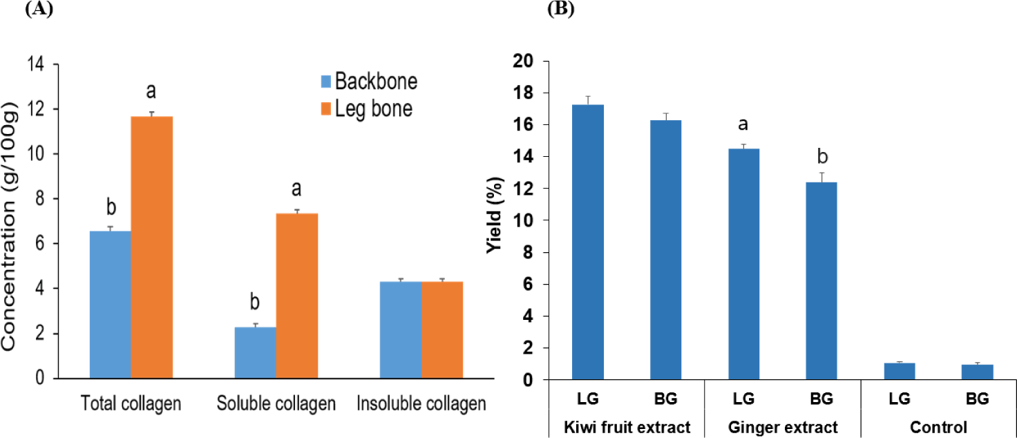
The collagen contents of the pork bones studied are shown in Fig. 2A. The total collagen content was higher in the leg bone (11.68 g/100g) than in the backbone (6.56 g/100g) (p < 0.05). Similarly, the soluble collagen content was significantly higher in the leg bone than in the backbone (p < 0.05). Previous studies have reported total collagen contents ranging from 0.2 g/100g to 2.0 g/100g, and soluble collagen contents ranging from 0.1 g/100g to 0.6 g/100g in beef and ovine meat [43,44]. Collagen is an essential and indispensable component for building the structure of connective tissues [8], accounting for about 30% of the total protein in the animal body. In vertebrates, collagen is commonly found in the skin, bones, tendons, and intramuscular connective tissues [8,12].
Amino acids are the primary building blocks of proteins. In this study, 16 amino acids, including eight essential amino acids (EAAs: valine, methionine, isoleucine, leucine, phenylalanine, lysine, histidine, and threonine) and eight non-essential amino acids (NAAs) were detected in both bone types (Table 5). In the leg bone, EAA levels varied depending on the amino acid, ranging from 8.21 mg/100g to 91.50 mg/100g. In the backbone, EAA content ranged from 14.42 mg/100g to 122 mg/100g. Lysine was the most abundant amino acid in both bone types. Lysine plays a key role in protein synthesis in the body, with minimum daily requirements of 4–7 mg/kg for children and 12–68 mg/kg for adults [45]. EAAs cannot be synthesized by the body and must be obtained through the daily diet. The total EAA content per 100g was approximately 403 mg in the leg bone and 554 mg in the backbone. Half of the individual EAAs, as well as the total EAA content, were significantly higher in the backbone than in the leg bone (p < 0.05). Among the NAAs, glycine was the most abundant, followed by proline in both bone types. Glycine, proline, and hydroxyproline are the three main amino acids involved in the structural units of collagen molecules [8]. Additionally, glutamic acid was the third most abundant amino acid after glycine and proline. From a taste perspective, glycine and proline contribute sweetness, while glutamic acid imparts an umami flavor to cooked meat products [46]. The composition and concentration of amino acids have also been analyzed and reported in bone broths, with notable variations depending on the animal species. Shaw and Flynn [47] reported that beef bone broth contained higher amino acid content (0.2–3.7 mg/g) than chicken or turkey bone broth. Similar to our findings, they also observed higher levels of glycine and proline compared to other amino acids.
The fatty acid composition of pork bones is presented in Table 6. In this study, 12 fatty acids, including three major saturated fatty acids (SFAs), four monounsaturated fatty acids (MUFAs), and five polyunsaturated fatty acids (PUFAs), were detected. Among them, palmitic acid (C16:0), oleic acid (C18:1n9), and linoleic acid (C18:2n6) were the most abundant SFA (27%–29%), MUFA (39%–40%), and PUFA (8%–10%) in both bone types. No significant differences were observed between the two bone types for most fatty acids, except for C18:2n6 and linolenic acid (C18:3n3), which were significantly higher in the backbone than in the leg bone (p < 0.05). The total SFA, MUFA, and PUFA contents in the backbone were 46.71%, 42.42%, and 10.87%, respectively, compared to 49.53%, 41.60%, and 8.87% in the leg bone. The n6/n3, MUFA/SFA, and PUFA/SFA ratios in the backbone were 38.57, 0.91, and 0.23, respectively, compared to 35.74, 0.85, and 0.18 in the leg bone. Studies on beef have reported SFA contents ranging from 41% to 44%, MUFA from 53% to 54%, and PUFA from 1% to 4% [48]. Researchers suggest that most fatty acids in bones originate from the bone marrow [31]. Comparable MUFA levels and lower PUFA levels than those observed in our study have been reported in the bone marrow of farmed deer [37].
Ginger and kiwifruit are recognized sources of cysteine proteinases, specifically zingibain and actinidin, respectively [20]. Vinegar, known as an acidic seasoning, is widely used in various culinary cultures [18]. Its low pH can cause collagen molecules to swell and weaken their cross-links, thereby facilitating enzymatic hydrolysis [13]. The yield of gelatin extracted with/or without plant-derived extracts is shown in Fig. 2B. Extraction of leg bones with ginger and kiwifruit extracts yielded 14.47% and 17.26%, while the extraction of backbones with the same extracts resulted in yields of 12.38% and16.27%, respectively. In contrast, the yields of gelatin from leg bone and backbone extracted without the addition of extracts were only 1.07 and 0.95%, respectively. The gelatin yields from leg bones and backbones were similar when extracted using kiwifruit extract, whereas a higher yield was found from leg bones than from backbones when extracted using ginger extract (p < 0.05). Overall, using kiwifruit extract resulted in higher gelatin yields (approximately 1%–2%) than those extracted with ginger extract. Cao et al. [12] reported a gelatin yield of 11.75% from bovine bones after pretreatment with citric acid for 21 h, followed by hydrolysis with pepsin for 5 h and extraction at 70°C for 7 h. The number of treatment steps required for collagen or gelatin extraction largely depends on the source of the raw material [8]. Based on our results, the use of proteolytic enzymes appears essential for gelatin extraction from challenging raw materials such as bones.
According to the results of morphological analysis, all the gelatin samples exhibited rough and coarse surfaces with numerous voids (Fig. 3). Collagen molecules naturally have a stable triple helix structure, which becomes unstable under acidic conditions [8]. It was observed that the original collagen structure was largely disrupted under the applied extraction conditions. The gelatin extracted using kiwifruit extract (Fig. 3B) exhibited more small strands, filaments, and voids compared to the sample extracted with ginger extract (Fig. 3A), suggesting that kiwifruit extract has a greater ability to cleave peptide bonds in collagen molecules during extraction. This finding partially explains the higher gelatin yields (approximately 1%–2%, Fig. 2B) from both types of bones when extracted with the kiwifruit extract. Consistent with our findings, Cao et al. [12] reported a rough, porous surface morphology in bovine bone gelatin extracted with various acids and pepsin at 70°C.
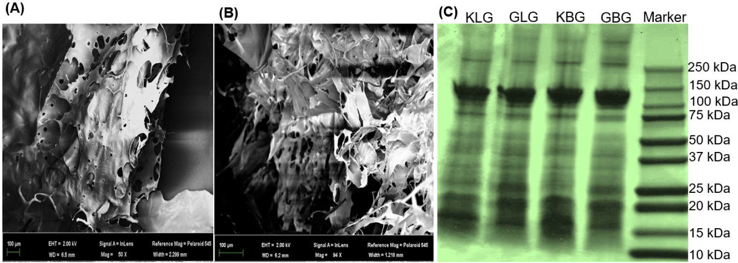
The SDS-PAGE results revealed a variety of gelatin peptides with different molecular weights resulting from enzymatic hydrolysis (Fig. 3C). For both bone types, the hydrolysis with ginger and kiwifruit extracts produced gelatin products ranging from 75 to 10 kDa. The appearance of bands between 130 and 110 kDa corresponds to the α-chains of native collagen molecules that were not hydrolyzed. Meanwhile, bands between 20 and 15 kDa were the predominant products, indicating the degradation of the α-chains of native collagen molecules after swelling with vinegar and hydrolysis with the extracts. Beyond their application in meat tenderization, enzyme extracts from kiwifruit and ginger have also been used to hydrolyze connective tissues during the extraction of bioactive substances from other materials [22,49]. Similarly, Cao et al. [12] used hydrochloric, acetic, and citric acids along with pepsin to extract gelatin from bovine bone, observing peptide bands primarily in the ranges of 100–70 kDa and 25–15 kDa.
The DPPH free radical scavenging and ferrous chelating activities of pork bone gelatin are presented in Tables 7 and 8, respectively. For both the synthetic antioxidant (BHT) and gelatin samples, the DPPH scavenging and ferrous chelating efficiencies increased with their concentration (p < 0.05). At the lowest concentration (0.1 mg/mL), gelatin samples showed DPPH scavenging and ferrous chelating activities of 28%–60% and 34%–44%, respectively, which were partly lower than those of BHT (78% and 58% in the DPPH and ferrous chelating assays, respectively). At the highest concentration tested (0.7 mg/mL), gelatin samples exhibited DPPH scavenging and ferrous chelating efficiencies of 76%–89% and 67%–81%, respectively. Interestingly, the gelatin extracted with kiwifruit exhibited higher antioxidant activity than that extracted with ginger at almost all concentrations for both bone types (p < 0.05). BHT is a synthetic antioxidant. Compared to its antioxidant efficiency at 0.7 mg/mL, the antioxidant activity of gelatin from leg bone or backbone extracted with kiwifruit was only about 10% lower in both the DPPH and ferrous chelating assays. Choi et al. [50] evaluated the ABTS free radical scavenging activity of porcine gelatin produced by hydrothermal processing, and reported approximately 80% antioxidant activity at 5 mg/mL. Recently, Hao et al. [11] prepared collagen peptides from pork bone using citric acid and Alkaline 2.4, and reported ferrous chelating activity above 80% at 1 mg/mL. Nurilmala et al. [51] found that gelatin peptides from tuna skin with molecular weights ranging from 10 to 30 kDa exhibited the highest antioxidant activity.
The mechanisms underlying the antioxidant activity of various compounds are now well understood, primarily by inhibiting the formation of free radicals or interrupting their propagation through free radical scavenging, metal ion chelation, lipid oxidation inhibition, and support of the body’s antioxidant defense systems [52,53]. The antioxidant efficacy of peptides can vary significantly depending on the type of proteolytic enzymes used during extraction and the molecular weights of the resulting peptides [7]. The human body possesses an intrinsic defense system to counteract free radicals or reactive oxygen species (ROS) generated during metabolic processes or as a result of pathological conditions. This defense relies on maintaining a balance between ROS production and antioxidant availability. An imbalance (characterized by excessive ROS and insufficient antioxidants) leads to oxidative stress, which can damage cellular proteins and DNA [54]. Furthermore, the efficiency of the body’s oxidative stress defense mechanisms tends to decline with age [55], making the dietary intake of antioxidants increasingly important for health maintenance. Based on the results of this study, the use of cooking vinegar in combination with plant-derived enzyme extracts (particularly those from kiwifruit) offers a promising approach for producing bioactive pork bone gelatins or nutritious bone-based foods that are both functional and safe for consumers.
CONCLUSION
For the first time, this study evaluated the quality changes of pork bones, along with their nutritional composition, and developed an effective method for extracting their bioactive gelatin. Although a low total microbial count was observed throughout the storage periods, the pork bones exhibited high levels of lipid oxidation and discoloration after 14 days of storage under aerobic packaging and refrigerated conditions. Pork bones were found to be rich in collagen content, essential amino acids, and monounsaturated and polyunsaturated fatty acids. The extraction using vinegar and plant-derived enzyme extracts produced gelatin products with multiple molecular weights and a heterogeneous surface structure. Gelatin extracted with kiwifruit extract exhibited higher antioxidant activity than that extracted with ginger, and was nearly comparable to the synthetic antioxidant at the same concentration. From the results obtained, it is concluded that pork bones exhibited high lipid oxidation levels and rapid discoloration during refrigerated storage. Additionally, cooking vinegar and plant-derived enzyme extracts have been proven to be effective natural alternatives to synthetic chemicals for extracting bioactive gelatin. This safe and efficient method shows great potential for both home-scale preparation of nutritious bone-based foods and industrial-scale recycling of meat by-products into valuable biomaterials. Further studies are needed to investigate alternative packaging methods (e.g., skin and modified atmosphere packaging) under various temperature conditions (e.g., chilling and freezing) to determine the optimal storage conditions for maintaining the quality of meat by-products such as pork bones.


