SHORT COMMUNICATION

Molecular characterization and functionality of rumen-derived extracellular vesicles using a Caenorhabditis elegans animal model

Hyejin Choi1https://orcid.org/0000-0002-5977-2780, Daye Mun1https://orcid.org/0000-0002-3470-7632, Sangdon Ryu1https://orcid.org/0000-0001-5338-8385, Min-jin Kwak1https://orcid.org/0000-0001-9832-3251, Bum-Keun Kim2https://orcid.org/0000-0002-9752-741X, Dong-Jun Park2https://orcid.org/0000-0001-9452-9391, Sangnam Oh3,*https://orcid.org/0000-0002-2428-412X, Younghoon Kim1,*https://orcid.org/0000-0001-6769-0657
Author Information & Copyright
1Department of Agricultural Biotechnology and Research Institute of Agriculture and Life Science, Seoul National University, Seoul 08826, Korea
2Korea Food Research Institute, Wanju 55365, Korea
3Department of Functional Food and Biotechnology, Jeonju University, Jeonju 55069, Korea
*Corresponding author: Sangnam Oh, Department of Functional Food and Biotechnology, Jeonju University, Jeonju 55069, Korea. Tel: +82-63-220-3109, E-mail: osangnam@jj.ac.kr
*Corresponding author: Younghoon Kim, Department of Agricultural Biotechnology and Research Institute of Agriculture and Life Science, Seoul National University, Seoul 08826, Korea. Tel: +82-2-880-4808, E-mail: ykeys2584@snu.ac.kr

© Copyright 2023 Korean Society of Animal Science and Technology. This is an Open-Access article distributed under the terms of the Creative Commons Attribution Non-Commercial License (http://creativecommons.org/licenses/by-nc/4.0/) which permits unrestricted non-commercial use, distribution, and reproduction in any medium, provided the original work is properly cited.

Received: Dec 01, 2022; Revised: Dec 16, 2022; Accepted: Dec 18, 2022

Published Online: May 31, 2023

Abstract

The rumen fluids contain a wide range of bacteria, protozoa, fungi, and viruses. The various ruminal microorganisms in the rumen provide nutrients by fermenting the forage they eat. During metabolic processes, microorganisms present in the rumen release diverse vesicles during the fermentation process. Therefore, in this study, we confirmed the function of rumen extracellular vesicles (EVs) and their interaction with the host. We confirmed the structure of the rumen EVs by transmission electron microscope (TEM) and the size of the particles using nanoparticle tracking analysis (NTA). Rumen EVs range in size from 100 nm to 400 nm and are composed of microvesicles, microparticles, and ectosomes. Using the Caenorhabditis elegans smart animal model, we verified the interaction between the host and rumen EVs. Exposure of C. elegans to rumen EVs did not significantly enhance longevity, whereas exposure to the pathogenic bacteria Escherichia coli O157:H7 and Staphylococcus aureus significantly increased lifespan. Furthermore, transcriptome analysis showed gene expression alterations in C. elegans exposed to rumen EVs, with significant changes in the metabolic pathway, fatty acid degradation, and biosynthesis of cofactors. Our study describes the effect of rumen EV interactions with the host and provides novel insights for discovering biotherapeutic agents in the animal industry.

Keywords: Ruminal fluids; Extracellular vesicles; Biotherapeutic agents; Caenorhabditis elegans

INTRODUCTION

Ruminants are an important source of most meat and dairy products consumed by people worldwide [15]. Unlike monogastric animals, the ruminant animals consumed and fermented the feeds containing plant materials in the rumen, an anaerobic chamber in which various microorganisms are digested and absorbed [68]. The rumen fluids in the anaerobic fermentation chamber mainly contain a wide range of anaerobic bacteria, protozoa, fungi, methanogenic archaea, and phages [9]. During fiber degradation metabolism, various rumen microorganisms release a variety of metabolites and vesicles through anaerobic fermentation [10,11].

Extracellular vesicles (EVs) are nanosized vesicles released by all cells, including bacteria [12,13]. EVs are lipid bilayer-enclosed structures containing proteins, lipids, and nucleic acids. And they belong to a large family of exosomes (30–200 nm), microvesicles (100–350 nm), and apoptotic blebs (500–1000 nm) [14,15]. According to recent studies, EVs mediate cell and organ interactions because they contain DNA, RNA, proteins, and lipids and have the potential as biomarkers and targeted therapeutic mediators [16]. Recent studies reported that all kinds of bacteria could produce EVs. The existence of bacteria-derived EVs in body fluids such as milk, blood, sweat, urine, or feces suggests that these molecules have an impact on host cells and organs by directly activating host receptors, conveying different bioactive chemicals or integrating EVs into host cells [1720]. In particular, in the livestock industry, EVs are widely used as biomarkers for monitoring bovine mastitis, stress conditions, and different diseases in livestock [2123]. The Caenorhabditis elegans surrogate model has been widely used to understand conserved mechanisms in host-EV interactions. C. elegans is a multicellular animal model that has the potential to close the gap between in vitro and in vivo approaches by genetic tractability, simplicity of culture, and comparatively short lifespan [24,25]. Therefore, we used the genetically tractable model host C. elegans to investigate the effect of EVs on the host.

To date, numerous studies on the function of the various microorganisms in the rumen and their interactions have been conducted, but EVs released by rumen microorganisms and their interactions with the host are still poorly understood. Consequently, this study aimed to identify rumen EVs (REVs) and investigate REVs-host interactions using a C. elegans animal model.

MATERIALS AND METHODS

Isolation of ruminal extracellular vesicles

The isolation of REVs was performed using the standard differential ultracentrifugation method described in previous studies with some modifications [26]. In brief, bovine rumen was centrifuged at 5,000×g at 4°C for 15 min to remove large particles. The supernatant was centrifuged at 7,000×g at 4°C for 30 min to remove any remaining debris and cells. Then, the supernatant was passed through a 0.45 μm filter and a 0.22 μm filter to remove residual cell debris. The clear supernatant was centrifuged at 150,000×g at 4°C for 60 min using an ultracentrifuge. And we collected pelletized EVs, resuspended them in phosphate-buffered saline (PBS), and kept them at −80°C. For the preparation of sonication REVs samples (SREVs) as negative control, EVs were sonicated to 50 w (1 min for 8 cycles) using an ultrasonic homogenizer on ice before use for the experiment. The experimental protocol for this study was reviewed and approved by the Institutional Animal Care and Use Committee of Jeonju University (approval jjIACUCU-2022-0409-A1).

Characterization of extracellular vesicles

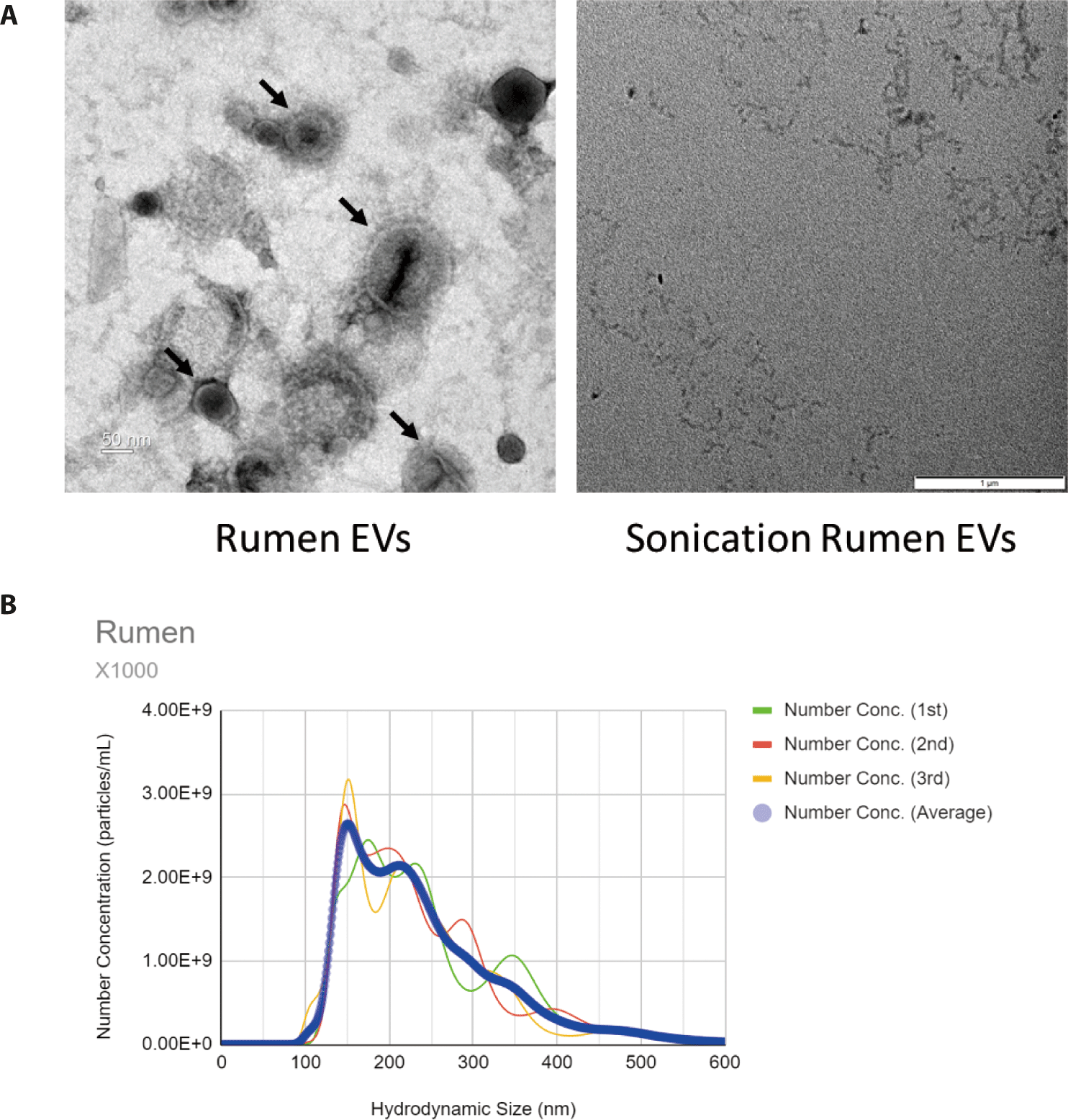
To characterize EVs, transmission electron microscopy (TEM) and nanoparticle tracking analysis (NTA) were performed. TEM investigations were performed according to a previously described method [27,28]. A 5 μL EV sample was placed onto a carbon-coated grid and allowed to settle for 60 sec. The samples were negatively stained with 2% uranyl acetate. The samples were visualized by TEM at NICEM (The National Instrumentation Center for Environmental Management, Seoul National University, Korea). EV size distribution and number were assessed by NTA using a NanoSight NS300 (Malvern). The data were analyzed using NTA software version 3.4 Analytical.

Maintenance and lifespan assay

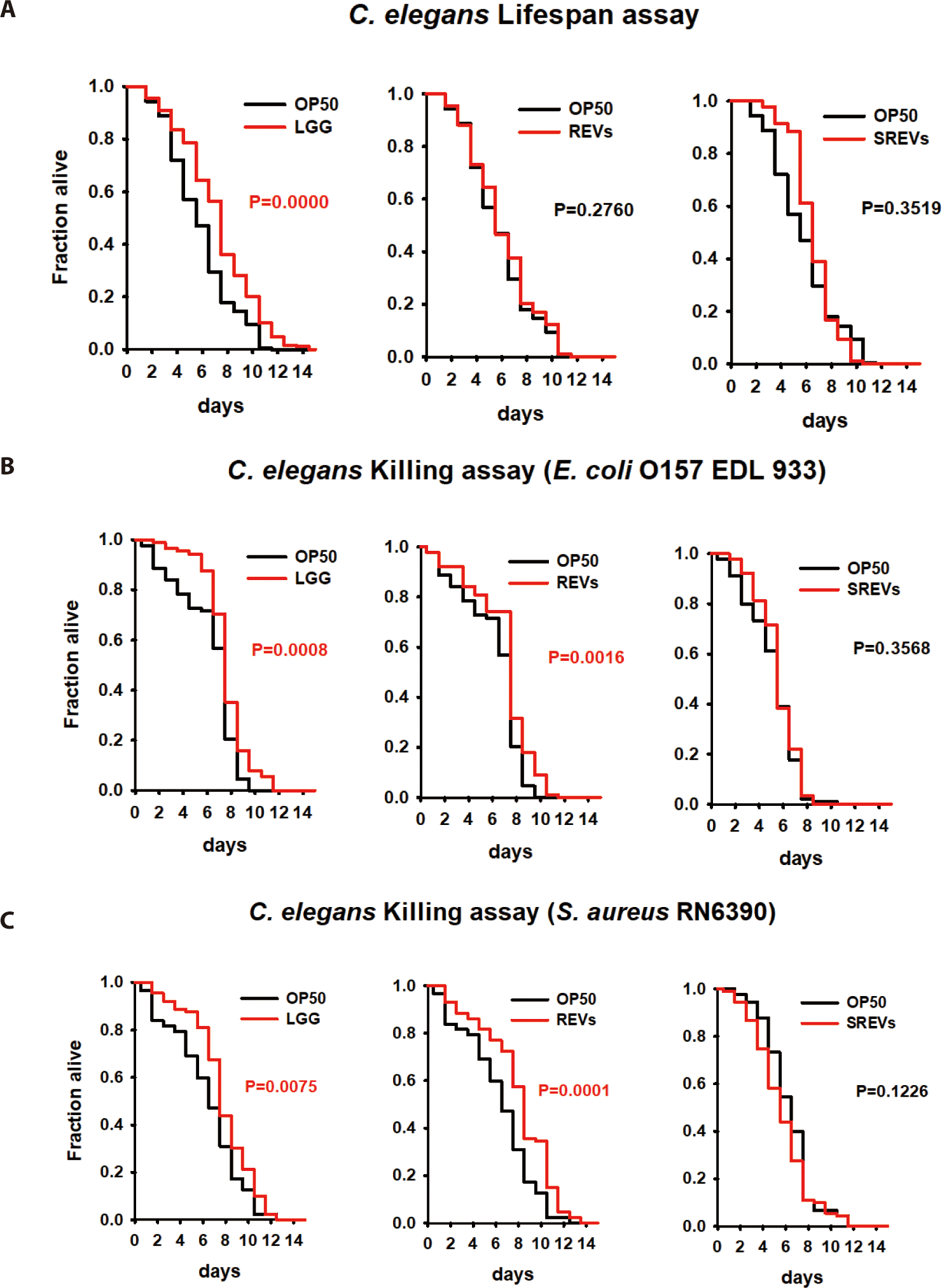
C. elegans was maintained on nematode growth medium (NGM) agar plates supplemented with Escherichia coli OP50 strain at 20°C by using a standard protocol [29]. For the lifespan experiment, we used the CF512 fer-15(b26)II;fem-1(hc17) IV (fer-15;fem-1 worms) strain. In the lifespan assay, the L4 young adult stage worms were exposed to OP50 with EVs (approximately 1.0×10¹⁰ beads), and the number of live and dead worms was recorded every day. For the killing assays, L4 young adult stage worms were preconditioned with OP50 and EVs (1×10¹⁰) for 24 h and then transferred to Escherichia coli O157:H7 EDL933 and Staphylococcus aureus RN6390 as pathogenic bacteria. Then, living worms were counted every day.

C. elegans RNA sequencing

For C. elegans transcriptome analysis, L4 young adult worms were exposed to EVs for 24 h. After 24 h, worms were harvested in M9 buffer and TRIzol for RNA isolation. After homogenization, RNA was extracted using a Qiagen RNeasy kit according to the manufacturer’s instructions. Total RNA concentration was measured using Quant-IT RiboGreen (Invitrogen, Waltham, MA, USA). To determine the quality of samples total RNA, samples are run on the TapeStation RNA screentape (Agilent Technologies, Santa Clara, CA, USA). For the construction of RNA libraries, we using high-quality RNA preparations (RIN > 7.0). Each sample’s library was individually prepared using the Illumina TruSeq Stranded Total RNA Library Prep Gold Kit (Illumina, San Diego, CA, USA). First, removing the rRNA from the total RNA. The remaining mRNA is broken up to little fragments using divalent cations at a high temperature. With the use of random primers and SuperScript II reverse transcriptase from Invitrogen, the cleaved RNA fragments are converted into first-strand cDNA. Next, Second-strand cDNA synthesis by utilizing DNA Polymerase I, RNase H, and dUTP. These cDNA fragments are next subjected to an end repair procedure, a single “A” base addition, and adapter ligation. The final cDNA library is made by purifying and enriching the results with polymerase chain reaction (PCR). Then, using KAPA Library Quantification kits for quantified libraries accroding to the quantitative PCR (qPCR) Quantification Protocol Guide (KAPA BIOSYSTEMS) and validated using TapeStation D1000 ScreenTape (Agilent Technologies). After the indexing of libraries, paired-end (2×100 bp) sequencing was conducted on a Illumina NovaSeq (Illumina).

Statistical analysis

The C. elegans lifespan and killing assay were analyzed according to the Kaplan–Meier (KM) method. A log-rank test between survival curves was performed using STATA software to examine the significance of differences (USA). A significant difference was defined as one that showed a significance (p-value 0.05) through all replicates.

RESULTS

Identification of extracellular vesicles in the bovine rumen

In the TEM image, membrane vesicles with various sizes were observed (Fig. 1A). No EV was observed in samples treated with sonication (Fig. 1A). NTA was used to examine the size, distribution, and concentration of EVs. EVs in the rumen have an average size of 157.4 nm, which is considered to include microvesicles, microparticles, ectosomes and exosomes (Fig. 1B).

jast-65-3-652-g1
Fig. 1. Characterization of EVs derived from rumen. (A) Transmission electron microscopy (TEM) images of REVs and SREVs (scale bar: left = 50 nm; right = 1 μm). (B) Analysis of REVs by nanoparticle tracking analysis (NTA) to identify EVs size distribution. EV, extracellular vesicle; REVs, rumen EVs; SREVs, sonication rumen EVs.
Download Original Figure
Rumen extracellular vesicles improve C. elegans resistance to pathogen infection

To evaluate the interactions between REVs and the host, we used the C. elegans animal model as an in vivo model. First, we investigated the effect of REVs on the lifespan of C. elegans. The lifespan of nematodes was not affected by exposure to REVs compared to the control E. coli OP50 alone (Fig. 2A). Next, we investigated whether REVs could increase the C. elegans defense response to pathogenic bacteria. We exposed C. elegans to E. coli OP50 with REVs and SREVs for 24 h, transferred the worms to E. coli O157:H7- and S. aureus-pathogenic bacteria-treated plates, and counted the viability every day. Contrary to the lifespan results, REVs significantly increased the worm’s lifespan when C. elegans was exposed to pathogenic bacteria (Fig. 2B). However, treatment with SREVs did not increase the lifespan of the worms. These findings imply that REVs enhance the survival of C. elegans from exposure to pathogenic bacteria.

jast-65-3-652-g2
Fig. 2. Effects of rumen EVs on lifespan and resistance to pathogenic bacterial infection in C. elegans. (A) Lifespan curves of L4-stage C. elegansfer-15;fem-1 fed REVs, SREVs and Lactobacillus rhamnosus GG (positive control). Survival statistics: p = 0.0000 for LGG, p = 0.2760 for REVs and p = 0.3519 for SREVs compared with worms exposed to E. coli OP50 (control). (B) Survival of C. elegansfer-15;fem-1 infected with E. coli O157:H7 EDL 933 after conditioning with the REVs, SREVs and LGG for 24 h. Survival statistics: p = 0.0008 for LGG, p = 0.0016 for REVs, p = 0.3568 for SREVs, respectively, compared with worms exposed to E. coli OP50 for 24 h. (C) Survival of C. elegans fer-15;fem-1 infected with S. aureus RN6390 after conditioning with REVs, SREVs and LGG for 24 h. Survival statistics: p = 0.0075 for LGG, p = 0.0001 for REVs, p = 0.1226 for SREVs, compared with worms exposed to E. coli OP50 for 24 h. C. elegans, Caenorhabditis elegans; E. coli, Escherichia coli; Staphylococcus aureus; EV, extracellular vesicle; REVs, rumen EVs; SREVs, sonication rumen EVs.
Download Original Figure
Analysis of Caenorhabditis elegans transcript changes induced by ruminal extracellular vesicles

Since REVs induced C. elegans protection from pathogenic bacteria, we performed an RNA sequencing assay to investigate the genes regulated by exposed REVs. RNA from worms exposed to REVs (treatment group) and worms exposed to SREVs (control) were sequenced. Here, we focused on investigating transcript level changes on the previously unreported effects of REV’s treatment. Gene expression levels modulated by REVs and SREVs treatments were shown (Fig. 3A). REVs treatment in worms could significantly increase 433 genes and decrease 643 genes compared to the SREVs-treated group. Notably, REVs upregulated genes related to an olfactory receptor, chitinase activity, and peptide receptor activity, and downregulated genes related to double-stranded DNA binding, adenylate cyclase activity, and phosphoprotein phosphatase activity (Fig. 3B).

jast-65-3-652-g3
Fig. 3. Transcriptional profiling of Caenorhabditis elegans treated with rumen EVs and sonication rumen EVs. (A) Volcano plot of transcriptome differences. (B) Gene expression relative to Control (SREVs). Gene Ontology (GO) enrichment analysis for (C) biological process, (D) molecular function and (E) cellular component. (F) Top 10 Kyoto Encyclopedia of Genes and Genomes (KEGG) pathway enrichment dot plot. EV, extracellular vesicle.
Download Original Figure

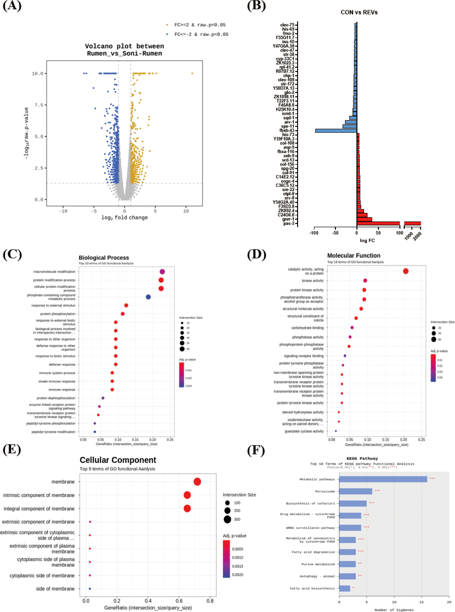
For an in-depth understanding, the results of enrichment analysis through the Gene Ontology (GO) Database for significant genes are shown as a dot plot. The REVs treatment in C. elegans could significantly regulate 69 GO terms. The 42 terms were for biological process (BP) (Fig. 3C), 18 for molecular function (MF) (Fig. 3D), and 9 for cellular component (CC) (Fig. 3E). The main GO terms in BP were GO:0044419 (biological process involved in interspecies interaction between organisms), GO:0098542 (defense response to other organism), and GO:0006955 (immune response). The most significantly enriched MFs were GO:0042302 (structural constituent of cuticle), GO:0004715 (nonmembrane spanning protein tyrosine kinase activity), and GO:0140096 (catalytic activity, acting on a protein). In CC, GO:0016021 (integral component of membrane), GO:0031224 (intrinsic component of membrane), and GO:0016020 (membrane) were significantly enriched. We further analyzed transcript changes using the Kyoto Gene and Genome (KEGG) pathway. Based on the KEGG pathway analysis, the REVs treatment in C. elegans mainly changed the metabolic pathway, peroxisome, and biosynthesis of cofactors pathway (Fig. 3F). In particular, metabolic pathway-related genes (ZK892.4, gst-8, gcy-20) and peroxisome-related genes (ZK892.4, F41E7.6) were upregulated by REVs, and biosynthesis of cofactor pathway-related genes (F17C8.9, sams-5, F52B11.2) was downregulated.

The top 5 genes increased by REV treatment are shown in Table 1. The expression of pas-3 (proteasome) and gnrr-1 (neuroactive ligand‒receptor interaction) were substantially increased. Among the five upregulated genes, substantially increased in genes related to proteasome (pas-3). Although not included in the top 5, genes associated with the immune response (F54D5.4, fbxa-116, sysm-1, irg-5, F55G11.2, tsp-1, C17H12.6, C34H4.1, Y43C5B.2, pals-23, T24C4.4, valv-1, C08E8.4) were significantly increased by REVs treatment in C. elegans. Conversely, REVs treatment significantly decreased the expression level of spe-1 (reproduction), arv-1 (lipid metabolic process), and sqd-1 (ortholog of human hepatocellular carcinoma) compared to SREVs treatment in C. elegans (Table 2).

Table 1. Top 5 significantly upregulated genes in rumen EV-treated Caenorhabditis elegans
Gene Gene description Fold change (REVs/SREVs)
pas-3 Proteasome subunit alpha type-4 2,050.93
gnrr-1 G_PROTEIN_RECEP_F1_2 domain-containing protein 35.49
C24G6.8 Probable peptidyl-tRNA hydrolase 2 24.02
Y67D8C.26 Unclassified noncoding RNA Y67D8C.26 23.49
F08G2.9 small nuclear RNA F08G2.9 17.51

EV, extracellular vesicle; REVs, rumen extracellular vesicles; SREVs, sonication rumen extracellular vesicles.

Download Excel Table
Table 2. Top 5 significantly downregulated genes in rumen EV-treated Caenorhabditis. elegans
Gene Gene description Fold change (REVs/SREVs)
fbxb-43 F-box domain-containing protein −97.29
spe-11 Spermatocyte protein spe-11 −33.41
arv-1 ARV1 homolog −27.58
sqd-1 Homologous to Drosophila SQD (squid) protein −17.46
Y62E10A.24 Ribonuclease P protein subunit p20 −17.07

EV, extracellular vesicle; REVs, rumen extracellular vesicles; SREVs, sonication rumen extracellular vesicles.

Download Excel Table

DISCUSSION

Many studies are being conducted on the rumen of ruminants. The rumen microbiome is an essential component of the ruminant gastrointestinal tract, and it consists of bacteria (1010–1011 cells/mL), methanogenic archaea (107–109 cells/mL), protozoa (104–106 cells/mL), anaerobic fungi (103–106 cells/mL) and bacteriophages (109–1010 particles/mL) [2,30]. The rumen microbiome mainly acts as a fermenter and can digest ingested fibrin, which is not available to monogastric animals. Recently, research has been conducted to improve livestock production by understanding host–rumen microbial interactions. Studies focus on investigating the effect on rumen metabolism, methanogenesis, and host microRNA expression [3033]. As the understanding between the host and the rumen has become more important, we attempted to identify the interaction with the host by isolating EVs released by various microorganisms present in the rumen.

Ruminal EVs were isolated using an ultracentrifuge, and particle shape and size were observed through TEM and NTA analysis. REVs were observed with various sizes ranging from 100–400 nm. This is a result of the release of EVs such as exosomes, microvesicles, microparticles and ectosomes through metabolic processes by various microorganisms in the rumen.

We used the C. elegans animal model to investigate the genetic changes of REVs on the host. REVs did not change the lifespan of C. elegans but increased the resistance of C. elegans to pathogenic bacteria. In transcriptome analysis, a significant increase in genes (gst-8, mtl-1) related to lifespan extension of C. elegans was not confirmed, so these findings are consistent with our findings. The C. elegans killing assay results showed that immune response genes (fbxa-116, sysm-1, irg-5, tsp-1, pals-23, and valv-1) were significantly increased. In particular, in previous studies, irg-5 is known as a necessary immune effector for host defense in pathogen infection and is a target of the p38 MAPK PMK-1 pathway [34,35]. Our findings demonstrate that treatment with REVs can stimulate C. elegans immune responses and induce more resistance to pathogenic bacteria.

According to the literature, early weaning of young ruminants has been reported to be essential for the rapid growth of ruminants, but early weaning can induce various stresses in young ruminants, which can cause growth retardation and suppression of the immune system [36,37]. Therefore, it is known that early settlement of microorganisms can be induced by transplanting rumen fluid to young ruminants for successful early weaning and rumen development, and digestive disorders of ruminants can be treated by transplanting rumen fluid [38,39]. Interestingly, consistent with our results, REVs upregulated the C. elegans membrane, developmental process, and structure-related genes in GO analysis. Similarly, according to KEGG analysis, treatment with REVs significantly upregulated the standard proteasome subunit pas-3 gene, which affects the embryonic viability, growth, fertility, and motility of C. elegans [35,40]. Particularly, upregulated chitinase activity by REVs treatment could indicate a relationship with increased host’s immune and defense functions. Chitinase activity can also reflect the increment of host digestibility, immunity, and protective systems, suggesting that it may play a role similar to previous studies of increasing the growth and development of young ruminants via rumen transplantation [41,42].

Currently, numerous continuing studies are elucidating the functionality and role of livestock-derived EVs in livestock industry fields [4346]. As the first to isolate rumen derived EVs, this study showed that REVs increased pathogen resistance by regulating nematode immune genes. Furthermore, it could suggest that REVs may promote worm growth by significantly increasing genes involved in worm metabolism and development. Despite significant differences between ruminants and C. elegans, REVs stimulated genes involved in nematode metabolism and development, similar to the effects of ruminant transplantation in ruminants previously studied. These results suggest that REVs may bring growth and health benefits to ruminants. These data establish the basis for further investigation into the role of REVs. However, since these are the results of using the C. elegans model, additional studies are needed to deepen our understanding of the role of rumen EVs.

Competing interests

No potential conflict of interest relevant to this article was reported.

Funding sources

This research was supported by a National Research Foundation of Korea Grant, funded by the Korean government (MEST) (NRF-2021R1A2C3011051) and by the Main Research Program (E0211100-03) of the Korea Food Research Institute (KFRI)funded by the Ministry of Science and ICT (Korea).

Acknowledgements

Not applicable.

Availability of data and material

Upon reasonable request, the datasets of this study can be available from the corresponding author.

Authors’ contributions

Conceptualization: Choi H, Oh S, Kim Y.

Data curation: Choi H, Oh S, Kim Y.

Formal analysis: Choi H, Mun D, Ryu S, Kwak MJ, Kim BK, Park DJ, Oh S, Kim Y.

Writing - original draft: Choi H, Mun D, Ryu S, Kwak MJ, Park DJ, Oh S, Kim Y.

Writing - review & editing: Choi H, Mun D, Ryu S, Kwak MJ, Kim BK, Park DJ, Oh S, Kim Y.

Ethics approval and consent to participate

The experimental protocol for this study was reviewed and approved by the Institutional Animal Care and Use Committee of Jeonju University (approval jjIACUCU-2022-0409-A1).

REFERENCES

1.

Jami E, Mizrahi I. Composition and similarity of bovine rumen microbiota across individual animals. PLOS ONE. 2012; 7e33306

2.

Malmuthuge N, Guan LL. Understanding host-microbial interactions in rumen: searching the best opportunity for microbiota manipulation. J Anim Sci Biotechnol. 2017; 8:8

3.

Ababakri R, Dayani O, Khezri A, Naserian AA. Influence of flaxseed with rumen undegradable protein level on milk yield, milk fatty acids and blood metabolites in transition ewes. J Anim Sci Technol. 2021; 63:475-90

4.

Lim DH, Kim TI, Park SM, Ki KS, Kim Y. Evaluation of heat stress responses in Holstein and Jersey cows by analyzing physiological characteristics and milk production in Korea. J Anim Sci Technol. 2021; 63:872-83

5.

Um KH, Park BK. Study on the rumen fermentation, growth performance and carcass characteristics according to the supplementation of lupin flake in Hanwoo steers. J Anim Sci Technol. 2022; 64:1077-91

6.

McGovern E, McGee M, Byrne CJ, Kenny DA, Kelly AK, Waters SM. Investigation into the effect of divergent feed efficiency phenotype on the bovine rumen microbiota across diet and breed. Sci Rep. 2020; 10:15317

7.

Baek YC, Choi H, Jeong JY, Lee SD, Kim MJ, Lee S, et al. The impact of short-term acute heat stress on the rumen microbiome of Hanwoo steers. J Anim Sci Technol. 2020; 62:208-17

8.

Song Y, Fiaz M, Kim DW, Kim J, Kwon CH. Increasing forage yield and effective weed control of corn-soybean mixed forage for livestock through using by different herbicides. J Anim Sci Technol. 2019; 61:185-91

9.

Mizrahi I. Rumen symbioses.In In: Rosenberg E, DeLong EF, Lory S, Stackebrandt E, Thompson F, editors.ediotrs The prokaryotes: prokaryotic biology and symbiotic associations. BerlIn: Springer-Verlag Berlin Heidelberg. 2013; p p. 533-44

10.

Ceconi I, Ruiz-Moreno MJ, DiLorenzo N, DiCostanzo A, Crawford GI. Effect of urea inclusion in diets containing corn dried distillers grains on feedlot cattle performance, carcass characteristics, ruminal fermentation, total tract digestibility, and purine derivatives-to-creatinine index. J Anim Sci. 2015; 93:357-69

11.

Wang L, Zhang G, Li Y, Zhang Y. Effects of high forage/concentrate diet on volatile fatty acid production and the microorganisms involved in VFA production in cow rumen. Animals. 2020; 10:223

12.

Yáñez-Mó M, Siljander PRM, Andreu Z, Bedina Zavec A, Borràs FE, Buzas EI, et al. Biological properties of extracellular vesicles and their physiological functions. J Extracell Vesicles. 2015; 4:27066

13.

Ñahui Palomino RA, Vanpouille C, Costantini PE, Margolis L. Microbiota–host communications: bacterial extracellular vesicles as a common language. PLOS Pathog. 2021; 17e1009508

14.

Li P, Kaslan M, Lee SH, Yao J, Gao Z. Progress in exosome isolation techniques. Theranostics. 2017; 7:789-804

15.

Quan SY, Nan XM, Wang K, Zhao YG, Jiang LS, Yao JH, et al. Replacement of forage fiber with non-forage fiber sources in dairy cow diets changes milk extracellular vesicle-miRNA expression. Food Funct. 2020; 11:2154-62

16.

Macia L, Nanan R, Hosseini-Beheshti E, Grau GE. Host- and microbiota-derived extracellular vesicles, immune function, and disease development. Int J Mol Sci. 2020; 21:107

17.

Kang CS, Ban M, Choi EJ, Moon HG, Jeon JS, Kim DK, et al. Extracellular vesicles derived from gut microbiota, especially Akkermansia muciniphila, protect the progression of dextran sulfate sodium-induced colitis. PLOS ONE. 2013; 8e76520

18.

Pérez-Cruz C, Delgado L, López-Iglesias C, Mercade E. Outer-inner membrane vesicles naturally secreted by gram-negative pathogenic bacteria. PLOS ONE. 2015; 10e0116896

19.

O’Donoghue EJ, Krachler AM. Mechanisms of outer membrane vesicle entry into host cells. Cell Microbiol. 2016; 18:1508-17

20.

Kim SY, Yi DY. Analysis of the human breast milk microbiome and bacterial extracellular vesicles in healthy mothers. Exp Mol Med. 2020; 52:1288-97

21.

Karayel-Hacioglu I, Duran-Yelken S, Alkan F. Molecular detection of picornaviruses in diarrheic small ruminants at a glance: enterovirus, hunnivirus, and kobuvirus in Türkiye. Kafkas Univ Vet Fak Derg. 2022; 28:499-506

22.

Gebremedhn S, Ali A, Gad A, Prochazka R, Tesfaye D. Extracellular vesicles as mediators of environmental and metabolic stress coping mechanisms during mammalian follicular development. Front Vet Sci. 2020; 7:602043

23.

Saenz-de-Juano MD, Silvestrelli G, Bauersachs S, Ulbrich SE. Determining extracellular vesicles properties and miRNA cargo variability in bovine milk from healthy cows and cows undergoing subclinical mastitis. BMC Genom. 2022; 23:189

24.

Yoo J, Lee J, Zhang M, Mun D, Kang M, Yun B, et al. Enhanced γ-aminobutyric acid and sialic acid in fermented deer antler velvet and immune promoting effects. J Anim Sci Technol. 2022; 64:166-82

25.

Clark LC, Hodgkin J. Commensals, probiotics and pathogens in the Caenorhabditis elegans model. Cell Microbiol. 2014; 16:27-38

26.

Yun B, Kim Y, Park DJ, Oh S. Comparative analysis of dietary exosome-derived microRNAs from human, bovine and caprine colostrum and mature milk. J Anim Sci Technol. 2021; 63:593-602

27.

Ghosh K, Senevirathne A, Kang HS, Hyun WB, Kim JE, Kim KP. Complete nucleotide sequence analysis of a novel Bacillus subtilis-infecting bacteriophage BSP10 and its effect on poly-gamma-glutamic acid degradation. Viruses. 2018; 10:240

28.

Liu Y, Zhao L, Wang M, Wang Q, Zhang X, Han Y, et al. Complete genomic sequence of bacteriophage P23: a novel Vibrio phage isolated from the Yellow Sea, China. Virus Genes. 2019; 55:834-42

29.

Choi HJ, Shin D, Shin M, Yun B, Kang M, Yang HJ, et al. Comparative genomic and functional evaluations of Bacillus subtilis newly isolated from Korean traditional fermented foods. Foods. 2020; 9:1805

30.

Yue ZB, Wang J, Liu XM, Yu HQ. Comparison of rumen microorganism and digester sludge dominated anaerobic digestion processes for aquatic plants. Renew Energy. 2012; 46:255-8

31.

Nagaraja TG, Titgemeyer EC. Ruminal acidosis in beef cattle: the current microbiological and nutritional outlook. J Dairy Sci. 2007; 90:E17-38

32.

Hook SE, Steele MA, Northwood KS, Dijkstra J, France J, Wright ADG, et al. Impact of subacute ruminal acidosis (SARA) adaptation and recovery on the density and diversity of bacteria in the rumen of dairy cows. FEMS Microbiol Ecol. 2011; 78:275-84

33.

McCann JC, Luan S, Cardoso FC, Derakhshani H, Khafipour E, Loor JJ. Induction of subacute ruminal acidosis affects the ruminal microbiome and epithelium. Front Microbiol. 2016; 7:701

34.

Troemel ER, Chu SW, Reinke V, Lee SS, Ausubel FM, Kim DH. p38 MAPK regulates expression of immune response genes and contributes to longevity in C. elegans. PLOS Genet. 2006; 2e183

35.

Foster KJ, Cheesman HK, Liu P, Peterson ND, Anderson SM, Pukkila-Worley R. Innate immunity in the C. elegans intestine is programmed by a neuronal regulator of AWC olfactory neuron development. Cell Rep. 2020; 31:107478

36.

Williams PEV, Frost AI. Feeding the young ruminant. BSAP Occas Publ. 1992; 15:109-18

37.

Hamid MMA, Moon J, Yoo D, Kim H, Lee YK, Song J, et al. Rumen fermentation, methane production, and microbial composition following in vitro evaluation of red ginseng byproduct as a protein source. J Anim Sci Technol. 2020; 62:801-11

38.

Laflin SL, Gnad DP. Rumen cannulation: procedure and use of a cannulated bovine. Vet Clin North Am Food Anim Pract. 2008; 24:335-40

39.

Kim HS, Whon TW, Sung H, Jeong YS, Jung ES, Shin NR, et al. Longitudinal evaluation of fecal microbiota transplantation for ameliorating calf diarrhea and improving growth performance. Nat Commun. 2021; 12:161

40.

Papaevgeniou N, Chondrogianni N. The ubiquitin proteasome system in Caenorhabditis elegans and its regulation. Redox Biol. 2014; 2:333-47

41.

Suzuki M, Morimatsu M, Yamashita T, Iwanaga T, Syuto B. A novel serum chitinase that is expressed in bovine liver. FEBS Lett. 2001; 506:127-30

42.

Tabata E, Kashimura A, Kikuchi A, Masuda H, Miyahara R, Hiruma Y, et al. Chitin digestibility is dependent on feeding behaviors, which determine acidic chitinase mRNA levels in mammalian and poultry stomachs. Sci Rep. 2018; 8:1461

43.

Mun D, Oh S, Kim Y. Perspectives on bovine milk-derived extracellular vesicles for therapeutic applications in gut health. Food Sci Anim Resour. 2022; 42:197-209

44.

Burns GW, Brooks KE, Spencer TE. Extracellular vesicles originate from the conceptus and uterus during early pregnancy in sheep. Biol Reprod. 2016; 94:56

45.

Quan S, Nan X, Wang K, Jiang L, Yao J, Xiong B. Characterization of sheep milk extracellular vesicle-miRNA by sequencing and comparison with cow milk. Animals. 2020; 10:331

46.

Ding Q, Jin M, Kalds P, Meng C, Wang H, Zhong J, et al. Comparison of microRNA profiles in extracellular vesicles from small and large goat follicular fluid. Animals. 2021; 11:3190