INTRODUCTION
Haimen goats are renowned across the nation for their remarkable lambing rates, superior meat quality, and the exceptional properties of both their skin and fur. However, the development of the goat breeding industry faces significant challenges, including the underdeveloped breeding infrastructure, limited distribution of elite breeds, and the indiscriminate introduction of foreign varieties. Addressing these issues by advancing research into goat breeding mechanisms and enhancing efficient reproduction and control techniques holds substantial practical importance for the sustainable growth of the goat industry. Currently, a suite of breeding technologies has been extensively adopted in goat production. Techniques such as estrus synchronization, artificial insemination, superovulation, and embryo transfer are being synergistically applied, offering vital technical support for the enhancement of goat breeding, breed improvement, and the expansion of production on a large scale [1–3].
The estrous cycle of livestock fundamentally involves the alternation between the follicular phase and the luteal phase, where the growth and development of follicles, formation, and regression of corpora lutea are regulated by endogenous neurohormones and influenced by exogenous hormones [4,5]. Livestock synchronized estrus typically involves the use of progesterone (P4) releasing intravaginal device (PRID) to synchronize individuals in different estrous cycles to enter the luteal phase concurrently. After being retained for 9–14 days, the PRID removal prompts the treated goat to rapidly enter the follicular phase, thereby achieving synchronized estrus [6–8]. Prostaglandin (PG), this steroid hormone dissolves corpora lutea while also regulating the release of gonadotropins and promoting ovulation. PG is typically administered at the time of intravaginal insert removal to assist in ensuring synchronized estrus [9].
Previous studies have used equine chorionic gonadotropin (eCG) to induce estrus and superovulation in livestock [10,11]. The eCG possesses dual functions of follicle-stimulating hormone (FSH) and luteinizing hormone with a relatively long half-life [12]. However, due to individual response variability and dosage control challenges, adverse reactions such as ovarian cysts and oocyte aging may occur. Therefore, FSH is preferred in livestock superovulation, due to its shorter half-life, and decreasing doses every 12 h have shown better results [13,14]. Research indicates that FSH can promote follicular development, rescue follicular atresia, and significantly increase ovulation and ovarian follicle numbers in livestock [15,16]. A study comparing single eCG and FSH injections found that the FSH led to more ovulation and high-quality embryos in mice, indicating FSH superior superovulation effect [17,18]. In actual livestock production applications, a mixture of eCG and FSH treatment is more widely used, as they can complement each other to enhance superovulation efficiency [19]. However, in model animals like mice, eCG can replace FSH to achieve normal superovulation effects and reduce hormone costs [20].
Research has shown a close correlation between elevated FSH levels in livestock and changes in ovarian function, such as stimulating ovarian growth and follicular development. However, excessive FSH can increase large follicles, premature ovarian aging, and decreased oocyte quality [21]. FSH does not significantly affect the initial development of primordial follicles (PF), but it plays a crucial role as a survival factor in the transition of PF to secondary follicles [22,23]. FSH exerts its functions by binding to follicle-stimulating hormone receptors (FSHR) expressed exclusively on granulosa cells (GCs), playing an important role in inducing GCs proliferation, differentiation, and steroidogenesis [24–26]. Additionally, FSH is of significant importance in regulating follicular development, oocyte maturation, and the ovulation process.
Based on the aforementioned research findings, this study investigated the impact of combined eCG/FSH treatment on goat follicular development, focusing on plasma hormone levels, ovarian tissue morphology, and GCs function. This study aims to provide a solid theoretical foundation for refining and applying estrus synchronization and superovulation methodologies in goats, contributing valuable insights into the biological mechanisms underlying multiple follicular development and ovulation cycles.
MATERIALS AND METHODS
All the hormones or analogues used in this study were provided by Ningbo Sansheng Biological Technology, with experimental consumables supplied by Guangzhou Jet Bio-Filtration, unless otherwise mentioned. Two- to three-year-old healthy multiparous goats were selected at Haimen Goat Industry Research Institute in Jiangsu. The reproductive tracts of all goats were healthy as monitored by ultrasonography. There were no purulent or mucoid vaginal secretions, no clinical or subclinical endometritis, no signs of reproductive system disease or microbial contamination, and no ovarian cysts. All protocols involving the use of animals adhered to the approved Guidelines for Animal Experiments of Nanjing Agricultural University and received approval from the Animal Care and Use Committee of Nanjing Agricultural University (Approval ID: SYXK2022-0031).
As shown in Fig. 1, twelve Haimen goats with similar physical conditions were selected for superovulation and divided into two groups: eCG_FSH and FSH. This division aimed to investigate the effects of the eCG_FSH and FSH injection regimens on superovulation in Haimen goats. The superovulation protocol commenced with the insertion of PRID into the does’ vaginas for 12 days, starting on day 1. On the morning of day 13, PRID removal and PG injection were performed. Hormone administration commenced on the afternoon of day 10, with seven injections totaling 325 IU FSH over 4 days, administered in decreased doses twice a day (75, 75/50, 50/25, 25/25). The key distinction between the groups was the administration of 100 IU eCG on the first day of hormone injections (day 10). On day 14, estrus identification was conducted by externally observing ewes and using buck for estrus detection. Natural mating was carried out upon estrus confirmation. Multiple breeding sessions were employed to ensure further successful mating, accompanied by the injection of Luteinizing Hormone Releasing Hormone A3. Fertilized embryos were flushed in the fallopian tubes by surgical method on day 17 [27,28]. The 8-cell stage embryos were collected 3 days after donor fertilization and examined for quality under a stereomicroscope. Sixty embryos from each group were transferred into 20 recipient fallopian tubes. Pregnancy was detected 45 days later, and the number of newborn lambs was counted 150 days later.
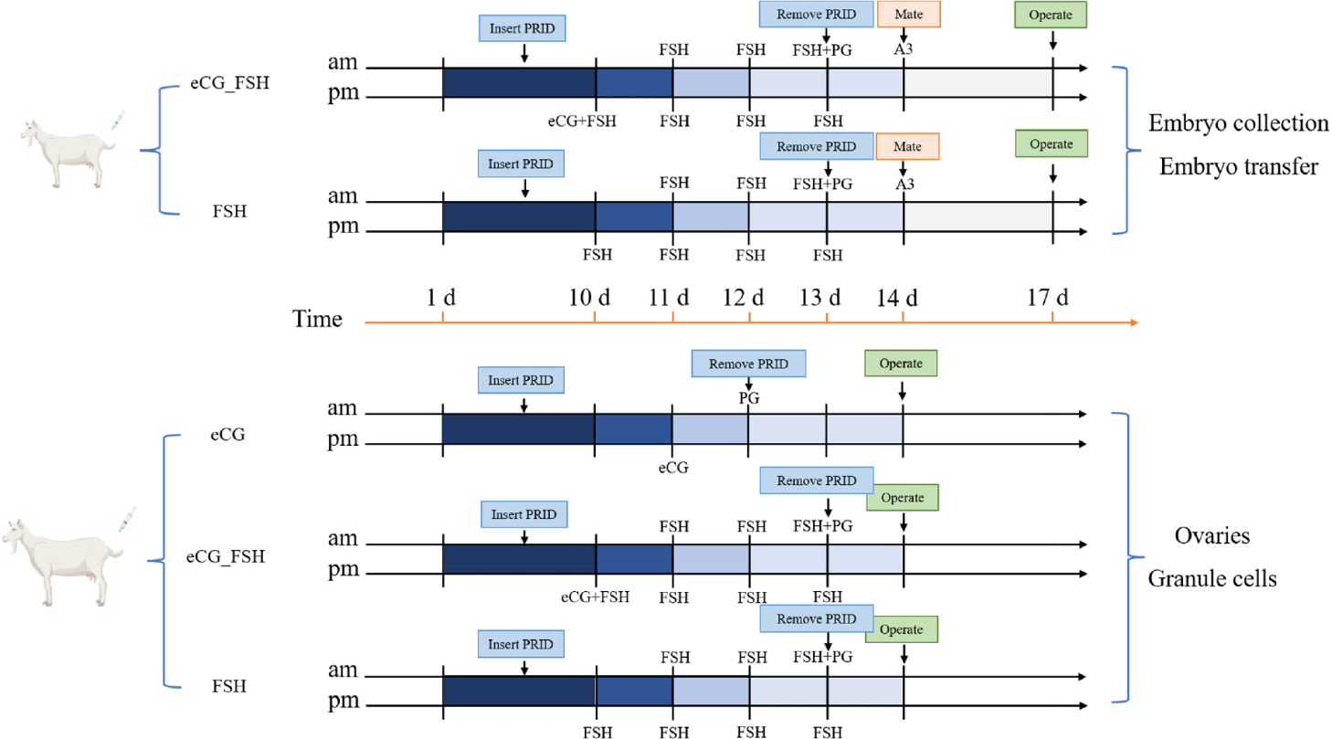
Additionally, nine Haimen goats with comparable physical conditions were divided into three groups: eCG, eCG_FSH, and FSH. This study aimed to investigate the effects of these injection regimens on follicular development. The superovulation procedures for the eCG_FSH and FSH groups mirrored those aforementioned. The eCG group received PRID for 11 days, with eCG administration on day 11 and PG injection following PRID removal on day 12. Estrus identification was performed on day 14, followed by a surgical procedure to extract ovaries, and GCs for further analysis.
Hematoxylin and Eosin (HE) staining was utilized to observe the morphological characteristics of follicular tissues in goat ovaries across various estrus cycle stages. In brief, ovaries fixed in 4% paraformaldehyde were processed and embedded in paraffin using standard protocols. Subsequently, 5 μm thick sections of the embedded ovaries were prepared and subjected to HE staining, where cell nuclei were stained blue and cytoplasm red. The stained sections were examined under a light microscope (Nikon) for histological analysis.
The classification of follicle grade was based on previous studies [29]. PF are characterized by an oocyte in the middle, surrounded by a layer of flattened follicular epithelial cells, without follicular membrane and follicular cavity, mainly distributed in the outer layer of the ovarian cortex. Primary follicle consists of an oocyte surrounded by a single layer of cuboidal follicular cells, lacking a follicular membrane and cavity, and are mainly distributed in the periphery of the ovarian cortex. Secondary follicles are composed of oocytes and surrounding layers of GCs, with follicular membrane and no follicular cavity; Tertiary follicles are formed by oocytes and surrounding layers of GCs, with follicular membrane and follicular cavity. Follicles that have not yet developed a follicular lumen, including those up to the secondary follicle stage, are collectively referred to as preantral follicles, while tertiary and subsequent follicles are classified as antral follicles.
Immunohistochemistry was conducted on goat ovaries following a previously established protocol [30]. Primary antibodies used included rabbit anti-FSHR (22665-1-AP, 1:200, ProteinTech) and estrogen receptor (ER; 22665-1-AP, 1:200, ProteinTech), while goat anti-rabbit IgG (AP132P, 1:1000, Millipore) served as the secondary antibody. Negative control sections were exposed to Tris-buffered saline instead of primary antibodies. Diaminobenzidine (AR1026, Boster) was employed for staining, and all sections were examined using a light microscope (Nikon). FSHR protein expression positivity of ovarian follicles was evaluated by quantifying the % area of immunopositivity of each follicular area with Image J.
Blood samples were collected via ethylenediaminetetraacetic acid anticoagulation from the jugular vein, followed by centrifugation at 500×g for 30 minutes. The supernatant was then collected for analysis of P4, FSHR, and estrogen (E2) using goat Enzyme-linked immunosorbent assays (ELISA) kits (Kmsbiotech) following the manufacturer’s instructions. The intra-assay and inter-assay coefficients of variation were 10% and 15%, respectively. The detailed steps are as follows: samples, standards, and horseradish peroxidase (HRP)-labeled detection antibodies were sequentially added to microplate wells pre-coated with hormone antibodies. After incubation and thorough washing, substrate 3,3’,5,5’-tetramethylbenzidine (TMB) was added for color development. TMB, catalyzed by peroxidase, converted into blue, and subsequently into yellow under acidic conditions. Absorbance (OD value) was measured at a wavelength of 450 nm using an enzyme label analyzer to calculate sample concentrations.
To acquire GCs, the surgical method was employed to open the abdominal cavity of goats to locate the position of the ovaries. The fluid of the antral follicle was aspirated using a 1ml syringe, followed by washing three times with DPBS and centrifugation at 300×g for 5 minutes, and total RNA was extracted using the Trizol (Accurate Biology, AG21101). Agarose gel electrophoresis was performed to analyze the integrity of sample RNA and the presence of DNA contamination. Nano Photometer spectrophotometer was utilized to assess RNA purity, while Qubit 2.0 Fluorometer was employed for accurate quantification of RNA concentration.
The obtained total RNA was subjected to rRNA depletion, followed by enrichment of eukaryotic mRNA containing poly A tails using magnetic beads with Oligo (dT). Fragmented mRNA was used as a template to synthesize the first strand of cDNA in the M-MuLV reverse transcriptase system. Subsequently, RNA chains were degraded using RNaseH, and the second strand of cDNA was synthesized in the DNA polymerase I system. Purified double-stranded cDNA underwent end repair, A-tailing, and adapter ligation, followed by size selection of cDNA fragments of approximately 200 bp using AMPure XP beads. Polymerase chain reaction (PCR) amplification was conducted, and the resulting PCR products were purified again using AMPure XP beads. Finally, the cDNA libraries were sequenced on the Illumina sequencing platform by Genedenovo Biotechnology.
Quality control of the raw reads obtained from sequencing was conducted using fastp to filter out low-quality data and obtain clean reads. The HiSAT2 software was utilized to map the sequencing reads to the goat genome (NCBI_GCF_001704415.2). Transcripts were reconstructed using String tie, and RSEM was employed to calculate the expression levels of all genes in each sample. Differentially expressed genes (DEGs) were identified using DESeq2 with an absolute fold change > 2 and false discovery rate < 0.05. Subsequently, Gene Ontology (GO) analysis, Kyoto Encyclopedia of Genes and Genomes (KEGG) pathway analysis, and Gene Set Enrichment Analysis (GSEA) were performed to analyze the DEGs.
Statistical analyses of hormone concentrations were subjected to one-way analyses of variance (ANOVA) run using Duncan’s test. The basic data (number of antral follicles, number of ovulations, number of corpus luteum, and quantitative analysis of immunopositivity of FSHR) were conducted employing a two-tailed Student’s t-test. The analysis was performed using SPSS v25.0 (IBM). The symbol * indicates a significant difference (p < 0.05). Data are expressed as the mean ± SEM from a minimum of three independent experiments (n ≥ 3).
RESULTS
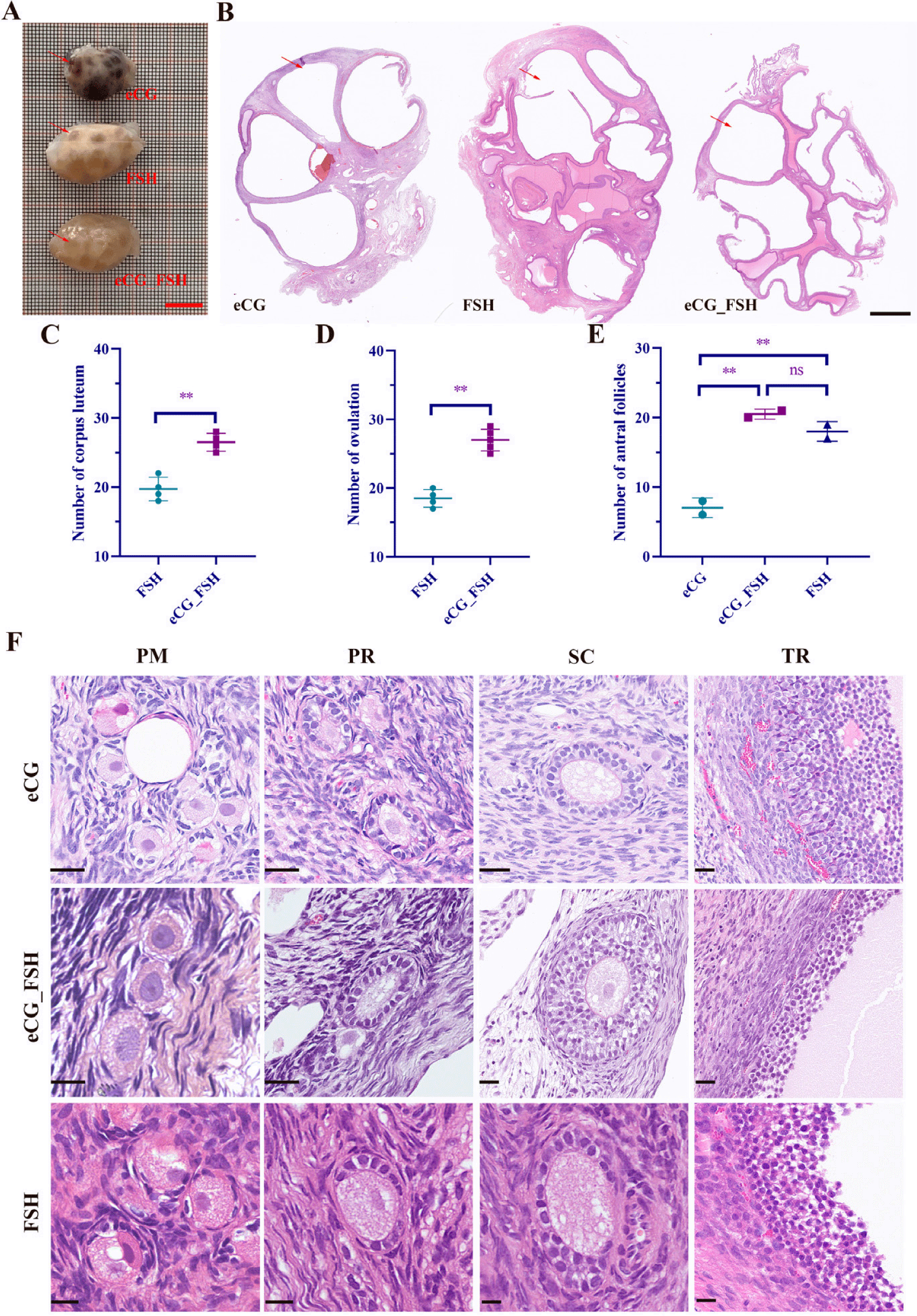
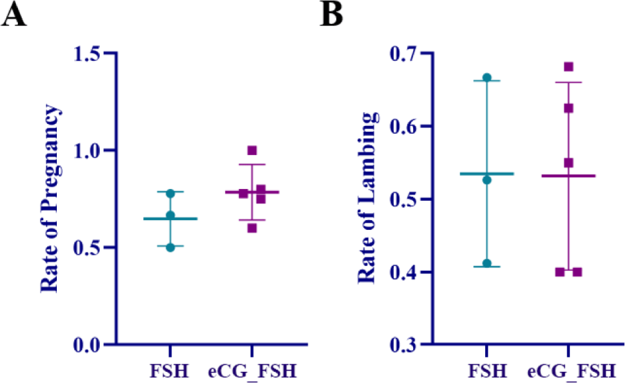
To further investigate the effects of the eCG/FSH treatment on superovulation, we statistically analyzed the number of corpora lutea, ovulations, and antral follicles in goats. Ovaries subjected to superovulation were collected for HE staining (Figs. 2A and 2B). The fertilized embryo was flushed out of the oviducts two days after mating, the results showed significantly higher ovulation and corpus luteum numbers in the eCG_FSH group compared to the FSH group (Figs. 2C and 2D). The numbers of antral follicles in the eCG_FSH and FSH groups were significantly higher than those in the eCG group, with no significant difference observed between the eCG_FSH and FSH groups (Fig. 2E). Furthermore, the regimen of eCG, eCG_FSH, and FSH exhibited normal follicles development at various stages without significant differences (Fig. 2F). In addition, the embryo transfer results showed no difference in pregnancy rate (Fig. 3A) and lambing rate (Fig. 3B) between the eCG group and the FSH group.
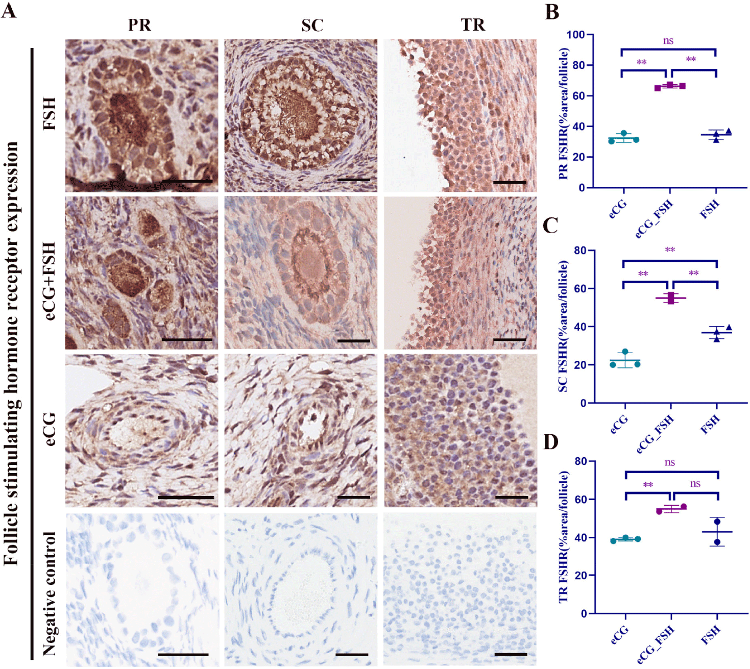
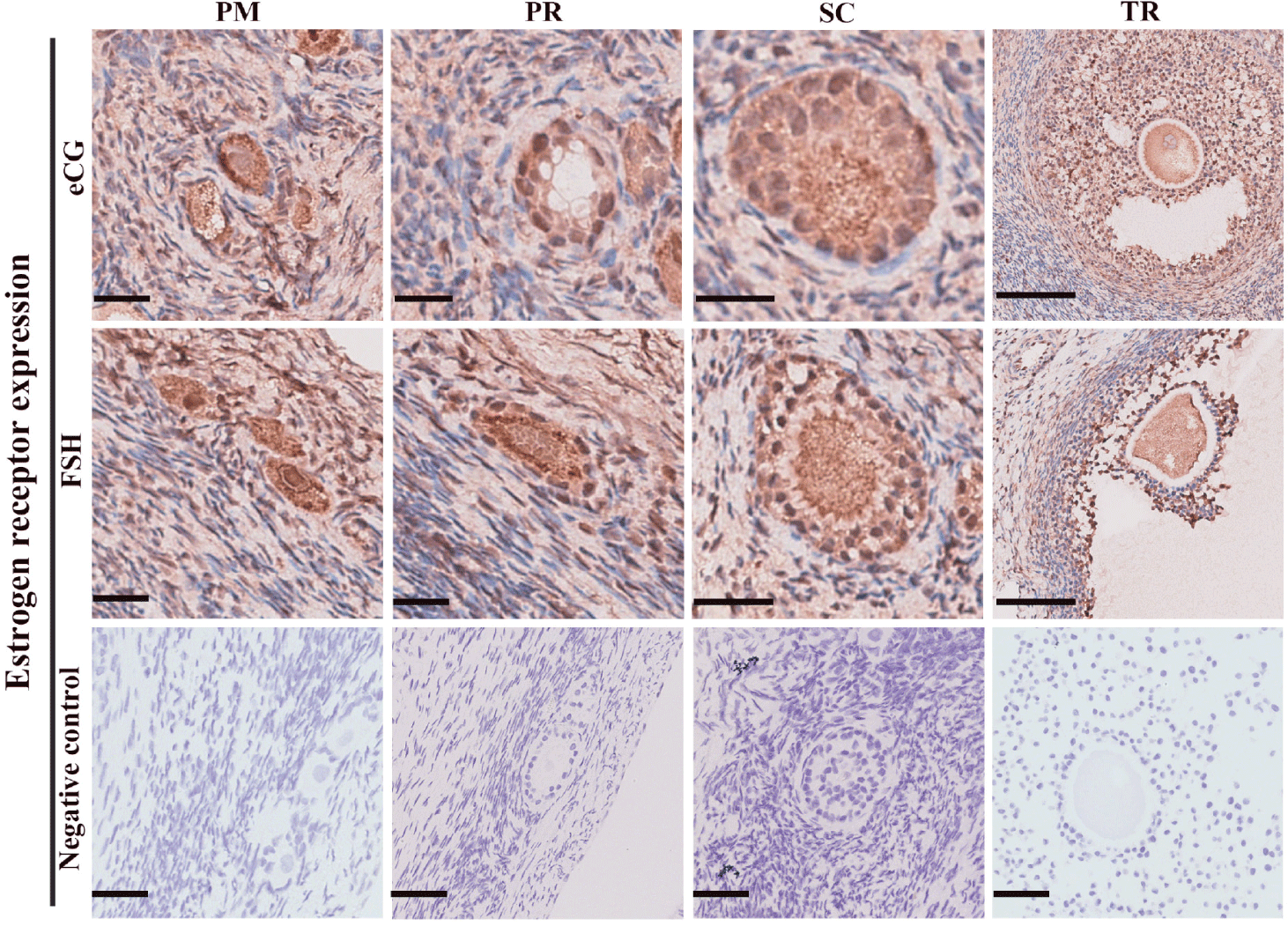
To further understand the effects of the eCG/FSH treatment on goat follicular development, we examined FSHR and ER expression levels. Our results demonstrated that FSHR expression was primarily localized in the cytoplasm of ovarian GCs, compared to the negative control group (Fig. 4A). As shown in Figs. 4B, 4C, and 4D, our immunohistochemistry results revealed that FSHR expression in follicles at the primary, secondary, and tertiary stages was significantly higher in the eCG_FSH group compared to the eCG group; FSHR expression in follicles at the primary and secondary stages was significantly higher in the eCG_FSH group compared to the FSH group; FSHR expression in follicles at the secondary stage was significantly higher in the FSH group compared to the eCG group. Similarly, ER expression was also predominantly localized in the cytoplasm of ovarian GCs compared to the control group (Fig. 5).
The plasma E2, FSHR, and P4 Levels were detected by ELISA. As shown in Table 1, E2 levels in plasma in the FSH group showed an increasing trend from the time of PRID removal but were not significantly different and maintained a peak until the time of embryo flushing, whereas E2 levels in the eCG_FSH group were significantly higher from the time of estrus and significantly lower at 24 h after estrus. Plasma levels of FSHR in the FSH group peaked before PRID removal and declined significantly during estrus, with a tendency to rebound 24 h after estrus, but declined before embryo flushing; while FSHR levels in the eCG_FSH group continued to increase after PRID removal until a significant difference was observed at 24 h post estrus, and remained at a peak until just before embryo flushing. In the FSH group, plasma levels of P4 were significantly decreased after PRID removal and only slightly increased 24 h after estrus, but remained low until embryo flushing. In contrast, P4 levels in the eCG_FSH group decreased significantly after withdrawal of the bolus increased significantly at 24 h post-estrus, and remained elevated until implantation.
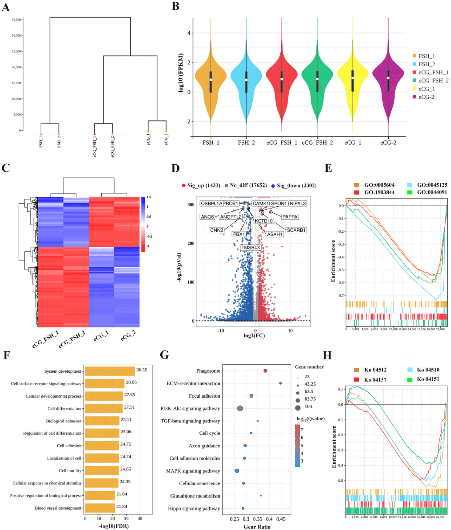
Hierarchical clustering analysis of transcriptome sequencing revealed that samples from the same group clustered together, showing distinct expression patterns between groups (Fig. 6A). Based on the Transcripts Per Million values of all genes, expression distribution plots were generated to illustrate the expression distribution of transcripts across different samples (Fig. 6B). Heatmaps were used to present DEGs expression patterns of the eCG_FSH and eCG groups (Fig. 6C). Comparing eCG_FSH with eCG, we identified 1,433 up-regulated and 2,302 down-regulated DEGs, and the volcano map demonstrates key genes with large fold change difference, such as PBX1, ASAH1, TMSB4X, SCARB1, PAPPA (Fig. 6D). GO and KEGG analyses were performed to evaluate the function of the DEGs. GO enrichment analysis revealed that these DEGs were predominantly associated with the cellular development process, biological adhesion, cell motility and cellular response to chemical stimulus (Fig. 6F); KEGG enrichment analysis showed that these DEGs were mainly enriched in ECM-receptor interaction, focal adhesion, TGF-β signaling pathway and MAPK signaling pathway (Fig. 6G). To further confirm the functional annotation results, GSEA analysis was performed. This analysis revealed the enrichment of these genes in the membrane biogenesis, regulation of cellular response to transforming growth factor beta stimulus, focal adhesion and mitophagy-animal (Figs. 6E and 6H).
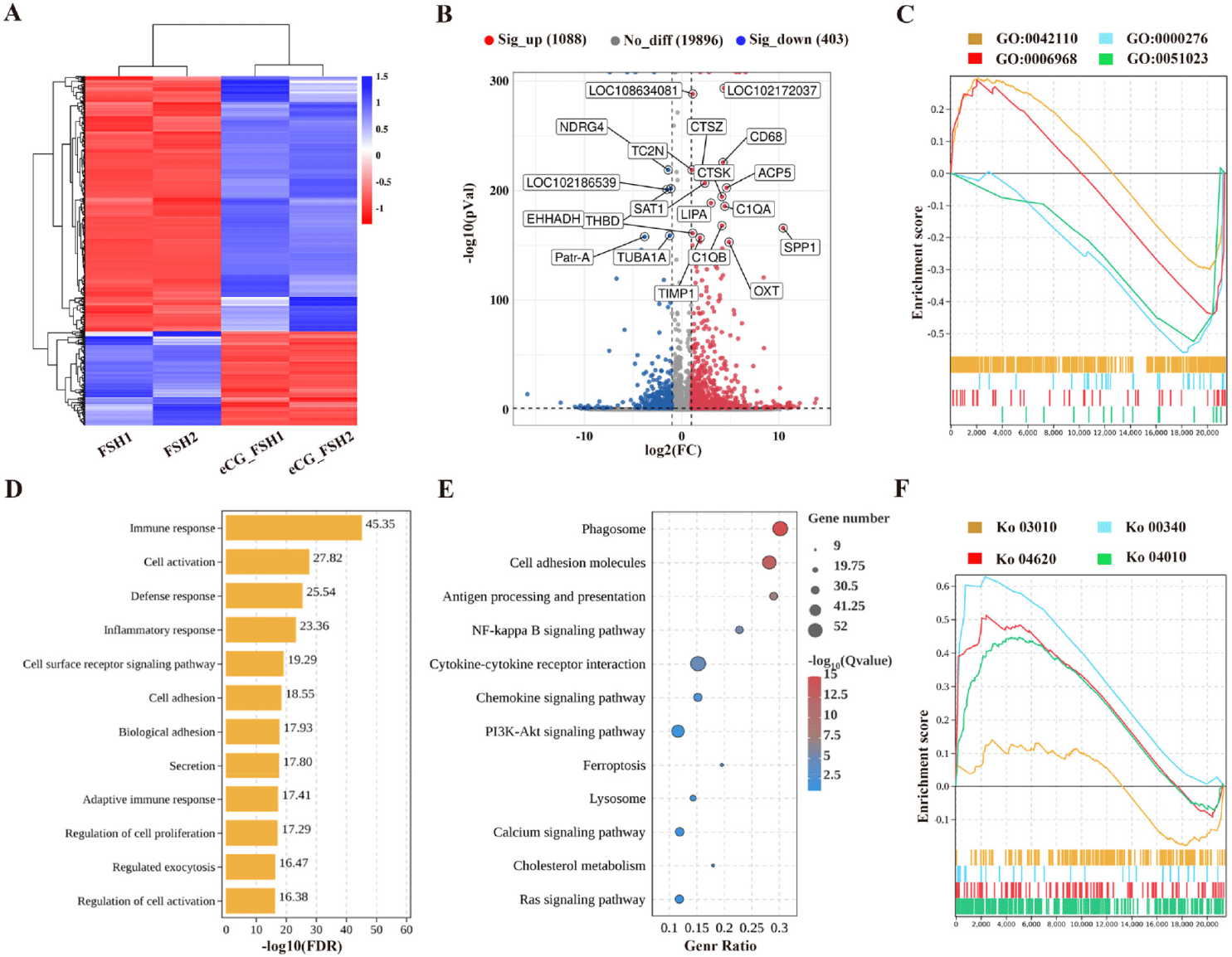
Heatmaps were used to present DEGs expression patterns of the eCG_FSH and FSH groups (Fig. 7A). Comparing eCG_FSH with FSH, we identified 403 up-regulated and 1,088 down-regulated DEGs, and the volcano map demonstrates key genes with large fold change difference, including SAT1, THBD, TUBA1A, TIMP1, OXT, and SPP1 (Fig. 7B). Functional annotation through GO and KEGG analyses was conducted to elucidate the roles of these DEGs. GO enrichment analysis revealed that these DEGs were predominantly associated with immune response, cell surface receptor, cell adhesion and secretion (Fig. 7D); KEGG enrichment analysis showed that these DEGs were mainly enriched in cell adhesion molecules, NF-kappa B signaling pathway and PI3K signaling pathway (Fig. 7E). To further confirm the functional annotation results, GSEA analysis was performed. This analysis revealed the enrichment of these genes in the mitochondrial proton translocation adenosine triphosphate (ATP) synthesis coupling factor, the cytosolic decay response, ribosomes, and the MAPK signaling pathway (Figs. 7C and 7F).
DISCUSSION
To improve the reproductive ability of purebred Haimen goats, and swiftly increase the offspring of outstanding females, we accelerated the purebred breeding of Haimen goats by employing superovulation and embryo transfer at the base of the goat farm facility. The results of showed that the combined eCG_FSH regimen significantly increased the number of ovulations compared with FSH alone, with a more consistent and stable ovulation effect. Subsequent investigations delved into the impact of eCG on superovulation in goats, examining factors such as plasma hormone levels, ovarian tissue morphology, and follicular GC function. This study provides a theoretical basis for the standardized use of hormones in goat production and a technical reference for the production of large-scale farms, promoting the sustainable advancement of the goat industry.
We conducted an investigation into the levels of FSHR, E2, and P4 before and after PRID removal in the eCG_FSH and FSH regimens to elucidate their relationships with follicular development, ovulation, and ovarian response. Our findings revealed distinct patterns in FSHR levels between the two treatment groups. In the FSH group, FSHR levels peaked before PRID removal and subsequently decreased significantly, stabilizing thereafter. Conversely, in the eCG_FSH group, FSHR levels exhibited a progressive increase from before PRID removal to 24 h after estrus, reaching a peak at a later stage compared to the FSH group. This delay in the FSHR peak suggests that eCG administration may prolong the expression of FSHR levels. Immunohistochemical analysis of ovarian tissues corroborated these findings, demonstrating significantly higher FSHR expression levels in the eCG_FSH group compared to both the FSH and eCG groups, consistent with prior studies [25,31,32]. E2 and P4, pivotal steroid hormones secreted by the ovary, play crucial roles in the female reproductive endocrine system and estrous cycle, significantly influencing pregnancy establishment and embryonic development [33]. In the FSH group, E2 levels peaked post-PRID removal and gradually stabilized, while in the eCG_FSH group, E2 levels peaked during estrus, then rapidly declined and stabilized. Similarly, in the FSH group, P4 levels reached their lowest during estrus, then stabilizing, while in the eCG_FSH group, P4 levels also declined after PRID removal and then gradually increased. These results align with previous studies, emphasizing the strong correlation between elevated serum E2 concentrations and estrus in animals, followed by a significant decrease post-estrus [34].
Due to the prolonged half-life of eCG, residual eCG in the body can potentially impact follicular maturation and ovulation, leading to sustained secretion of E2 by the ovaries and subsequent elevation in plasma E2 levels [35]. Conversely, repeated administration of eCG in goats may result in the development of hormone tolerance within the ovaries, thereby compromising their reproductive performance [36]. To mitigate these unfavorable effects, we opted to adhere to a lower eCG dosage based on previous recommendations, utilizing only 100 IU of eCG in our experiment. Research has indicated that elevated serum E2 concentration during pre-estrus promotes the expression of the P4 receptor, which in turn inhibits luteolysis in cows [37,38]. Furthermore, studies have elucidated the pivotal roles of E2 levels and peripheral P4 concentrations in regulating changes in endometrial diameter or thickness and governing various uterine functions throughout the normal estrous cycle [39–41]. Moreover, P4 plays a crucial role in early embryonic development, embryo attachment, and the maintenance of pregnancy within the female organism [42,43].
In our study, we identified several key genes exhibiting significant fold change differences between the eCG_FSH group and the eCG group in goat follicular GCs, including PBX1, PAPPA, SCARB1, and TMSB4X, which play crucial roles in regulating follicle development. PBX1 is expressed in GCs across all stages of follicular development, predominantly localized in the nucleus [44]. Pregnancy-associated plasma protein-A (PAPP-A) serves as the primary protease for insulin-like growth factor binding protein-4 (IGFBP-4) in follicular fluid. Both eCG and FSH significantly influence PAPPA expression in a dose-dependent manner, consequently modulating follicle development [45–47]. GCs express SCARB1, which facilitates high-density lipoprotein uptake, crucial for maintaining normal ovarian cholesterol homeostasis and steroid synthesis within the luteal cells [48,49]. TMSB4X, driving GCs differentiation, is enriched in pathways regulating ATP-dependent activity [50]. KEGG enrichment analysis revealed that these DEGs are predominantly associated with ECM-receptor interaction, focal adhesion, the transforming growth factor (GF)-β signaling pathway, and the MAPK signaling pathway. Notably, the TGF-β signaling pathway plays a pivotal role in various aspects of ovarian function and is indispensable for oocyte maturation [51]. Moreover, members of the TGF-β superfamily, including GDF9, activin, and several BMPs, influence FSH function via FSHR in GCs. Studies have demonstrated that these factors stimulate FSHR expression and enhance FSHR mRNA stability in GCs [52,53]. In practical production, we observed that the goats treated with a combination of eCG and FSH exhibited earlier estrus, larger ovarian volume, a higher number of antral follicles and ovulations compared to the eCG group. These results suggest that FSH may play a significant role in promoting follicle development, ovarian growth, and estrogen synthesis by regulating key genes such as PBX1, PAPPA, and SCARB1, as well as signaling pathways like TGF-β and MAPK signaling pathways.
In the comparison between the eCG_FSH group and the FSH group of follicular GCs in goats, we identified key genes with significant differences in expression, such as THBD, TIMP1, and OXT. Studies have indicated that following treatment of gonadotropin-primed immature mice with an ovulatory dose of human chorionic gonadotropin (hCG), there were marked increases in THBD expression in GCs and cumulus cells of preovulatory follicles [54]. Tissue inhibitor of metalloproteinase 1 (TIMP1) plays a vital role in various aspects of follicular growth and development, ovulation, luteinization, and embryo development in mammals [55]. Oxytocin (OXT) influences ovarian steroidogenesis by promoting ovarian luteinizing hormone synthesis through enhanced BMP-15 activity [56]. KEGG enrichment analysis revealed that these DEGs were predominantly enriched in pathways related to cell adhesion molecules, the NF-kappa B signaling pathway, and the PI3K/Akt signaling pathway. Previous studies have demonstrated that FSH can inhibit the expression of BimEL via the PI3K/Akt signaling pathway, inducing apoptosis in GCs [57]. Additionally, activation of the PI3K/Akt signaling pathway has been found to be crucial for oogenesis and follicular development [58,59]. Analysis of transcriptome sequencing data from eCG_FSH and FSH regimens goat ovarian GCs revealed that the eCG/FSH treatment primarily influences follicular development by modulating hormone receptor expression, hormone secretion, GCs function, and oocyte maturation. The results indicate that the combined treatment of eCG and FSH significantly increased the number of corpora lutea, ovulations, and antral follicles in goats, compared to treatment with eCG alone. This suggests that eCG may play a crucial role in promoting follicle development and corpus luteum formation by regulating key genes such as THBD, TIMP1, and OXT, as well as signaling pathways like PI3K/Akt and cell adhesion molecules.
CONCLUSION
In summary, both the eCG_FSH and FSH regimens proved effective in enhancing superovulation in goats. The addition of 100 IU eCG resulted in elevated concentrations of FSHR, P4, and E2, significantly increasing ovulation numbers and the presence of large follicles in goats, and maintaining normal follicular morphology throughout various stages of development. Furthermore, transcriptome analysis of follicle GCs showed FSH may play a significant role in promoting follicle development, ovarian growth, and estrogen synthesis by regulating key genes such as PBX1, PAPPA, and SCARB1, as well as signaling pathways like TGF-β and MAPK signaling pathways. eCG may promote follicle development and corpus luteum formation by regulating key genes such as THBD, TIMP1, and OXT, as well as signaling pathways like PI3K/Akt and cell adhesion molecules. This study comprehensively analyzed the effects of eCG/FSH on the reproductive performance of Haimen goats during synchronized estrus, providing a foundation for further investigations into the mechanisms regulating reproduction in domestic animals.