INTRODUCTION
Increasing livestock productivity significantly affects the nutrition and environment of animals. Dietary probiotics are widely used as alternatives to antibiotics to promote pig health and productivity [1]. The gut flora of pigs profoundly affects host health [2]. Supplementing pig feed with probiotics improves growth performance, gut health, and immunity [3]. Intestinal microorganisms influence numerous immune tissues and cells in the gut, which in turn affect systemic host homeostasis [4,5]. Specifically, expression of immune-related genes in the gut and associated lymphoid tissues has been studied, highlighting the relations between the gut microbiota and immune function [6]. Additionally, pro-biotic administration alleviates colitis and increases mucosal expression through the action of tight junction proteins, such as Zonula occludens-1 (ZO-1) and Occludin [7].
Among probiotics, Bacillus strains are known to improve the intestinal health of various hosts, including pigs. These strains exhibit antibacterial and antioxidant properties and positively influence immune function [8,9]. They are also effective in preventing and alleviating intestinal inflammation [10,11]. Originating in the soil, Bacillus strains have been found in the intestines of humans and pigs, where they act as probiotics [12–14]. Specifically, Bacillus subtilis and Bacillus licheniformis are noted for their ability to form biofilms, enabling them to persist in the intestine by producing anaerobic spores that can withstand gastrointestinal stress [15]. These unique characteristics indicate that both species are valuable feed additives.
Feeding Bacillus subtilis to pigs increases average daily gain and feed efficiency while reducing the incidence of diarrhea [16]. Moreover, Bacillus subtilis enhances pig growth and disease resistance by promoting digestive enzyme activity and increasing villus height in the ileum [17]. Bacillus subtilis also suppresses harmful bacteria in the large intestine, strengthens the probiotic microbial community, and modulates metabolic activity, collectively improving growth efficiency in pigs [18]. Bacillus subtilis and Lactobacillus can alleviate colitis and enhance mucosal expression by regulating the expression of tight junction proteins, including ZO-1 and Occludin [19]. Fermented Bacillus subtilis products have similarly been shown to boost immunity in the small intestine, contributing to growth efficiency [20].
Bacillus licheniformis also supports digestion by producing proteases and amylases [21,22]. It detoxifies zearalenone, a mycotoxin found in feed [23], and produces surfactin, which exhibits potent antibacterial activity against Clostridium perfringens, a common cause of diarrhea in pigs [24]. Fermented products containing Bacillus licheniformis positively affect pig gut microbiota and reduce the frequency of diarrhea [25]. Additionally, the combined administration of Bacillus licheniformis and other beneficial probiotics has been associated with improved immunity and reduced diarrheal symptoms [26].
Recent studies have confirmed that feeding both Bacillus licheniformis and Bacillus subtilis directly to pigs increases digestibility, enhances Lactobacillus populations in feces, and supports growth performance [27,28]. Despite these reports, the effects of Bacillus licheniformis and Bacillus subtilis on intestinal tissue and villi in relation to intestinal immunity and the function of weaning pigs have not been clearly identified. Therefore, this study aimed to determine the contribution of dietary supplementation with these two bacterial species to villus structure, particularly with respect to tight junctions in the intestines of weaning pigs, in the context of serological immune indicators.
MATERIALS AND METHODS
In this experiment, 28-day-old piglets (n = 50 per group; total = 200) were randomly selected, excluding underweight individuals and assigned to four groups: CTRL (basal diet), BS (basal diet + Bacillus subtilis), BL (basal diet + Bacillus licheniformis), or BSL (basal diet + both Bacillus strains). All piglets were provided with the same starter diet, which did not contain antibiotics, prebiotics, or probiotics. The handling procedures followed the ethical guidelines established by the Institutional Animal Care and Use Committee, and the study was conducted with approval under protocol WG-IACUC-2024-003. The experimental feeding regimen consisted of two phases: Phase 1 and Phase 2 diets were supplied as the basal diet for 14 days each, while feed additives were continuously administered for 28 days. Resistant maltodextrin (2%, Daesang, Korea) was included in BS, BL and BSL groups as a prebiotic to function synergistically with probiotics, thus constituting a synbiotic formulation. Throughout the experimental period, piglets had unrestricted access to both feed and water. The detailed composition of the basal diet and the additional feed additives are presented in Tables 1 and 2, respectively.
1) Supplied per kg of diet: 16,000 IU vitamin A (palmitate), 2.00 mg vitamin B1 (thiamin), 5.00 mg vitamin B2 (riboflavin), 2.00 mg vitamin B6 (pyridoxine), 0.03 mg vitamin B12 (cyanocobalamin), 25.00 mg niacin, 0.40 mg folic acid, 0.05 mg biotin, 5.00 mg ethoxyquin, 2,000 IU vitamin D3 (cholecalciferol), 75.00 mg vitamin E (dl-α-tocopheryl acetate), and 2.00 mg vitamin K3 (menadione).
| Item | CTRL | Treat 1 (BS) | Treat 2 (BL) | Treat 3 (BSL) |
|---|---|---|---|---|
| Bacillus subtilis (CFU/g) | - | 1 × 10⁸ | - | 1 × 10⁸ |
| Bacillus licheniformis (CFU/g) | - | - | 1 × 10⁸ | 1 × 10⁸ |
| Dietary fiber (resistant maltodextrin) | - | 2% | 2% | 2% |
At both the beginning and the end of the experiment, body weight changes were measured in 40 piglets, with 10 animals assigned to the control (CTRL) group and 10 to each treatment group (BS, BL, and BSL). Additionally, a total of 24 piglets (six per group: CTRL, BS, BL, and BSL) were selected for molecular biological analysis. Blood samples were collected in the morning under fasting conditions. The blood for complete blood count (CBC) analysis was collected from the anterior inferior vena cava, placed in EDTA K3 tubes (AB medical), and stirred at room temperature for 2 h. Blood for serum analysis was centrifuged at 3,000×g for 10 min at 4°C to separate serum, and stored at 4°C. The collected whole blood (1 mL) and serum (600 µL) were analyzed for alanine aminotransferase (ALT), aspartate aminotransferase (AST), creatinine, cortisol, and CBC at Samkwang Medical Research Institute. All piglets were humanely euthanized using electrical stunning (250 V, 0.5 A) for 5–6 seconds. Ileum tissue samples were immediately excised and sectioned into 2.0-cm-thick slices using phosphate-buffered saline (PBS; Welgene).
Serum was analyzed using enzyme-linked immunosorbent assay (ELISA) kits to quantify interferon-γ (IFN-γ, MyBioSource), interleukin 6 (IL-6, MyBioSource), and tumor necrosis factor-alpha (TNF-α, MyBioSource), following the manufacturer’s guidelines. Absorbance readings were taken at 450 nm, with a reference wavelength of 570 nm, using an Epoch spectrophotometer (BioTek). Six serum samples from each group (total: 24 samples) were tested, with measurements conducted at least three times. Until analysis, serum samples were preserved at –80°C.
Ileum tissue samples were fixed in 4% paraformaldehyde at 4°C for 48 hours. The samples were then cut into 1.0 cm sections, rinsed with distilled water for 30 minutes, and sequentially treated with ethanol at concentrations of 70%, 80%, 90%, 95%, and 100% (each for 90 minutes) to ensure dehydration while being gently agitated at 24°C. The dehydrated tissues were embedded in paraffin wax for 2 hours and subsequently formed into blocks at room temperature. Using a Leica Biosystems microtome (Nussloch, Germany), the paraffin-embedded ileum tissues were sectioned into 4-µm-thick slices, dried in a slide dryer at 40°C for 12 hours, and mounted onto slides.
For hematoxylin and eosin (H&E) staining and immunohistochemical analysis, the ileum sections were deparaffinized by immersing them in xylene three times for 5 minutes each, followed by rehydration through a graded ethanol series (100%, 90%, 80%, and 70%) and distilled water for 10 minutes at each step. H&E staining was performed at room temperature using hematoxylin for 1 minute and eosin for 30 seconds. After staining, the sections were dehydrated with ethanol (95% and 100%, each for 5 minutes) and then cleared in xylene for 5 minutes. Stained samples were visualized using a microscope (Olympus IX73; Olympus). Five ileum samples from each group (total 20 slides) were selected, and at least three samples were included per replicate. The paraffin blocks were stored at –80°C until further analysis.
Rehydrated slides underwent heat-induced epitope retrieval by treatment with 10 mM sodium citrate buffer (pH 6.0) at 100°C for 15 minutes. Primary antibodies (detailed in Table 3) were diluted according to the manufacturer’s instructions and incubated with ileum tissue sections 16 hours at 4°C. After washing with PBS, the sections were treated with Alexa Fluor 488- and 568-conjugated secondary fluorescent antibodies for 1 hour at room temperature. Following additional PBS washes, nuclei were counterstained with 4′,6-diamidino-2-phenylindole (DAPI; 1 µg/mL). Immunostained ileum tissues were imaged using a fluorescence microscope, and fluorescence intensity was quantified with ImageJ software. Three ileum samples from each group (total 12 slides) were selected, with at least three samples included per replicate.
| Antibody | Manufacturer | Catalog number | Dilution (usage) |
|---|---|---|---|
| β-Actin | Santa Cruz | SC-47778 | 1:2000 (WB) |
| Occludin | Invitrogen | 71-1500 | 1:1000 (WB), 1:300 (IHC) |
| Zonula occludens-1 | Invitrogen | 40-2200 | 1:300 (IHC) |
For qPCR analysis, total RNA was extracted using the AccuPrep® Universal RNA Extraction Kit (Bioneer) with on-column DNase treatment (Qiagen), following the manufacturer’s instructions. The extracted RNA was then reverse transcribed into complementary DNA using the RevertAid First Strand cDNA Synthesis Kit (Thermo Fisher Scientific) at a concentration of 1 μg/20 μL.
QPCR was performed using a QuantStudio 1 system (Applied Biosystems) with SYBR Green Master Mix (Thermo Fisher Scientific). The reaction conditions were set as follows: initial denaturation at 94°C for 1 minute, followed by 40 cycles of 94°C for 10 seconds, 57°C for 10 seconds, and 72°C for 20 seconds. Gene expression levels were normalized to the endogenous β-actin. Primers were designed using Primer3 software (http://frodo.wi.mit.edu) and are detailed in Table 4. Three RNA samples from each group (total 12 samples) were analyzed per group, with each sample tested in triplicate.
Ileal tissue was lysed using radioimmunoprecipitation assay buffer (Thermo Fisher Scientific) supplemented with a protease inhibitor mixture (Roche) to extract proteins. A total of 30 µg of ileal protein was separated on a 4%–20% sodium dodecyl sulfate-polyacrylamide gel (SDS-PAGE; Bio-Rad) and subsequently transferred onto a 0.45-µm polyvinylidene difluoride membrane.
Primary antibodies were diluted in 1% bovine serum albumin buffer containing 0.05% Tween-20, following the manufacturer’s instructions, and incubated with the membranes at 4°C for 16 hours. After washing with TBS-0.05% Tween, the membranes were incubated with HRP-conjugated anti-mouse and anti-rabbit secondary antibodies for 1 hour at 24°C. Protein detection was performed using Pierce ECL substrate (Thermo Fisher Scientific), and images were captured using the iBright™ Imaging System (Thermo Fisher Scientific). β-Actin was used as quantitative CTRL for protein normalization. The primary antibodies used for western blotting are listed in Table 3. Three protein samples from each group (total 12 samples) were analyzed, with all assays conducted in triplicate.
All data are presented as the mean ± standard error of the mean from independent experiments conducted at least three times in triplicate. Statistical analysis was performed using one-way ANOVA followed by Tukey’s post hoc test. All analyses were conducted using SPSS version 29 for Windows (IBM). Statistical significance was defined as follows: *p < 0.05, **p < 0.01, ***p < 0.001.
RESULTS
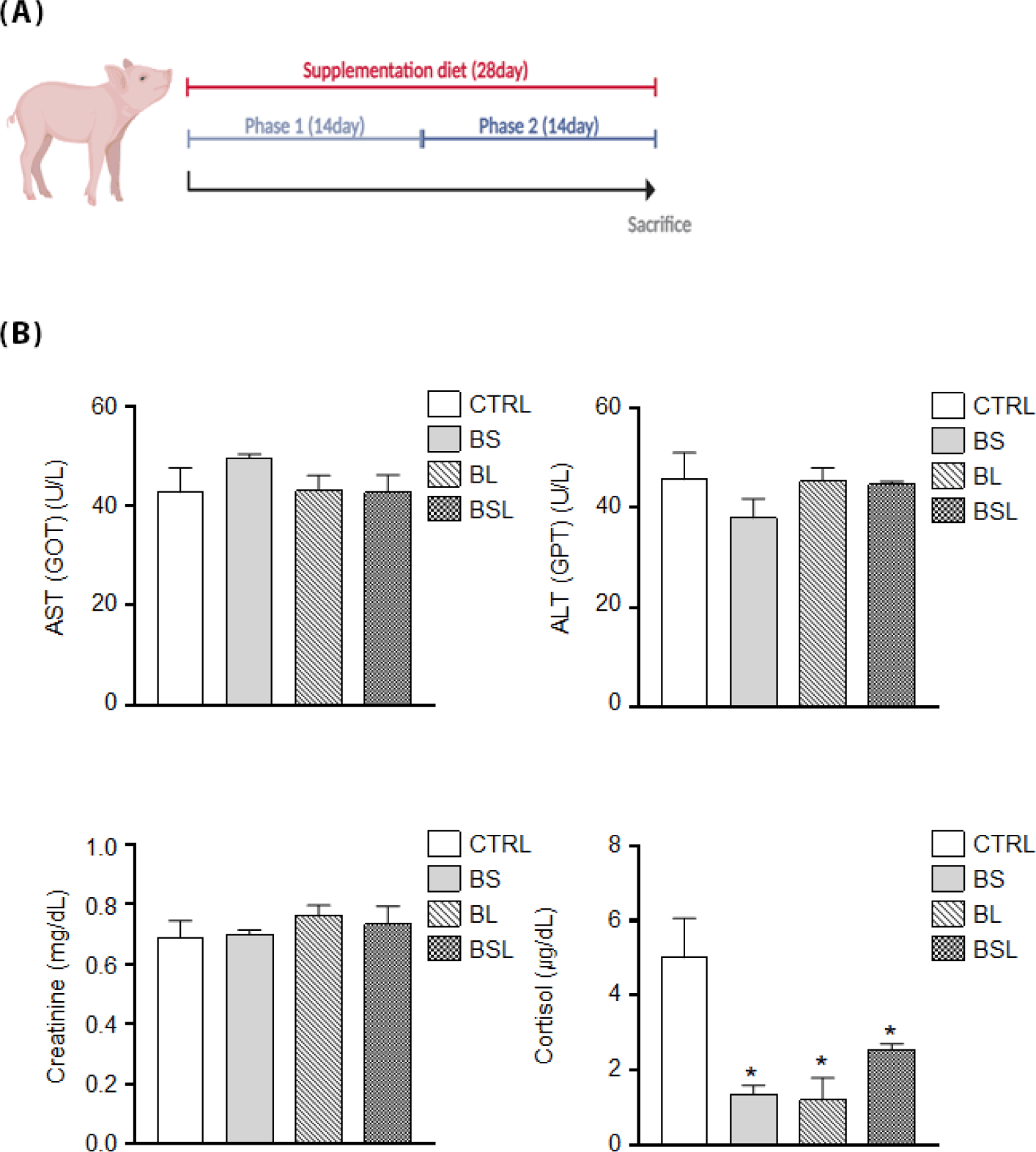
A schematic design of the 28-d supplementation period is presented (Fig. 1A). The body weight of pigs showed no significant difference between the CTRL and test groups (Table 5). To examine the effects of dietary supplementation on organ stress, serum ALT, AST, creatinine, and cortisol levels were evaluated after 28 d (Fig. 1B). ALT and AST levels showed no significant differences. Similarly, creatinine levels were not significantly different across all groups. However, the cortisol levels in BS, BL, and BSL groups (p < 0.05) were significantly lower than those in the CTRL group.
| Item | CTRL | BS | BL | BSL | ||||
|---|---|---|---|---|---|---|---|---|
| Mean | SEM | Mean | SEM | Mean | SEM | Mean | SEM | |
| Initial BW (kg) | 6.36 | 0.03 | 6.40 | 0.04 | 6.39 | 0.03 | 6.37 | 0.02 |
| Final BW (kg) | 15.7 | 0.26 | 15.93 | 0.09 | 16.15 | 0.15 | 16.20 | 0.17 |
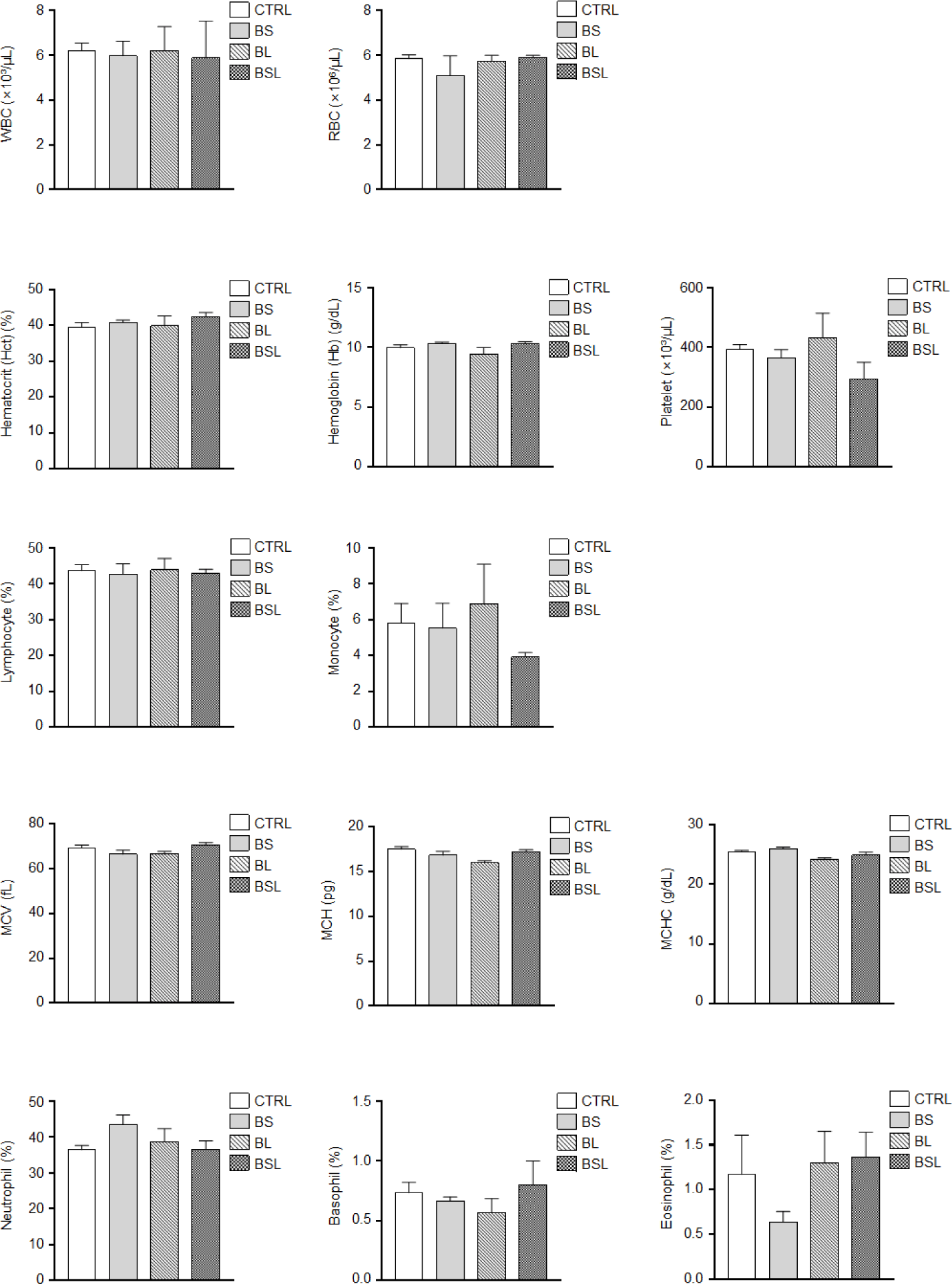
To evaluate the effect of Bacillus supplementation on hematological characteristics, a complete blood count was conducted for the CTRL, BS, BL, and BSL groups (Fig. 2). There were no significant differences observed in white blood cell (WBC) counts. Similarly, red blood cell (RBC), hematocrit, and hemoglobin levels were consistent across all groups. The platelet counts also showed no notable differences among the groups. Regarding WBC composition, the lymphocyte and monocyte percentage showed no significant differences among all groups. The percentages of neutrophils, basophils and eosinophils were comparable across all groups with no significant. Additionally, RBC quality indicators, including mean corpuscular volume (MCV), mean corpuscular hemoglobin (MCH), and mean corpuscular hemoglobin concentration (MCHC), showed no significant differences among groups.
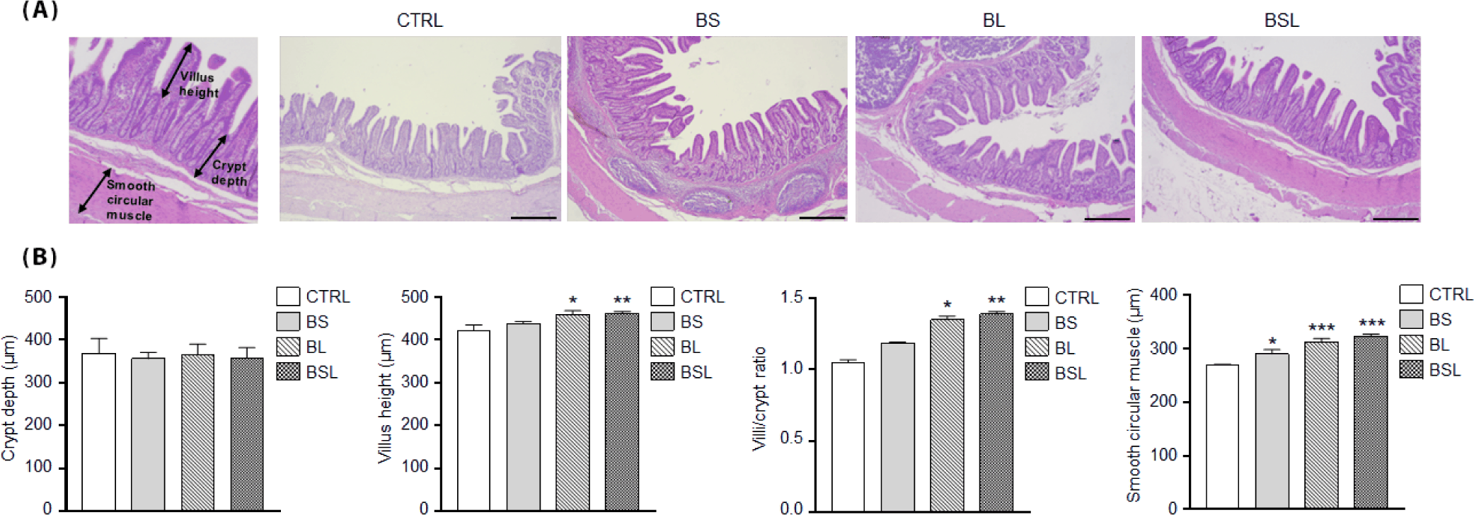
To examine the effects of Bacillus supplementation on the small intestine, the crypt depth, villus height, smooth circular muscle thickness, and villus/crypt ratio were measured using H&E staining (Fig. 3A). H&E staining revealed differences in villus height and smooth muscle thickness between the CTRL and supplemented groups. Crypt depth showed no significant differences among the groups (Fig. 3B). BS did not show significant differences in villus height and villus/crypt ratios compared to CTRL. However, the villus height showed increased in the BL and BSL supplemented groups. Notably, BL (p < 0.05) and BSL (p < 0.01) groups showed significantly increased villus height and villus/crypt ratios, with BSL showing the highest values (Fig. 3B). The thickness of the smooth circular muscle was significantly greater in the BS (p < 0.05), BL (p < 0.001), and BSL (p < 0.001) groups compared with that in the CTRL group (Fig. 3B).
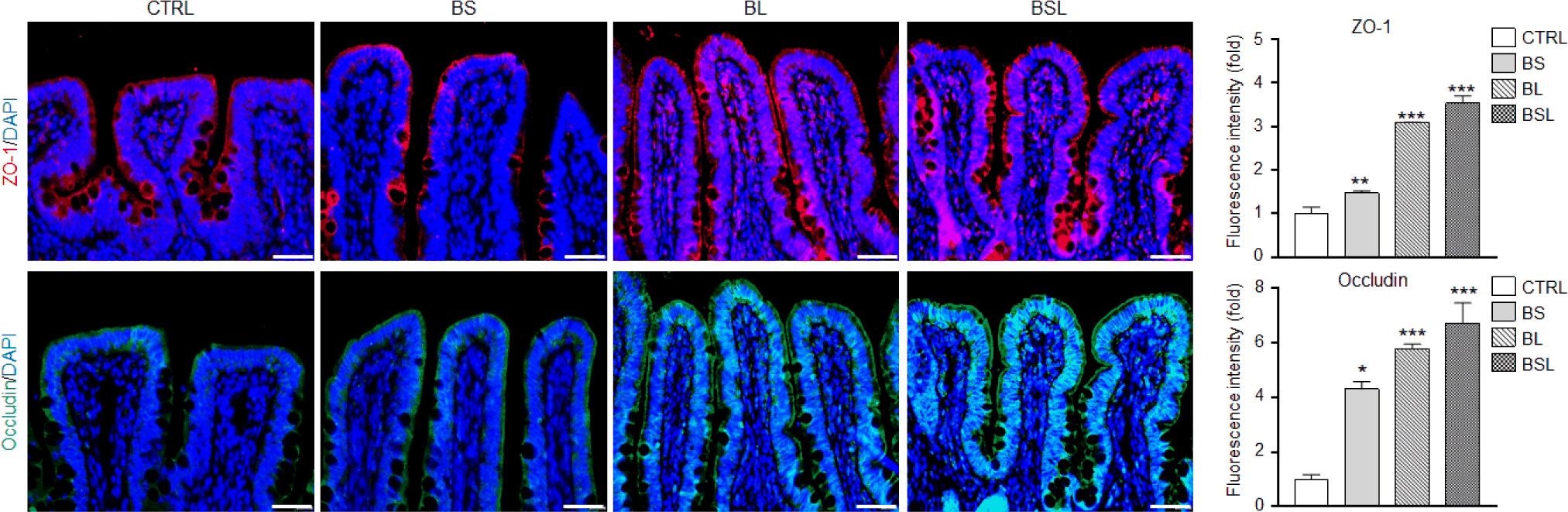
To further investigate the improvement of scaffold protein in intestinal epithelial cells to maintain barrier integrity and regulate paracellular permeability, immunostaining for ZO-1 and Occludin was conducted, respectively (Fig. 4). Immunofluorescence images showed that ZO-1 expression was enhanced in all Bacillus-supplemented groups. Similarly, Occludin expression increased in the supplemented groups. Furthermore, intensity levels showed that ZO-1 expression was significantly increased in the BS (p < 0.01), BL (p < 0.001), and BSL groups (p < 0.001) compared with that in the CTRL. The intensity of Occludin also significantly increased at BS (p < 0.05) and was more pronounced at BL (p < 0.001) and BSL (p < 0.001).
Inflammatory cytokine analysis was conducted to evaluate immune responses in CTRL and Bacillus-supplemented groups. The concentration of IFN-γ showed no significant difference among the CTRL, BS, BL, and BSL groups. Similarly, no notable changes were observed in TNF-α levels across all groups. IL-6 concentrations were also not significantly different (Fig. 5A).
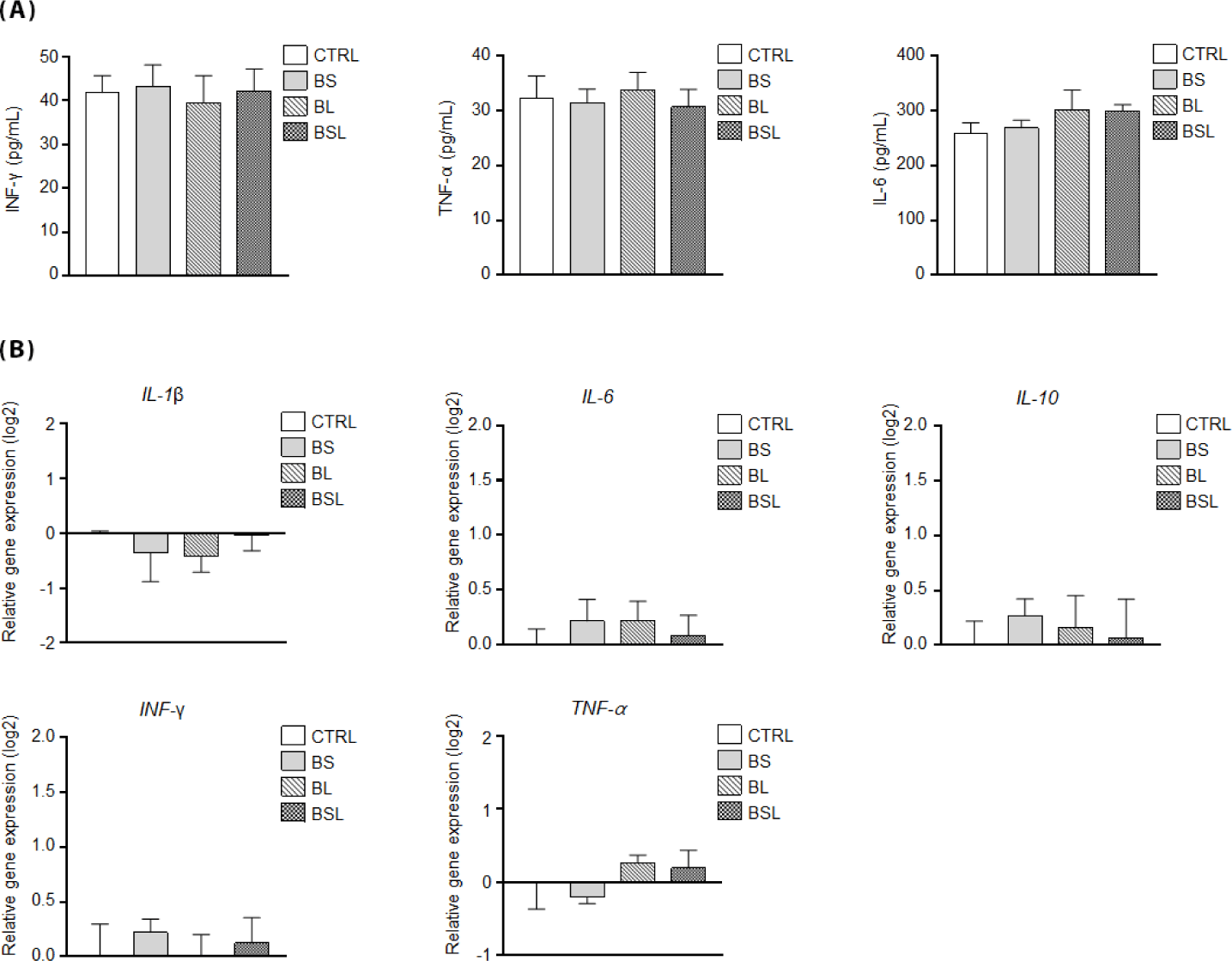
Subsequently, the relative mRNA expression of inflammatory cytokines was analyzed in the ileum, interleukin-1 beta (IL-1β), IL-6, interleukin 10 (IL-10), IFN-γ, and TNF-α (Fig. 5B). The proinflammatory cytokines IL-1β and IL-6 showed no significant changes in expression among the CTRL, BS, BL, and BSL groups. The anti-inflammatory cytokine IL-10 was also consistently expressed, with no notable differences between the groups. Similarly, IFN-γ and TNF-α did not exhibit significant differences.
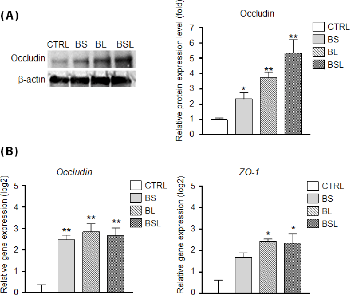
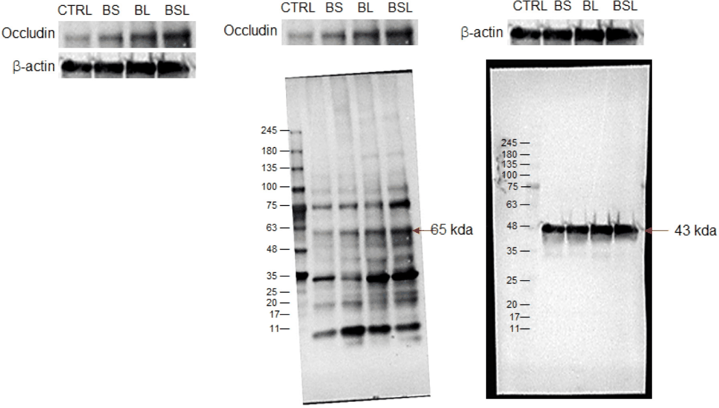
Tight junction-related protein and gene expression in the ileum were examined to determine the influence of Bacillus supplementation. Occludin expression was significantly increased in all Bacillus-supplemented groups (Fig. 6A). Notably, the quantified protein data showed a significant increase at BS (p < 0.05), with more pronounced increases observed at BL (p < 0.01) and BSL (p < 0.01). Correspondingly, the mRNA expression of occludin was significantly increased in all Bacillus-supplemented groups (p < 0.01, Fig. 6B). Moreover, the expression of ZO-1 was also elevated in all Bacillus-supplemented groups, particularly in BL (p < 0.05) and BSL (p < 0.05), which showed significant upregulation (Fig. 6B). Uncropped full western blot images were displayed in Fig. 7.
DISCUSSION
The weaning period is a critical phase in piglet development, significantly influencing growth performance and overall livestock productivity. During this stage, disruptions in gut microbiota balance can lead to intestinal infections, resulting in impaired growth and, in severe cases, increased mortality [3]. Traditionally, large-scale antibiotic administration has been employed to mitigate such infections. However, recent research efforts have focused on replacing antibiotics with probiotics to promote a healthier gut environment [1]. Thus, the application of Bacillus-based probiotics to maintain intestinal microbial homeostasis and intestinal development during the weaning period is of considerable interest.
The supplementation of probiotics in weaning piglets has been associated with enhanced disease resistance and improved growth performance [16]. Furthermore, studies have reported increased feed efficiency and a reduction in diarrhea incidence following probiotic administration [29]. Although no significant differences in body weight or feed intake were observed, probiotic-treated piglets exhibited beneficial effects on intestinal immune responses and gut microbiota composition [30]. Although studies on feeding Bacillus to pigs have shown inconsistent results regarding growth performance, many have consistently reported that nutrient digestibility is enhanced through improved intestinal microflora and gut health [16,18,29,30]. This suggests that growth performance is influenced by multiple factors and cannot be determined solely by improvements in gut health. In this study, although no statistically significant differences were observed between the CTRL and all the supplemented groups, body weight was the highest in the BSL group compared to the CTRL. The average body weight levels in this study were similar to findings from other studies comparing CTRL and probiotic-treated groups with significantly difference [31].
Studies involving long-term probiotic intake have reported decreased ALT, AST, and creatinine levels [32]. However, this study found no signs of toxicity or functional decline in the liver or kidneys of the BS, BL, and BSL groups. This absence of toxicity may be attributed to the focus of this study on the weaning period, which was limited to 28 d. Interestingly, cortisol, a key indicator of stress, was significantly reduced in all Bacillus-supplemented groups. Bacillus-supplementation during the weaning period may help alleviate stress from both internal and external environmental factors. This is consistent with findings that probiotic administration lowers cortisol levels and reduces mortality in weaned pigs [33]. While no significant differences in WBC composition were observed between the negative CTRL and pigs supplemented with Bacillus subtilis, pigs fed E. coli exhibited increased monocyte and neutrophil counts [29]. These results suggest that, in the absence of a toxin or pathogen challenge, blood characteristics may not display marked changes. Among the hematological characteristics, no significant differences were observed in blood components. The numerical reduction in BSL monocytes was likely the result of improved immunity owing to the synergistic effects of Bacillus licheniformis and Bacillus subtilis.
Transcriptome analysis has demonstrated that probiotics regulate cytokine expression [34]. Additionally, a study involving Bacillus supplementation in pigs reported an increase in immune indicators because of the antibacterial and antioxidant properties of probiotics [8,9]. However, in a study where probiotics were fed to weaning pigs during the same period, no significant differences in IL-6 and IL-10 levels in the blood were observed [31]. Similarly, our study did not observe significant changes in blood inflammatory cytokine levels, and the expression of both proinflammatory and anti-inflammatory cytokine genes in the ileum showed no differences compared to the CTRL. This minimal immune response across all groups, including the CTRL group, likely reflects a well-managed breeding environment. The reduction of immune responses in weaning pigs is typically more pronounced when probiotics are administered following intestinal diseases caused by enterotoxigenic E. coli [35].
Maintaining gastrointestinal health and promoting development during weaning is crucial for pigs [36]. During the neonatal period, gut microbiota influences the stem cell niche of intestinal cells and plays a critical role in villus formation [37]. In the small intestine, villus height and villus/crypt ratio are key indicators of nutrient absorption capacity [38]. Consistent with studies demonstrating that Bacillus supplementation increases villus height in the ileum [17], our results showed increased villus height in all supplemented groups. Notably, BSL exhibited a statistically significant increase, suggesting that the combination of Bacillus licheniformis and Bacillus subtilis was more effective than individual treatments. Concerning smooth circular muscle thickness, studies have reported that probiotics are associated with intestinal motility [39] and that they relieve intestinal muscle-related diseases, particularly in the large intestine [40]. Similarly, our findings indicate that the thickness of the smooth circular muscle increased in all supplemented groups, with the combined supplementation of Bacillus licheniformis and Bacillus subtilis showing the greatest effect. These results indicate that the combination of Bacillus licheniformis and Bacillus subtilis was more effective than individual treatments in improving intestinal morphology. Bacillus subtilis plays a crucial role in suppressing pathogenic bacteria in the intestine while promoting the growth of probiotic microbial communities, thereby contributing to a balanced gut environment [18]. Similarly, Bacillus licheniformis exhibits detoxification and antibacterial properties that help neutralize harmful compounds and inhibit pathogen growth [23,24].
Due to these characteristics, many studies on Bacillus supplementation have consistently demonstrated beneficial changes in gut microbiota composition during the weaning period in pigs [16,29,30]. These findings strongly suggest that in this study, the combined supplementation of Bacillus licheniformis and Bacillus subtilis contributed to the formation of a beneficial gut microbiota in weaning piglets. Our results indicate that Bacillus supplementation promotes intestinal development caused by the increase in beneficial microbiota. The improvements in body weight gain and feeding efficiency appear to be a consequence of this well-developed intestinal environment in the weaning period. This is supported by previous research that intestinal development during the weaning period is influenced by a favorable gut microbiota composition [37]. Additionally, these complementary roles suggest a synergistic interaction between the two strains, leading to an enhanced gut microbiota composition and improved overall intestinal health.
This morphological improvement in the intestine was further confirmed by the increased expression of ZO-1 and Occludin protein. They are essential tight junction proteins in the intestinal epithelium that regulate the proliferation and survival of epithelial cells while supporting nutrient absorption and waste secretion in the intestinal mucosa [41]. Probiotics can improve tight junction proteins, such as ZO-1 and Occludin, thereby supporting a healthy intestinal environment [7]. In line with previous findings of increased tight junctions following Bacillus subtilis supplementation [19], we observed enhanced protein expression of ZO-1 and Occludin in BS. Notably, the highest levels of these proteins were observed in the groups supplemented with Bacillus licheniformis and Bacillus subtilis. These results highlight that Bacillus strain supplementation enhanced intestinal health, as evidenced by the increased villus height and villus/crypt ratio in the small intestine during the weaning period. This suggests a synergistic effect between the two strains in enhancing intestinal barrier function. While the mRNA expression of occludin and ZO-1 increased compared with that in the CTRL, the greatest improvement was evident in western blot analyses, which revealed the highest Occludin protein levels in the ileum of pigs supplemented with both Bacillus licheniformis and Bacillus subtilis. Taken together, Bacillus strain supplementation can lower cortisol levels in weaning piglets without inducing significant inflammatory or immune responses. Additionally, by potentially promoting beneficial gut microbiota during the weaning period, that contributes to the development of intestinal structure and the enhancement of intestinal barrier integrity. These results further confirm the synergistic interaction between these two probiotic strains in supporting intestinal health.
CONCLUSIONS
This study demonstrates that the combined supplementation of Bacillus licheniformis and Bacillus subtilis synergistically enhances intestinal morphology, including villus height, villus/crypt ratio, and smooth circular muscle thickness, while significantly improving tight junction integrity through the increased expression of ZO-1 and Occludin. Although the body weight differences were not statistically significant, the combined supplementation group showed the highest increase, suggesting the potential benefit of combined probiotics. These findings highlight the potential of probiotics as effective feed additives for promoting intestinal health and barrier function in weaning pigs.