INTRODUCTION
As global meat consumption rises, traditional food production faces limitations due to environmental and public health concerns [1,2]. Cultured meat has emerged as a promising alternative, offering ethical and sustainable benefits by cultivating animal cells without slaughter [3,4]. Its development integrates technologies such as cell line establishment, bioreactor cultivation, and scaffold engineering [1]. A key challenge in cultured meat production is replicating the texture of real meat. This requires 3D scaffolds that support cell attachment, proliferation, and differentiation [5]. Unlike 2D systems, 3D scaffolds promote meat-like tissue formation and improve sensory qualities [6,7]. Common scaffold materials include collagen, gelatin, fibrin, and silk fibroin, valued for their biocompatibility and mechanical strength [6,8]. Among them, collagen and gelatin have been most studied [9]. However, scaffold performance can vary depending on protein composition, crosslinking, pore structure, and mechanical properties, all of which influence cell behavior [10,11]. Thus, careful scaffold design is essential to support efficient cell growth and tissue development in cultured meat systems [12,13].
Plant-derived proteins offer economic advantages over animal-based or synthetic scaffolds due to their lower production costs, scalability, and existing food-grade status. Furthermore, addressing sensory properties such as texture and edibility can help improve consumer acceptance, especially for hybrid or fully edible cultured meat systems. Recent studies have investigated the potential of food-grade proteins, such as soy and whey proteins, as alternative scaffold materials for cultured media [14,15]. The structural and chemical properties of plant-based proteins vary depending on their composition and processing methods, which can significantly affect their effectiveness as scaffolds in cellular agriculture. In addition to soy, legumes such as peas and faba beans are rich in globulin-type storage proteins, which contain functional groups that can promote cell adhesion. Pea protein is highly digestible, biocompatible, and exhibits excellent water absorption, creating a moist environment favorable for cell viability [16]. Moreover, its low allergenic potential makes it suitable for edible applications. Faba bean protein offers additional advantages, including high protein content, thermal stability, and strong gelling ability, which enhance scaffold integrity and may further promote cell attachment [17].
However, while previous studies have evaluated single plant-based proteins such as soy or pea individually, limited research has systematically investigated the combined use of multiple plant proteins in scaffold design. This study addresses this gap by investigating the synergistic effects of mixed plant-based protein scaffolds (soy, pea, and faba bean) and their influence on structural, mechanical, and biological properties. Understanding the functional interplay among these proteins offers new insights into compositional tuning strategies for optimizing scaffold properties in cultured meat applications. The incorporation of food-derived proteins into the scaffold design is a crucial step toward ensuring the safety and regulatory compliance of cultured meat products for commercial applications. Therefore, this study aimed to evaluate the structural and functional characteristics of various plant-based protein scaffolds with a particular focus on their physicochemical properties.
MATERIAL AND METHOD
In this study, protein materials including soybean protein (ES Food Ingredients), pea protein (Baekse Food), and faba bean protein (Cham Goods), were used for scaffold preparation. Potato starch (Ever Healthcare Food) and gellan gum (ES Food Ingredients) were also used.
The scaffold was prepared using the method described by Lin et al. [18], with some modifications. The mixing ratios of the protein-polysaccharide scaffolds are presented in Table 1. To investigate potential interactions and synergistic effects among plant-based protein components, both equal and varied ratios were used in the formulations. These combinations were determined based on preliminary experiments that evaluated gel formation, structural stability, and handling properties. In addition, relevant literature was consulted to support the selection of functionally compatible protein blends suitable for scaffold fabrication [19,20]. Each mixture was incubated in a water bath at 50°C for 30 min. The prepared solution was then poured into petri dishes (30 × 15 mm) to a height of 7.5 mm and frozen at –20°C for 24 h. The frozen samples were then immersed in a precooled (4°C) 8% (w/w) CaCl2 in 90% ethanol solution and maintained at –20°C for an additional 24 h to induce ionic cross-linking. After freezing, the scaffolds were cut into uniform sizes using an 8 mm punch. The cut scaffolds were then washed with 70% ethanol and triple-distilled water, followed by freeze-drying operated at –80°C for 48 h. The freeze-dried scaffolds were hydrated in muscle cell culture media and incubated for three days to assess their structural stability.
To investigate the surface porosity of the scaffolds, the samples were coated with platinum and mounted on carbon tape. Scanning electron microscopy (SEM; EM-30, Coxem) was used to capture images. The pore sizes on the scaffold surface were analyzed using ImageJ software (National Institutes of Health [NIH]).
The water absorption rate of the fabricated scaffolds was measured by immersing the samples in distilled water and incubating them in a water bath at 37°C. After 12, 24, 48, and 72 h, the scaffolds were removed, the excess surface water was blotted with filter paper, and the scaffolds were weighed [21]. The water absorption rate was calculated at each time point using the following equation:
To assess the degradation rate of the scaffolds, samples were immersed in phosphate-buffered saline (PBS, pH 7.4) at 37°C for up to 72 h. After immersion, the scaffolds were gently rinsed with distilled water to remove residual salts and were subsequently freeze-dried. The dry weight of the scaffolds was measured and the degradation rate was calculated using the following equation:
where W₁ is the dry weight of the scaffold after immersion for a specific time point, and W₀ is the initial dry weight of the scaffold before immersion.
To evaluate the hardness and chewiness of the scaffolds, texture analysis was conducted using the method described by Song et al. [22] with modifications. A texture analyzer (TA.XT2 plus, Stable Micro Systems) was used to assess the mechanical properties of the samples. Prior to the analysis, the scaffolds were immersed in distilled water for 24 h. A cylindrical probe with a 5 mm diameter was employed, and the test parameters were set as follows: pre-test speed of 2 mm/s, test speed of 1 mm/s, and post-test speed of 2 mm/s. A force of 5 g was applied to evaluate the hardness and chewiness of the scaffolds.
The growth medium consisted of 20% fetal bovine serum (Gibco) and 1% penicillin-streptomycin antibiotics (Gibco) in DMEM/F-12 (1:1) medium (Dulbecco’s Modified Eagle Medium/Nutrient Mixture F-12, Gibco). C2C12 myoblasts were cultured in growth medium at 37°C in 5% CO2 and humidified air (95%) for 3 days. Cells were harvested when confluence reached 70% and cell suspensions were prepared at a concentration of 5 × 10⁶ cells/mL. A 20 µL cell suspension was seeded onto each scaffold, resulting in a final density of 1 × 10⁵ cells per scaffold. After seeding, the scaffolds were incubated at 37°C in a 5% CO2 environment for 1 h to allow cell attachment. Following the attachment phase, 1 mL of growth medium was added to each scaffold, and the samples were incubated on a shaker for 3 days.
Cell viability was assessed using the water-soluble tetrazolium salt (WST) assay. At the end of the incubation period, 100 μL of WST solution (EZ-Cytox, DoGenBio) was added to each well and incubated at 37°C in 5% CO2 and humidified air (95%). After 4 h, 100 μL of the supernatant was transferred to a 96-well plate, and the absorbance was measured at 450 nm using a microplate reader (SpectraMax Plus 384, Molecular Devices).
The results are expressed as mean values ± SD. Statistical analyses were performed using one-way analysis of variance (ANOVA) in SPSS Ver. 20.0 (SPSS). Pairwise comparisons of means between samples were conducted using Tukey’s multiple comparison test, with statistical significance set at p < 0.05.
RESULTS AND DISCUSSION
The appearance and surface characteristics of the scaffolds manufactured with plant-based protein materials at various mixing ratios are shown in Fig. 1. All the scaffolds exhibited a light yellow color, regardless of the protein composition. Although the surface roughness increased after freezing and thawing, no major differences in the overall appearance were observed across the samples. Additionally, soybean protein, pea protein, faba bean protein, and protein-material-blended scaffolds maintained a diameter of approximately 8 mm after freeze drying, indicating minimal tissue shrinkage during freeze drying. According to Lin et al. [18], scaffolds containing soybean protein exhibit wrinkles on the surface and an ivory color, which intensifies with increasing protein content. Although the scaffolds generally exhibited a light-yellow hue, this trend was also observed in our study.
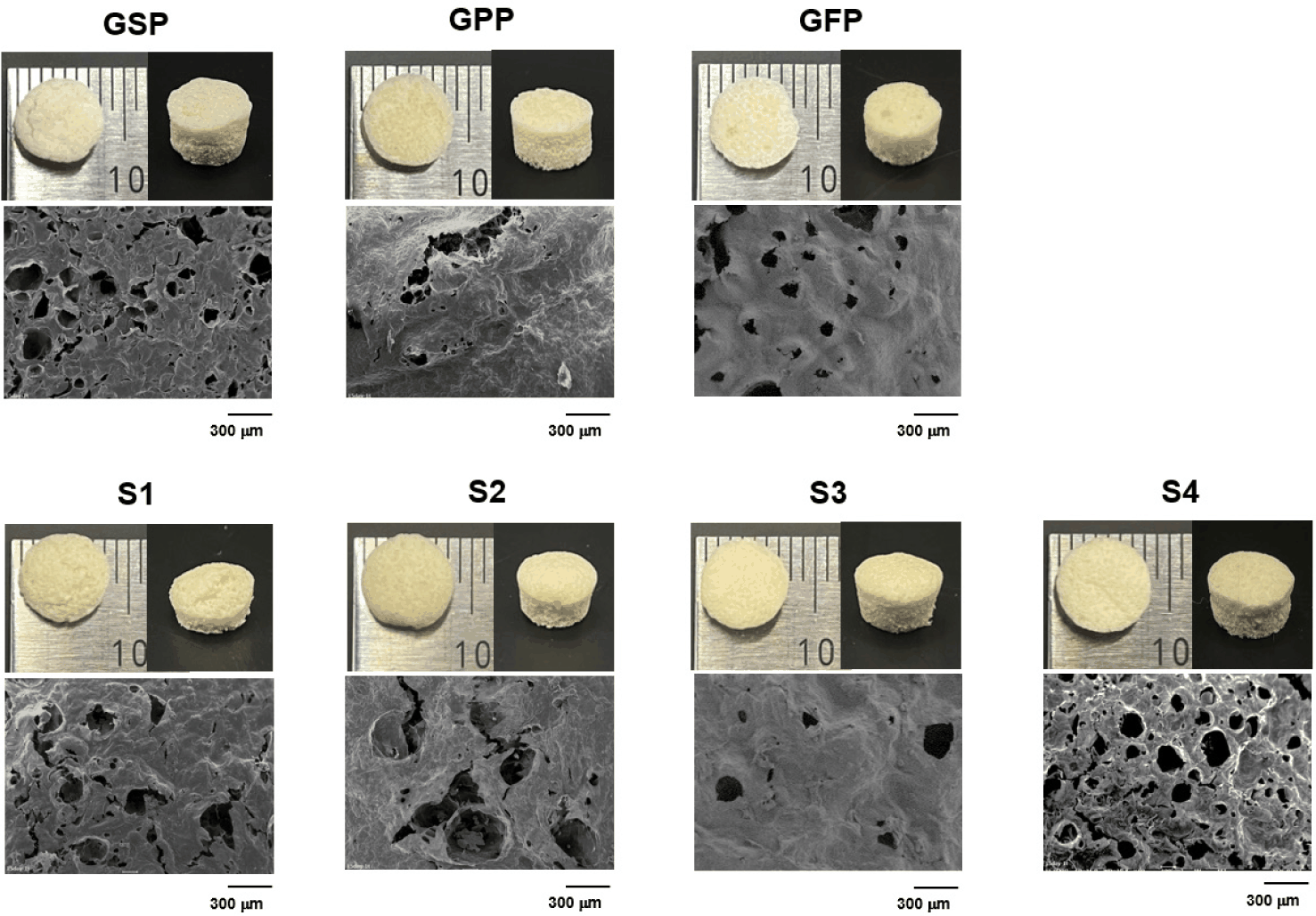
SEM analysis revealed clear differences in the microstructures of the scaffold surfaces depending on the protein material. Gel-based soybean protein scaffold (GSP) exhibited a higher number of surface pores than the other protein-based scaffolds, whereas gel-based pea protein scaffold (GPP) displayed fewer pores and a smoother surface. Gel-based faba bean protein scaffold (GFP) also exhibited a porous structure, but it was less pronounced than the soybean protein-based scaffold, with slight bulging of the tissue around the pores, showing distinct surface characteristics. Furthermore, the mixed protein scaffolds (S1–S4) exhibited varying pore sizes and densities depending on the mixing ratio. S2, in which some pea proteins replaced soybean and faba bean proteins, showed an increase in porosity, whereas S3 showed a decrease in porosity. The average pore size of the GSP was 117.8 µm, compared to 72.59 µm for GPP, and 94.80 µm for GFP. Among the mixed protein scaffolds, S2 exhibited the largest pores (113.5 µm), followed by S1 (107.1 µm), S3 (89.9 µm), and S4 (79.67 µm), suggesting that the microstructure of the scaffold is influenced by the protein composition and mixing ratio.
Pore size and distribution are critical factors in cell culture. Smaller pores can aid cell attachment and migration, whereas larger pores enhance nutrient and oxygen diffusion and promote cell proliferation and viability [23,24]. O’Brien et al. [25] reported a linear relationship between cell attachment and the specific surface area, suggesting that cell viability is influenced by pore size. The ideal scaffold porosity is considered to be between 30%–90%, with an optimal pore size range of 50–150 µm [26], aligning with the pore sizes observed in both the single and mixed protein scaffolds in this study. These findings indicate that the scaffolds used in this study have potential applications in cell culture. However, they exhibited non-uniform pore sizes, likely due to uncontrolled ice crystal formation during the freezing process. According to Chen et al. [27], scaffolds frozen at –30°C and –80°C exhibited pores with irregular polygonal shapes, whereas those treated with liquid nitrogen developed narrower pores characterized by elongated grooves. Xia et al. [28] reported that in soybean protein–carrageenan–sodium alginate mixtures, high Ca2+ concentrations induced rapid external gelation, hindering diffusion and resulting in irregular pore formation. In contrast, crosslinking with a CaCl2/KCl mixture allowed K+ to stabilize the network, promoting uniform crosslinking, more regular pore structures, and improved cell growth. This suggests that optimizing the freezing process or developing pore-controlling technologies is necessary to ensure a uniform pore distribution in the scaffolds.
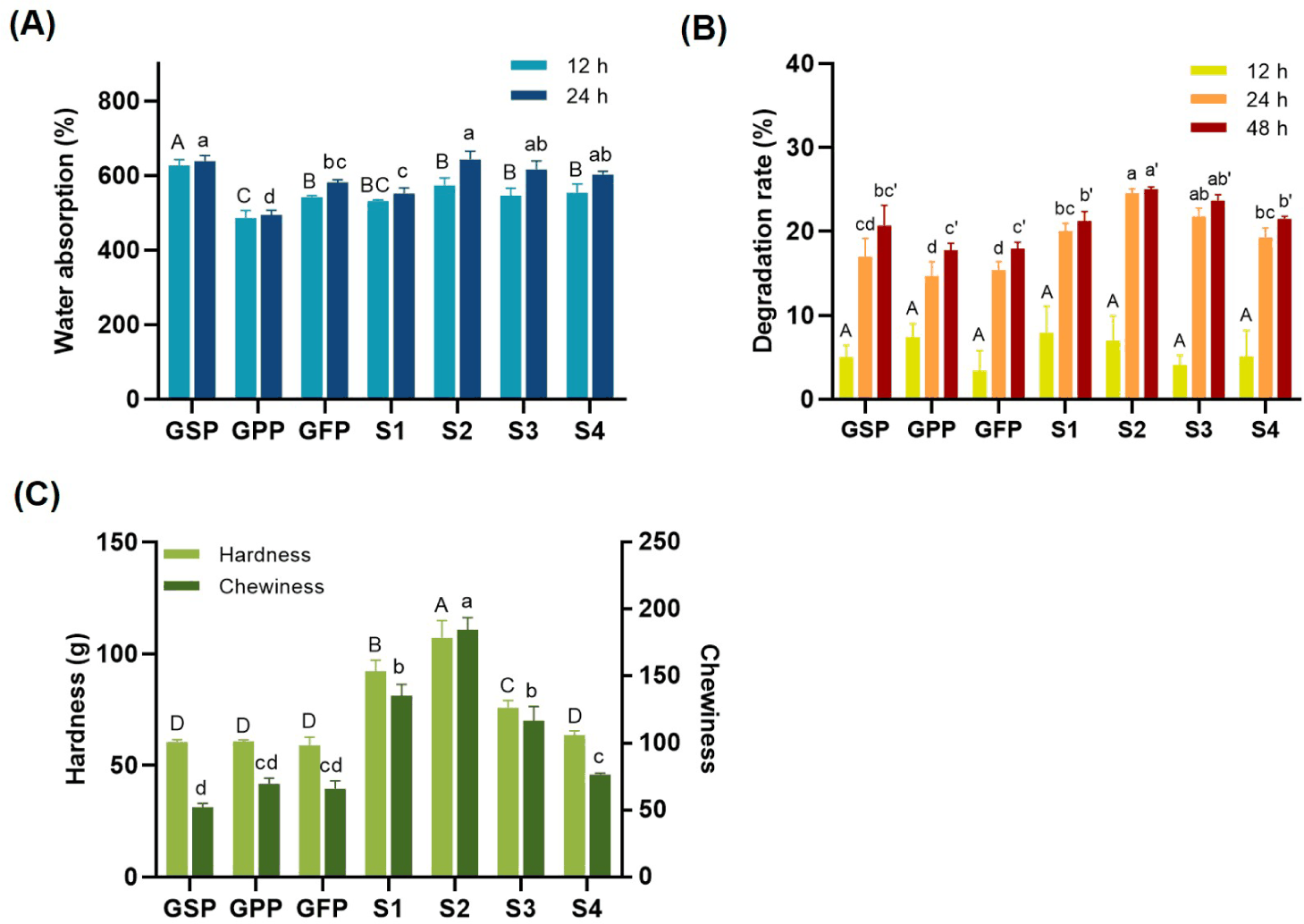
The water absorption rate is a key indicator that reflects the ability of a scaffold to absorb water over a certain period and is essential for evaluating its physical stability and shape retention (Fig. 2A). The water absorption behavior of scaffolds made from different plant protein sources was investigated. The GSP scaffold, composed of soybean protein, absorbed more than 600% of its initial weight after 12 h of immersion. In contrast, the GPP and GFP scaffolds, derived from pea and faba bean proteins, exhibited absorption rates of 480%–580%, indicating lower water uptake than that of GSP. This difference is attributed to the varying porous structures formed by different protein materials. A previous study also demonstrated that scaffolds containing soybean protein exhibited significantly higher water absorption (2,300%–2,500%) over 7 days, compared to those containing pea protein (1,100%–1,200%), highlighting the strong water-binding capacity of soybean-based materials [29].
Excessive water absorption may compromise the structural integrity of the scaffolds, leading to deformation, whereas insufficient absorption can hinder cell attachment, which is a critical factor in scaffold functionality. Therefore, selecting scaffolds with appropriate water absorption capacity is essential to support both cell attachment and proliferation [30]. The absorption rate is influenced by the protein type and composition. For instance, scaffolds containing glutenin, a major wheat protein, show water absorption rates ranging from 700% to 1,500%, depending on the glutenin content [5]. Additionally, the scaffold fabrication method significantly affects water absorption. Scaffolds made from soybean, mung bean, and faba bean proteins via extrusion exhibit lower water uptake, whereas those made from wheat protein display higher absorption rates [20]. Fang et al. [31] reported that scaffolds formed by dietary fiber–protein mixtures exhibit decreased water absorption upon crosslinking with CaCl2 or transglutaminase (TGase) owing to reduced porosity. The water absorption rate of uncrosslinked samples reached 413.94%, whereas cross-linked samples absorbed only approximately 300%.
For the mixed protein samples (S1–S4) composed of soybean, pea, and faba bean proteins, water absorption exceeded 500% after 12 h and surpassed 600% after 24 h in all samples except S1. This reduction compared to GSP is likely due to lower soybean protein content. However, partial substitution with pea or faba bean proteins enhanced the overall water absorption capacity, suggesting that hydration properties can be optimized by adjusting the protein composition. Consistent with our findings, Lin et al. [18] reported that scaffolds with higher protein contents exhibited enhanced water absorption owing to the presence of hydrophilic functional groups, even when the porosity was reduced. These results highlight the critical role of protein composition in determining the water absorption behavior of the scaffolds.
Understanding scaffold degradation is essential to evaluate the structural stability and functional lifespan of scaffolds under cell culture conditions. In cultured meat production, the degree of scaffold degradation varies depending on the intended application. In some cases, complete degradation may be desirable to avoid residual materials in the final product, particularly in fully edible systems. In other instances, partial or minimal degradation may be preferable for maintaining structural support throughout the culture period. Degradation is typically assessed by changes in physical properties, such as weight or morphology, and can be influenced by factors such as medium composition, pH, osmotic pressure, and enzymatic activity [6].
The degradation rate of the scaffolds over time is presented in Fig. 2B. At 12 h, all samples exhibited relatively low and comparable degradation rates, ranging from 3.42% to 7.91%, with no statistically significant differences observed among the groups. However, at 24 h and 48 h, distinct degradation patterns emerged depending on the scaffold composition. Scaffolds composed of mixed plant proteins (S1–S4) generally exhibited higher degradation rates than those composed of single proteins. Among them, S2 demonstrated the highest degradation rate, reaching 24.54% at 24 h, and 25.05% at 48 h. S1 and S3 also showed relatively high degradation levels, exceeding 21% at 48 h. In contrast, single-protein scaffolds, such as GSP, GPP, and GFP, exhibited lower degradation rates, ranging from 17.76% to 20.71% at the final time point. Scaffolds composed of mixed plant proteins (S1–S4) generally exhibited higher degradation rates than those composed of single proteins. These differences in degradation behavior may not be solely attributed to water permeability, as the single-protein scaffold exhibited higher water permeability but also showed a lower degradation rate than the mixed-protein scaffolds.
This apparent contradiction may be attributed to the reduced structural cohesion observed in mixed-protein scaffolds. The incorporation of proteins with differing conformational and electrostatic properties likely disrupts the formation of uniform and compact networks, thereby weakening intermolecular interactions and increasing susceptibility to hydrolytic degradation. Prior studies have demonstrated that heterogeneous protein matrices often exhibit diminished mechanical integrity and lower resistance to enzymatic or aqueous breakdown [32]. In contrast, single-protein scaffolds tend to form more homogenous and densely entangled networks, stabilized by stronger intramolecular bonding, which may account for their greater structural stability even under conditions of higher water absorption. This suggests that factors beyond water diffusion, such as network cohesiveness, protein–protein compatibility, and intrinsic structural stability, play critical roles in determining degradation resistance. These interactions may prevent the formation of tightly entangled scaffolds, rendering them more susceptible to hydrolytic degradation over time. In contrast, single-protein systems, such as GSP, may benefit from more uniform aggregation and stronger intramolecular interactions, forming a more stable network despite higher water uptake.
The structural stability of the scaffolds can also be significantly influenced by the composition, concentration, and processing methods of the raw materials. According to Chien and Shah [33], enzyme-treated soybean protein scaffolds (3% and 5% SPI) exhibited delayed degradation compared to untreated controls, especially under cell-seeded conditions, highlighting the role of enzyme-mediated crosslinking in enhancing scaffold stability. Measurement of weight loss in hydrogels with varying agar compositions revealed that hydrogels with less than 0.5% agar content completely degraded, exhibited very low stability. However, when the agar ratio increased to 3%, the hydrogels showed minimal degradation, which may hinder cell diffusion and proliferation in this platform [34]. Meanwhile, Ianovici et al. [32] reported that a higher protein content or enhanced crosslinking, such as in soybean-alginate scaffolds, may improve structural resilience but also result in slower degradation rates. In conclusion, the degradation behavior of plant protein-based scaffolds was significantly influenced by the scaffold composition, water permeability, and structural integrity. These results emphasize the importance of balancing the degradation rates with functional stability for specific applications, particularly in cultured meat production.
When considering scaffolds as edible materials, mechanical properties, such as hardness, chewiness, and elasticity, are critical factors that determine the overall texture and consumer acceptability of the final product. In this study, the textural properties of scaffolds fabricated from different plant protein sources were evaluated (Fig. 2C). The single-protein scaffolds (GSP, GPP, and GFP) exhibited hardness values of 60.48 g, 60.67 g, and 58.95 g, respectively, with no statistically significant differences observed among them. Pea proteins demonstrated slightly higher hardness and chewiness than soybean and faba bean proteins, which may be attributed to their lower porosity and distinctive hydrogel-forming properties. In contrast, the mixed-protein scaffolds (S1–S4), which incorporated varying ratios of soybean, pea, and faba bean proteins, displayed distinct mechanical characteristics. Notably, scaffolds S1, S2, and S3 exhibited a marked increase in hardness compared to the single-protein scaffolds, with values of 92.10, 107.15, and 75.79 g, respectively. These increases suggest that certain combinations of plant proteins may induce synergistic effects, potentially enhancing protein–protein interactions and forming a denser, more crosslinked gel network. However, scaffold S4, prepared with equal proportions (1:1:1) of the three protein sources, showed a hardness of 63.5 g—comparable to that of the single-protein scaffolds and not significantly different from GSP, GPP, or GFP. This finding suggests that evenly balanced protein mixtures may not offer the same reinforcing effect, possibly because of interference among the gelation behaviors of each protein component, which could disrupt optimal network formation.
Chewiness, another key mechanical property, exhibited more pronounced differences than hardness. The GSP, GPP, and GFP scaffolds showed chewiness values in the range of 52–70, indicating relatively soft and elastic textures without significant variation. In contrast, the mixed-protein scaffolds demonstrated considerable enhancement in chewiness. Among them, scaffold S2 exhibited the highest chewiness value of 184.6, which was nearly three times greater than that of the single-protein scaffolds. Scaffolds S1 and S3 also recorded chewiness values exceeding 100, indicating that specific combinations, particularly those with a higher proportion of peas or faba bean proteins, significantly contributed to enhanced mechanical resistance during mastication.
These changes in texture are likely driven by differences in protein–protein and protein–water interactions, gelation behavior, and the formation of three-dimensional gel networks [35,36]. Zhang et al. [37] demonstrated that the combination of peanut protein and wheat gluten improved the fiberization and elasticity of plant-based matrices through complementary structural roles and intermolecular interactions. These findings align with the current results, where mixed-protein scaffolds such as S2 and S3 exhibited enhanced chewiness, likely owing to synergistic interactions and optimized network formation. In contrast, S4, which incorporated equal proportions of soybean, pea, and faba bean proteins, did not show similar improvements, suggesting that uniformly blended protein systems hinder cohesive gelation because of competitive or antagonistic interactions. From this result, in single-protein scaffolds, homogeneous network formation is favored due to uniform gelation behavior, which leads to consistent crosslinking density and stable hydrogel networks. In contrast, mixed-protein scaffolds involve heterotypic interactions among proteins with distinct isoelectric points and structural flexibility, potentially disrupting the uniformity of gelation. This can result in heterogeneous pore formation and variable water uptake. Certain protein combinations, such as those in S2 and S3, may still achieve favorable network structures due to synergistic interactions that enhance intermolecular bonding and matrix stability.
Previous studies have reported that the hardness and chewiness values of pork neck meat are approximately 154.16 and 87.05 g, respectively [38]. Compared with these values, our S2 scaffold, which showed a hardness of 107.15 g and chewiness of 184.6, demonstrated comparable firmness and even superior chewiness. These findings suggest that the scaffold developed in this study indicates the potential of plant-based proteins to replicate the mechanical characteristics of actual meat. However, as textural transformation during cooking was not assessed in this study, further investigation is needed to evaluate the structural integrity and mechanical behavior of the scaffolds after thermal processing, which is critical for their practical application in real meat analogs.
These mechanical properties are not only important structurally but also influence consumer perception of cultured meat. Scaffolds like S2 and S3, with higher chewiness and moderate hardness, may better replicate the fibrous texture of cuts like flank or thigh meat, while softer scaffolds such as GFP or S4 may suit tenderloin-type or processed products. Since consumers expect plant- or cell-based alternatives to mimic both taste and texture, including bite and mouthfeel, the tunable textural profiles of our scaffolds suggest their potential for various cultured meat applications [39].
The cell proliferation on each scaffold was evaluated after 4 days of culture using the WST assay (Fig. 3A). The results showed clear differences in cell growth depending on the scaffold composition. Among the single-protein scaffolds, GFP supported the highest cell proliferation (122.94%), followed by GPP (96.60%), and GSP (62.86%). In contrast, the mixed-protein scaffolds exhibited lower proliferation rates: S1 (84.02%), S3 (64.86%), S2 (36.78%), and S4 (30.71%). These results indicated that single-protein scaffolds, particularly GFP, were more favorable for cell growth than mixed-protein formulations. According to Lin et al. [18], the viability of L929 cells cultured on starch-soybean protein-gellan gum composite scaffolds increased with higher soybean protein content. These findings highlight the potential of plant-based proteins in enhancing cell proliferation, suggesting their promising applications in tissue engineering and cellular agriculture. Previous findings further support our results and reinforce the applicability of plant-based proteins for cell growth in scaffold systems. However, conflicting results have been reported in previous studies, where protein-blended hydrogels did not show significant improvements in cell adhesion or proliferation compared with pure polysaccharide gels, indicating the limited supportive effects of proteins under unmodified conditions [19]. Therefore, it is important to consider not only the type of protein, but also the composition of materials, such as polysaccharides, in scaffold design and optimization.
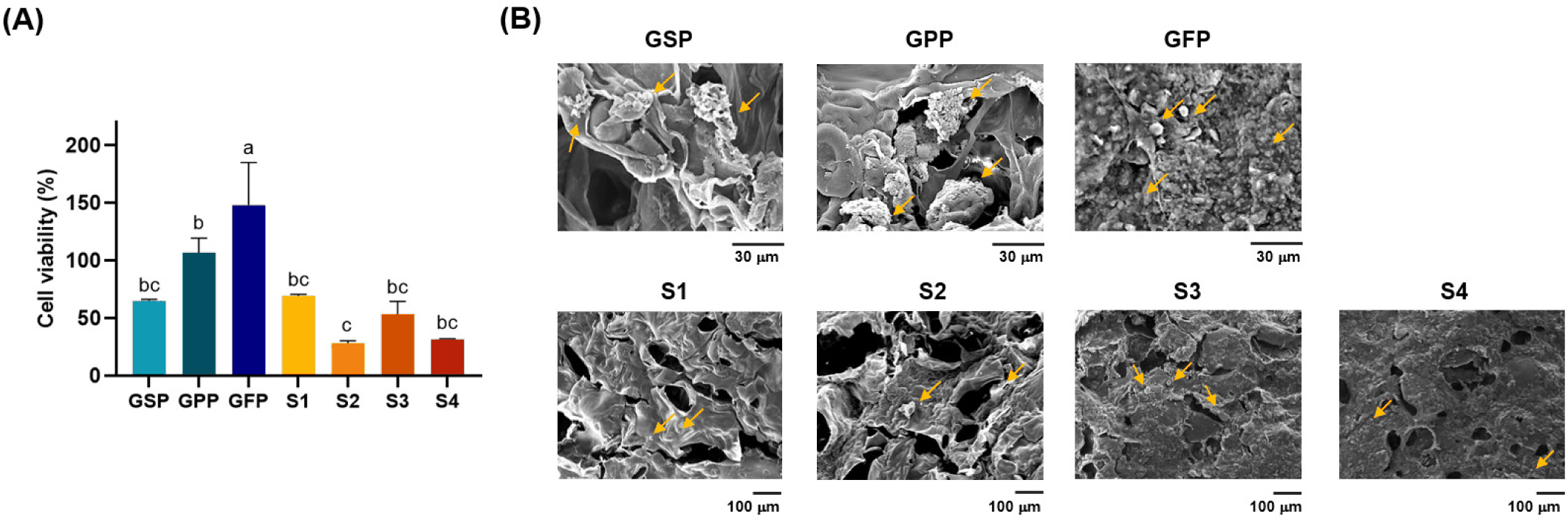
These quantitative findings are supported by the SEM images shown in Fig. 3B, which depict the morphology and distribution of the cells on each scaffold. Cells on the GFP and GPP surfaces were more abundant and evenly distributed, showing spread-out morphologies (yellow arrows) indicative of active attachment and proliferation. In particular, GFP presented a dense and relatively homogeneous surface where cells adhered well across the structure. In contrast, the S2 and S4 scaffolds displayed fewer adhered cells with mostly rounded morphologies, suggesting lower cell–scaffold interactions and biocompatibility. GSP S1 and S3 exhibited intermediate cell densities. Although cell attachment was observed (yellow arrows), the distribution was either more localized or less extensive than that of GFP or GPP. Scaffold composition and structure are known to affect cell viability and proliferation, with factors such as surface chemistry, porosity, and mechanical stiffness influencing cell–matrix interactions [5,40].
Among these factors, porosity and pore size play a particularly important role in modulating cellular responses by facilitating nutrient and oxygen diffusion, waste removal, and providing surface area for cell attachment. Previous studies have shown that scaffolds with interconnected pores ranging from 50 to 150 µm offer optimal conditions for cell infiltration and proliferation, whereas irregular or excessively large pores may impair cell–matrix interactions and reduce structural stability [25]. In the present study, the relatively uniform and moderate pore sizes of GFP and GPP scaffolds, combined with appropriate stiffness and protein–protein interactions, likely contributed to enhanced cell proliferation by offering a structurally favorable and biocompatible microenvironment [41,42].
CONCLUSION
This study demonstrated the feasibility of using plant-based proteins as edible scaffolds for cultured meat applications. Scaffolds made from a single protein source, particularly faba bean protein, support superior cell adhesion and proliferation, highlighting their potential as viable edible scaffold materials. Mixed-protein scaffolds exhibit enhanced mechanical properties and water absorption, suggesting that compositional tuning can optimize scaffold functionality. Although the degradation rates increased over time, the overall structural stability remained within a usable range for short-term cell culture. These findings indicate that the strategic combination of plant proteins can enhance both the biological performance and textural properties, offering a promising approach for developing sustainable, functional scaffolds in cellular agriculture. By replacing animal-derived components with cost-effective plant proteins, this strategy also contributes to improved sustainability and economic feasibility in cultured meat production.