RESEARCH ARTICLE

Wheat phytase can alleviate the cellular toxic and inflammatory effects of lipopolysaccharide

Jeongmin An1https://orcid.org/0000-0002-2357-0941, Jaiesoon Cho1,*https://orcid.org/0000-0002-4511-8032
Author Information & Copyright
1Department of Animal Science and Technology, Konkuk University, Seoul 05029, Korea.
*Corresponding author: Jaiesoon Cho, Department of Animal Science and Technology, Konkuk University, Seoul 05029, Korea. Tel: +82-2-450-3375, E-mail: chojs70@konkuk.ac.kr

© Copyright 2021 Korean Society of Animal Science and Technology. This is an Open-Access article distributed under the terms of the Creative Commons Attribution Non-Commercial License (http://creativecommons.org/licenses/by-nc/4.0/) which permits unrestricted non-commercial use, distribution, and reproduction in any medium, provided the original work is properly cited.

Received: May 08, 2020; Revised: Jul 09, 2020; Accepted: Nov 04, 2020

Published Online: Jan 31, 2021

Abstract

The objective of this study was to characterize the enzymatic hydrolysis of lipopolysaccharide (LPS) by wheat phytase and to investigate the effects of wheat phytase-treated LPS on in vitro toxicity, cell viability and release of a pro-inflammatory cytokine, interleukin (IL)-8 by target cells compared with the intact LPS. The phosphatase activity of wheat phytase towards LPS was investigated in the presence or absence of inhibitors such as L-phenylalanine and L-homoarginine. In vitro toxicity of LPS hydrolyzed with wheat phytase in comparison to intact LPS was assessed. Cell viability in human aortic endothelial (HAE) cells exposed to LPS treated with wheat phytase in comparison to intact LPS was measured. The release of IL-8 in human intestinal epithelial cell line, HT-29 cells applied to LPS treated with wheat phytase in comparison to intact LPS was assayed. Wheat phytase hydrolyzed LPS, resulting in a significant release of inorganic phosphate for 1 h (p < 0.05). Furthermore, the degradation of LPS by wheat phytase was nearly unaffected by the addition of L-phenylalanine, the inhibitor of tissue-specific alkaline phosphatase or L-homoarginine, the inhibitor of tissue-non-specific alkaline phosphatase. Wheat phytase effectively reduced the in vitro toxicity of LPS, resulting in a retention of 63% and 54% of its initial toxicity after 1–3 h of the enzyme reaction, respectively (p < 0.05). Intact LPS decreased the cell viability of HAE cells. However, LPS dephosphorylated by wheat phytase counteracted the inhibitory effect on cell viability. LPS treated with wheat phytase decreased IL-8 secretion from intestinal epithelial cell line, HT-29 cell to 14% (p < 0.05) when compared with intact LPS. In conclusion, wheat phytase is a potential therapeutic candidate and prophylactic agent for control of infections induced by pathogenic Gram-negative bacteria and associated LPS-mediated inflammatory diseases in animal husbandry.

Keywords: Lipopolysaccharide; Wheat phytase; Therapeutic candidate; Prophylactic agent; Inflammatory diseases

INTRODUCTION

Lipopolysaccharide (LPS) is a major cell wall component of Gram-negative bacteria and is a physiologically well-known endotoxin derived from them, and one of the representative pathogen associated molecular patterns (PAMPs) [13]. LPS infection has been associated with increased cloacal temperature, depression, lethargy, diarrhea, avoidance of feed, hypercholesterolemia, hypouricemia and hyperphosphatemia occurred in broiler chickens [4,5]. It also reduced leukocyte and thrombocyte counts and induced leukocytosis in piglets [6]. It further contributed to a diverse range of mild and severe inflammatory diseases in human and animals such as ulcerative colitis [7], acute pneumonia [8] and systemic sepsis [9]. Surprisingly, in animal husbandry, swine farms and poultry houses are vulnerable to LPS contamination [1,5], especially detected as a dust suspension at higher concentrations of 4.9 μg/m3 at the former [1].

Structurally, the LPS carries phosphorylated lipid A moiety, which accounts for the toxicity and immunostimulatory effects of the intact molecule [6]. Therefore, the degradation of LPS by phosphatase activity disrupts its function [6,10,11]. Phytase is a specific type of phosphatases exclusively hydrolyzing phytate that exists as primary phosphorus reserve in edible plants such as cereals, legumes and oilseeds and acts as an anti-nutritional factor to non-ruminants [12]. Phytase is a well-established and commercially available feed additive enhancing phosphate bioavailability in animals, and reducing phosphate pollution in the environment [12]. Intriguingly, the function of wheat phytase that is classified as a unique multiple inositol polyphosphate phosphatase (MINPP) is attractive due to its non-specific phosphatase activity against other phosphorylated substrates such as p-nitrophenyl phosphate and 2,3-bisphosphoglycerate, a negative allosteric regulator of hemoglobin, even if it is much less known than microbial counterpart [13,14].

Therefore, we hypothesized that wheat phytase can dephosphorylate LPS. Until now, little has been reported about the hydrolysis of LPS by phytases. The objective of this study was to characterize the enzymatic hydrolysis of LPS by wheat phytase and to investigate the effects of the wheat phytase-treated LPS on in vitro toxicity, cell viability and the release of a pro-inflammatory cytokine, interleukin (IL)-8 from target cells when compared with the intact LPS.

MATERIALS AND METHODS

Dephosphorylation assay for lipopolysaccharide

Wheat phytase (Sigma-Aldrich, Saint Louis, MO, USA) was reconstituted in endotoxin-free water (Sigma-Aldrich). The phosphatase activity of the enzyme (28.6 mU/mL) against the substrate, LPS (100 μg/mL) was determined in acetate buffer (pH 5.0) at 37°C for the given duration (15 min or 1 h) in the presence or absence of inhibitors (10 mM L-phenylalanine or L-homoarginine). In addition, LPS (100 μg/mL) was treated with different units of wheat phytase (14.3 and 57.2 mU/mL) in acetate buffer (pH 5.0) at 37°C for 1 h and the phosphatase activity of the enzyme against LPS was assayed with different concentrations (5 and 20 mM) of the inhibitors. The inorganic phosphate release was measured at optical density (OD) 635 nm using the malachite green-based PiColor Lock gold phosphate detection kit (Innova Biosciences, Cambridge, UK), according to the manufacturer’s instructions.

In vitro toxicity assay of lipopolysaccharide

LPS (20 ng/mL) was hydrolyzed with wheat phytase (11.44 × 10⁻³ mU/mL) for 1–3 h, respectively and different levels of LPS (10 and 40 ng/mL) were treated with the enzyme for 3 h. The residual toxicities of the LPS were assayed at OD 545 nm using ToxinSensor chromogenic LAL (limulus amebocyte lysate)-based endotoxin assay kit (GenScript, Piscataway, NJ, USA), according to the manufacturer’s instructions.

Maintenance of cell culture

Human aortic endothelial (HAE) cell and human colorectal adenocarcinoma HT-29 cell were purchased from American Type Culture Collection (ATCC, Manassas, VA, USA). These two cells were used as well-established systems to measure LPS cytotoxicity and gut-induced IL-8 secretion, respectively. The former was maintained in cascade biologics medium 200 (Gibco Life technologies, Carlsbad, CA, USA) containing low serum growth supplement kit (Gibco Life technologies), and the latter in McCoy’s 5A medium (Gibco Life technologies) containing 10% fetal bovine serum and 1% penicillin-streptomycin solution (Gibco Life technologies). These cells were cultured at 37°C in a humidified incubator with 95% air and 5% CO2.

Cell viability assay

HAE cells were initially seeded onto a 96-well plate at a concentration of 2 × 10⁴ cells per well and cultured until 80% confluency. LPS (2 mg/mL) was incubated at 37°C for 1 h with or without wheat phytase (28.6 mU/mL). Aliquots (10 μL) of the reaction mixtures were added to the cells with final concentration of 200 μg/mL LPS at 37°C for 24 h in the CO2 incubator. In addition, aliquots of the reaction mixtures were applied to the cells with final concentration of LPS (100, 200, and 400 μg/mL), respectively. The cell viabilities were measured at OD 450 nm using EZ-CYTOX kit (DogenBio, Seoul, Korea), according to the manufacturer’s instructions.

Interleukin-8 assay

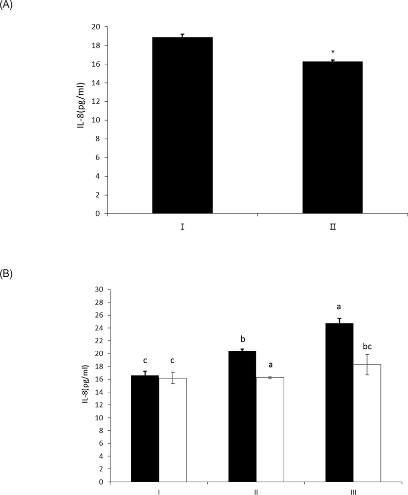
HT-29 cells were initially seeded onto a 96-well plate at a concentration of 2 × 10⁴ cells per well and cultured until 80% confluency. LPS (100 μg/mL) was incubated at 37°C for 2 h with or without wheat phytase (286 mU/mL) and aliquots (10 μL) of the reaction mixtures were added to the cells with final concentration of 10 μg/mL LPS at 37°C for 24 h in the CO2 incubator. In addition, aliquots of the reaction mixtures were applied to the cells with final concentration of LPS (5, 10, and 20 μg/mL), respectively. The levels of IL-8 secreted into the culture media were assayed at OD 450 nm using Cymax™ human IL-8 ELISA kit (Ab FRONTIER, Seoul, Korea), according to the manufacturer’s instructions.

Statistical analysis

Statistical significance between groups was determined by a one-way analysis of variance using PROC GLM (SAS 9.4, SAS Institute, Cary, NC, USA) followed by Duncan’s multiple range test or by Student’s t-test. A p-value < 0.05 was considered as statistically significant difference.

RESULTS

Dephosphorylation of lipopolysaccharide by wheat phytase

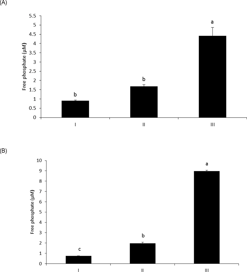
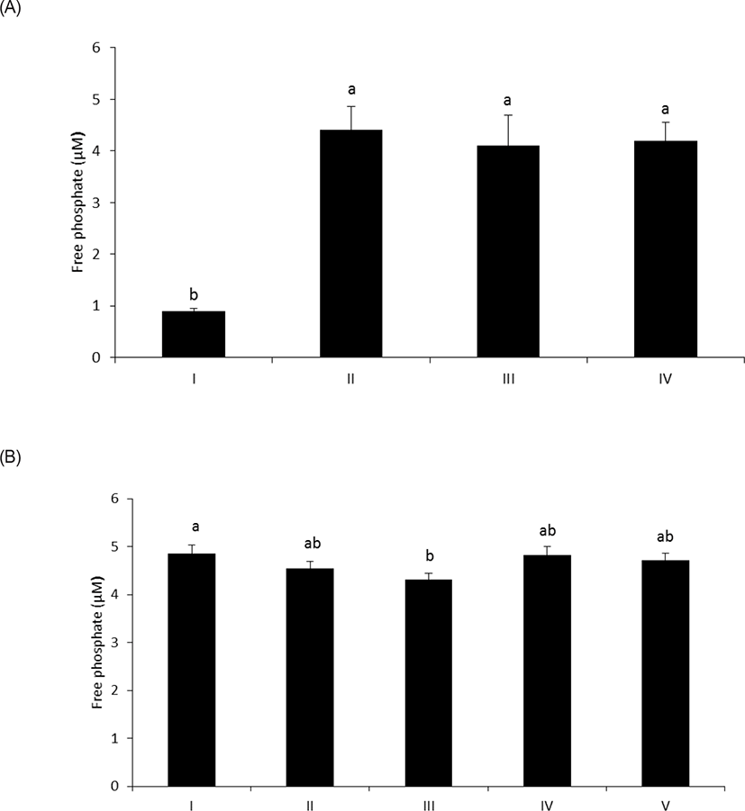
As shown in Fig. 1A, wheat phytase hydrolyzed LPS, significantly releasing inorganic phosphate for 1 h (p < 0.05). In addition, the enzyme dephosphorylated LPS in a dose-dependent fashion (p < 0.05) (Fig. 1B). Previously, alkaline phosphatase was regarded as a sole enzyme that degraded LPS, whether it is a tissue-specific or tissue-non-specific type [10,11,15]. Nevertheless, our novel finding is remarkable considering that wheat phytase has highly strict substrate specificity for phytate with little or no phosphatase activity against other physiologically-relevant phosphorylated conjugates such as simple sugar phosphates, ATP, adenosine diphosphate, adenosine monophosphate, guanosine triphosphate and phosphoenolpyruvate [13]. Furthermore, the degradation of LPS by wheat phytase was nearly unaffected by the addition of L-phenylalanine, the inhibitor of tissue-specific alkaline phosphatase or L-homoarginine, the inhibitor of tissue-non-specific alkaline phosphatase (Figs. 2A and 2B). Indeed, bovine intestinal alkaline phosphatase inhibited the dephosphorylation of LPS in the presence of L-phenylalanine, but not L-homoarginine [16], while murine uterine alkaline phosphatase inhibited the dephosphorylation of LPS in the presence of L-homoarginine, but not L-phenylalanine [15]. This present result suggests that the catalytic mechanism of LPS hydrolysis by wheat phytase may vary from that of alkaline phosphatases.

jast-63-1-114-g1
Fig. 1. Phosphatase activity of wheat phytase against LPS at different enzyme reaction times (A) or enzyme units (B). (A) LPS (100 μg/mL) was treated with wheat phytase (28.6 mU/mL) in acetate buffer (pH 5.0) at 37°C for the given duration. I: absence of enzyme reaction, II: enzyme reaction for 15 min, III: enzyme reaction for 1 h. Data were presented as mean and standard errors from three experiments. a,bMeans lacking common superscripts differ significantly (p < 0.05). (B) LPS (100 μg/mL) was treated with different units of wheat phytase in acetate buffer (pH 5.0) at 37°C for 1 h. I: absence of enzyme reaction, II: the enzyme (14.3 mU/mL), III: the enzyme (57.2 mU/mL). Data were presented as mean and standard errors from three experiments. a–cMeans lacking common superscripts differ significantly (p < 0.05). LPS, lipopolysaccharide.
Download Original Figure
jast-63-1-114-g2
Fig. 2. Effect of enzyme inhibitors (10 mM) such as L-phenylalanine and L-homoarginine on dephosphorylation of LPS (100 μg/mL) by wheat phytase (28.6 mU/mL) (A) and the phosphatase activity of the enzyme against the substrate with different concentrations (5 and 20 mM) of the inhibitors (B). (A) I: absence of enzyme reaction, II: enzyme reaction for 1 h in the absence of enzyme inhibitors, III: enzyme reaction for 1 h in the presence of L-phenylalanine, IV: enzyme reaction for 1 h in the presence of L-homoarginine. Data were presented as mean and standard errors from three experiments. a,bMeans lacking common superscripts differ significantly (p < 0.05). (B) I: enzyme reaction for 1 h in the absence of enzyme inhibitors, II: enzyme reaction for 1 h in the presence of 5 mM L-phenylalanine, III: enzyme reaction for 1 h in the presence of 20 mM L-phenylalanine, IV: enzyme reaction for 1 h in the presence of 5 mM L-homoarginine, V: enzyme reaction for 1 h in the presence of 20 mM L-homoarginine. Data were presented as mean and standard errors from three experiments. a,bMeans lacking common superscripts differ significantly (p < 0.05). LPS, lipopolysaccharide.
Download Original Figure
Change of in vitro toxicity of lipopolysaccharide by wheat phytase

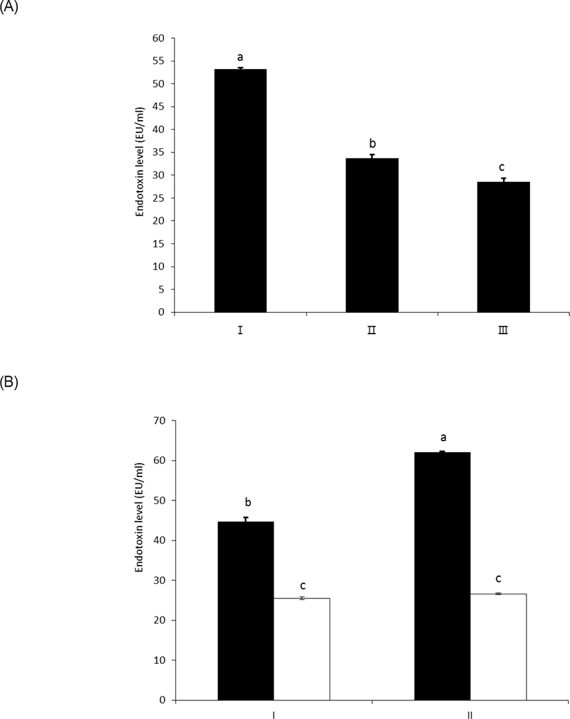
As shown in Figs. 3A and 3B, wheat phytase effectively reduced the in vitro toxicity of LPS, and 63% and 54% of its initial toxicity was retained after 1 h and 3 h of the enzyme reaction, respectively (p < 0.05) (Fig. 3A). In this regard, Koyama et al. [10] demonstrated that the structural destruction of LPS by enzymatic dephosphorylation was closely associated with LPS detoxification, due to defective binding to the effector cells.

jast-63-1-114-g3
Fig. 3. Effect of wheat phytase (11.44 × 10⁻³ mU/mL) on in vitro toxicity of LPS (20 ng/mL) (A) and the change of the toxicity at different levels of LPS (10 and 40 ng/mL) treated with the enzyme (B). (A) I: absence of enzymatic dephosphorylation of LPS, II: enzymatic dephosphorylation of LPS for 1 h, III: enzymatic dephosphorylation of LPS for 3 h. Data were presented as mean and standard errors from three experiments. a–cMeans lacking common superscripts differ significantly (p < 0.05). (B) I: (closed bar); absence of enzymatic dephosphorylation of LPS (10 ng/mL), (open bar); enzymatic dephosphorylation of LPS (10 ng/mL) for 3 h, II: (closed bar); absence of enzymatic dephosphorylation of LPS (40 ng/mL), (open bar); Enzymatic dephosphorylation of LPS (40 ng/mL) for 3 h. Data were presented as mean and standard errors from three experiments. a–cMeans lacking common superscripts differ significantly (p < 0.05). LPS, lipopolysaccharide.
Download Original Figure
Altered cell viability by lipopolysaccharide treated with wheat phytase

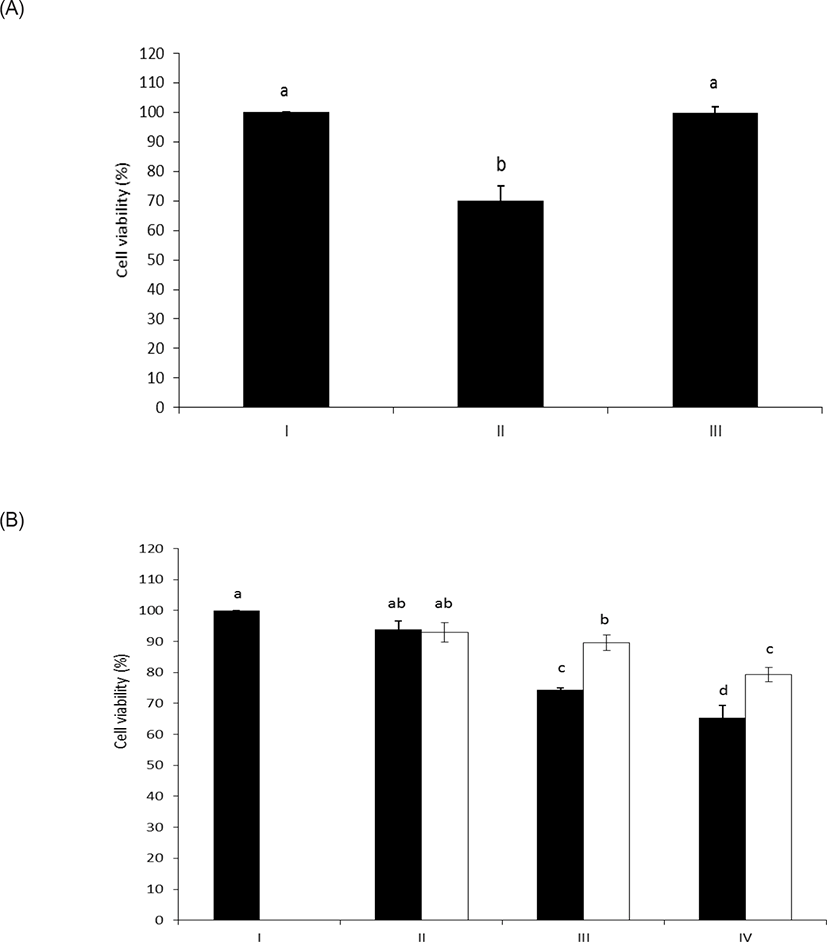
As shown in Figs. 4A and 4B, intact LPS clearly decreased the cell viability of HAE cells with unusual sensitivity [17]. However, the LPS dephosphorylated by wheat phytase counteracted the inhibitory effect on cell viability. Meanwhile, it was reported that the cell viability of HAE cells was enhanced following exposure to LPS and was attributed to the presence of alkaline phosphatase induced by IL-6 [10,18].

jast-63-1-114-g4
Fig. 4. Cell viability of HAE cells exposed to LPS treated with wheat phytase (A) and the change of cell viabilities at different levels of LPS treated with the enzyme (B). (A) I: no addition, II: addition of intact LPS (200 μg/mL), III: addition of LPS (200 μg/mL) dephosphorylated by wheat phytase (28.6 mU/mL). Data were presented as mean and standard errors from three experiments. a,bMeans lacking common superscripts differ significantly (p < 0.05). (B) I: no addition, II: (closed bar); addition of intact LPS (100 μg/mL), (open bar); addition of LPS (100 μg/mL) dephosphorylated by wheat phytase (28.6 mU/mL), III: (closed bar); addition of intact LPS (200 μg/mL), (open bar); addition of LPS (200 μg/mL) dephosphorylated by wheat phytase (28.6 mU/mL), IV: (closed bar); addition of intact LPS (400 μg/mL), (open bar); addition of LPS (400 μg/mL) dephosphorylated by wheat phytase (28.6 mU/mL). Data were presented as mean and standard errors from three experiments. a–dMeans lacking common superscripts differ significantly (p < 0.05). HAE, human aortic endothelial; LPS, lipopolysaccharide.
Download Original Figure
Changes in interleukin-8 release from HT-29 cells following lipopolysaccharide treatment with wheat phytase

LPS treated with wheat phytase decreased IL-8 secretion from intestinal epithelial cell line, HT-29 cells to 14% (p < 0.05) when compared with intact LPS (Fig. 5A). In particular, the decrease of IL-8 secretion from the cells was effective at 20 μg/mL of LPS treated with the enzyme (Fig. 5B). It was known that LPS specifically exerts its inflammatory effects via IL-8 in HT-29 cell [19], via appropriate interaction of phosphorylated lipid A moiety of LPS with toll-like receptor 4 (TLR4)-MD2 complex on the cell surface induced by interferon gamma (IFN γ) [20,21]. Thus, the dephosphorylation of LPS by wheat phytase may suppress the side-effects of excessive inflammation induced by IL-8 over-expression [22].

jast-63-1-114-g5
Fig. 5. Effect of LPS treated with wheat phytase on IL-8 secretion in HT-29 cells (A) and IL-8 release from the cells exposed at different levels of LPS treated with the enzyme (B). (A) I: addition of intact LPS (10 μg/mL), II: addition of LPS (10 μg/mL) dephosphorylated by wheat phytase (286 mU/mL). Data were presented as mean and standard errors from three experiments. (*p < 0.05: Student’s t-test). (B) I: (closed bar); addition of intact LPS (5 μg/mL), (open bar); addition of LPS (5 μg/mL) dephosphorylated by wheat phytase (286 mU/mL), II: (closed bar); addition of intact LPS (10 μg/mL), (open bar); addition of LPS (10 μg/mL) dephosphorylated by wheat phytase (286 mU/mL), III: (closed bar); addition of intact LPS (20 μg/mL), (open bar); addition of LPS (20 μg/mL) dephosphorylated by wheat phytase (286 mU/mL). Data were presented as mean and standard errors from three experiments. a–cMeans lacking common superscripts differ significantly (p < 0.05). LPS, lipopolysaccharide.
Download Original Figure

DISCUSSION

In animal husbandry, control of infection by pathogenic Gram-negative bacteria and related LPS-mediated inflammatory diseases can be addressed by the use of antibiotics and specific medications, or via application of immunostimulatory supplements such as probiotics and prebiotics. Indeed, antibiotics and certain medications such as the so-called chemical agents are associated with the increased resistance and abuse, harm and public dislike [2325]. The aforementioned immunostimulatory supplements exhibit indirect efficacy against the targets by boosting specific immune cells in the body [26], which may be mild, variable and even unclear, despite the wide range of products that are commercially available [27,28].

Previously, the occurrence of necrotizing enterocolitis known as a severe intestinal inflammatory disease in premature infants and rat pups was closely associated with the exposure of LPS and the lack of endogenous intestinal alkaline phosphatase activity [29]. The recovery of exogenous alkaline phosphatase activity by breast milk feeding in them helped to weaken LPS-induced inflammation and toxification [29]. Therefore, wheat phytase may act as an efficient reservoir of the phosphatase activity for LPS hydrolysis and detoxification in the intestine.

In our study, the mechanism of wheat phytase involved in the reduction of IL-8 secretion in HT-29 cells under LPS stimulus was still unclear at a cellular level. In this regard, the supplementation of exogenous intestinal alkaline phosphatase leaded to the activation of endogenous intestinal alkaline phosphatase for LPS hydrolysis in porcine intestine extracts through the enhancement of a canonical inflammatory regulator, nuclear factor kappa-light-chain-enhancer of activated B cells (NF-kB) [30], which may down-regulate IL-8 signaling.

Some studies suggested that the supplementation of phytase could have a direct effect on enhancing the immune performance of farm animals. For example, inorganic phosphate released from phytate by phytase can modulate the virulent phenotypes of pathogens in the intestine of weaned pigs [31], and the parameters such as the percentage of lymphocytes and the amount of mucosal immunoglobulin (Ig) A and serum Ig M were improved with phytase in broilers [32,33]. Therefore, inorganic phosphate hydrolized from LPS by wheat phytase may positively work on diminishing the intensity of inflammatory response in HT-29 cells.

Wheat phytase is a potential therapeutic candidate and prophylactic agent for control of infections induced by pathogenic Gram-negative bacteria and associated LPS-mediated inflammatory diseases. Additionally, it is a potential feed additive beyond its conventional role in improving phosphate availability and preventing phosphate pollution. The activity of wheat phytase is mediated via relatively direct, safe and enzymatic mechanisms. Future studies exploring the possibility of mass production of wheat phytase are warranted.

Competing interests

No potential conflict of interest relevant to this article was reported.

Funding sources

This research was supported by Basic Science Research Program through the National Research Foundation of Korea (NRF) funded by the Ministry of Education (2016R1D1A1B03930196).

Acknowledgements

Not applicable.

Availability of data and material

Upon reasonable request, the datasets of this study can be available from the corresponding author.

Authors’ contributions

Conceptualization: Cho J.

Data curation: An J.

Formal analysis: An J, Cho J.

Methodology: An J, Cho J.

Software: An J, Cho J.

Validation: An J, Cho J.

Investigation: An J, Cho J.

Writing - original draft: Cho J.

Writing - review & editing: An J, Cho J.

Ethics approval and consent to participate

This article does not require IRB/IACUC approval because there are no human and animal participants.

REFERENCES

1.

van Gucht S, van Reeth K, Pensaert M. Interaction between porcine reproductive-respiratory syndrome virus and bacterial endotoxin in the lungs of pigs: potentiation of cytokine production and respiratory disease. J Clin Microbiol. 2003; 41:960-6

2.

Estaki M, DeCoffe D, Gibson DL. Interplay between intestinal alkaline phosphatase, diet, gut microbes and immunity. World J Gastroenterol. 2014; 20:15650-6

3.

Lallès JP. Intestinal alkaline phosphatase: novel functions and protective effects. Nutr Rev. 2014; 72:82-94

4.

Xie H, Rath NC, Huff GR, Huff WE, Balog JM. Effects of Salmonella typhimurium lipopolysaccharide on broiler chickens. Poult Sci. 2000; 79:33-40

5.

Rauber RH, Perlin VJ, Fin CD, Mallmann AL, Miranda DP, Giacomini LZ, et al. Interference of Salmonella typhimurium lipopolysaccharide on performance and biological parameters of broiler chickens. Brazilian J Poultry Sci. 2014; 16:77-81

6.

Beumer C, Wulferink M, Raaben W, Fiechter D, Brands R, Seinen W. Calf intestinal alkaline phosphatase, a novel therapeutic drug for lipopolysaccharide (LPS)-mediated diseases, attenuates LPS toxicity in mice and piglets. J Pharmacol Exp Ther. 2003; 307:737-44

7.

Obana N, Takahashi S, Kinouchi Y, Negoro K, Takagi S, Hiwatashi N, et al. Ulcerative colitis is associated with a promoter polymorphism of lipopolysaccharide receptor gene, CD14. Scand J Gastroenterol. 2002; 37:699-704

8.

McClenahan D, Hellenbrand K, Atapattu D, Aulik N, Carlton D, Kapur A, et al. Effects of lipopolysaccharide and Mannheimia haemolytica leukotoxin on bovine lung microvascular endothelial cells and alveolar epithelial cells. Clin Vaccine Immunol. 2008; 15:338-47

9.

Doi K, Leelahavanichkul A, Yuen PST, Star RA. Animal models of sepsis and sepsis-induced kidney injury. J Clin Invest. 2009; 119:2868-78

10.

Koyama I, Matsunaga T, Harada T, Hokari S, Komoda T. Alkaline phosphatases reduce toxicity of lipopolysaccharides in vivo and in vitro through dephosphorylation. Clin Biochem. 2002; 35:455-61

11.

Chen KT, Malo MS, Moss AK, Zeller S, Johnson P, Ebrahimi F, et al. Identification of specific targets for the gut mucosal defense factor intestinal alkaline phosphatase. Am J Physiol Gastrointest Liver Physiol. 2010; 299:G467-75

12.

Dersjant-Li Y, Awati A, Schulze H, Partridge G. Phytase in non-ruminant animal nutrition: a critical review on phytase activities in the gastrointestinal tract and influencing factors. J Sci Food Agric. 2015; 95:878-96

13.

Dionisio G, Holm PB, Brinch-Pedersen H. Wheat (Triticum aestivum L.) and barley (Hordeum vulgare L.) multiple inositol polyphosphate phosphatases (MINPPs) are phytases expressed during grain filling and germination. Plant Biotechnol J. 2007; 5:325-38

14.

Kilaparty SP, Singh A, Baltosser WH, Ali N. Computational analysis reveals a successive adaptation of multiple inositol polyphosphate phosphatase 1 in higher organisms through evolution. Evol Bioinform. 2014; 10:239-50

15.

Lei W, Ni H, Herington J, Reese J, Paria BC. Alkaline phosphatase protects lipopolysaccharide-induced early pregnancy defects in mice. PLOS One. 2015; 10e0123243

16.

Moss AK, Hamarneh SR, Mohamed MMR, Ramasamy S, Yammine H, Patel P, et al. Intestinal alkaline phosphatase inhibits the proinflammatory nucleotide uridine diphosphate. Am J Physiol Gastrointest Liver Physiol. 2013; 304:G597-604

17.

Matsunaga T, Nakajima T, Sonoda M, Kawai S, Kobayashi J, Inoue I, et al. Reactive oxygen species as a risk factor in verotoxin-1-exposed rats. Biochem Biophys Res Commun. 1999; 260:813-9

18.

Nakazato H, Deguchi M, Fujimoto M, Fukushima H. Alkaline phosphatase expression in cultured endothelial cells of aorta and brain micro vessels: induction by interleukin-6-type cytokines and suppression by transforming growth factor betas. Life sci. 1997; 61:2065-72

19.

Angrisano T, Pero R, Peluso S, Keller S, Sacchetti S, Bruni CB, et al. LPS-induced IL-8 activation in human intestinal epithelial cells is accompanied by specific histone H3 acetylation and methylation changes. BMC Microbiol. 2010; 10:172

20.

Akashi S, Saitoh S, Wakabayashi Y, Kikuchi T, Takamura N, Nagai Y, et al. Lipopolysaccharide interaction with cell surface Toll-like receptor 4-MD-2: higher affinity than that with MD-2 or CD14. J Exp Med. 2003; 198:1035-42

21.

Suzuki M, Hisamatsu T, Podolsky DK. Gamma interferon augments the intracellular pathway for lipopolysaccharide (LPS) recognition in human intestinal epithelial cells through coordinated up-regulation of LPS uptake and expression of the intracellular Toll-like receptor 4-MD-2 complex. Infect Immun. 2003; 71:3503-11

22.

Skov L, Beurskens FJ, Zachariae CO, Reitamo S, Teeling J, Satijn D, et al. IL-8 as antibody therapeutic target in inflammatory diseases: reduction of clinical activity in palmoplantar pustulosis. J Immunol. 2008; 181:669-79

23.

Brustolim D, Ribeiro-dos-Santos R, Kast RE, Altschuler EL, Soares MBP. A new chapter opens in anti-inflammatory treatments: the antidepressant bupropion lowers production of tumor necrosis factor-alpha and interferon-gamma in mice. Int Immunopharmacol. 2006; 6:903-7

24.

Rice JA, Carrasco-Medina L, Hodgins DC, Shewen PE. Mannheimia haemolytica and bovine respiratory disease. Anim Health Res Rev. 2007; 8:117-28

25.

Allen HK, Levine UY, Looft T, Bandrick M, Casey TA. Treatment, promotion, commotion: antibiotic alternatives in food-producing animals. Trends Microbiol. 2013; 21:114-9

26.

Liu CF, Tseng KC, Chiang SS, Lee BH, Hsu WH, Pan TM. Immunomodulatory and antioxidant potential of Lactobacillus exopolysaccharides. J Sci Food Agric. 2011; 91:2284-91

27.

Callaway TR, Edrington TS, Anderson RC, Harvey RB, Genovese KJ, Kennedy CN, et al. Probiotics, prebiotics and competitive exclusion for prophylaxis against bacterial disease. Anim Health Res Rev. 2008; 9:217-25

28.

Buddington RK, Sangild PT. Companion animals symposium: development of the mammalian gastrointestinal tract, the resident microbiota, and the role of diet in early life. J Anim Sci. 2011; 89:1506-19

29.

Yang Y, Rader E, Peters-Carr M, Bent RC, Smilowitz JT, Guillemin K, et al. Ontogeny of alkaline phosphatase activity in infant intestines and breast milk. BMC Pediatr. 2019; 19:2

30.

Melo ADB, Silveira H, Bortoluzzi C, Lara LJ, Garbossa CAP, Preis G, et al. Intestinal alkaline phosphatase and sodium butylate may be beneficial in attenuating LPS-induced intestinal inflammation. Genet Mol Res. 2016; 15

31.

Heyer CME, Weiss E, Schmucker S, Rodehutscord M, Hoelzle LE, Mosenthin R, et al. The impact of phosphorus on the immune system and the intestinal microbiota with special focus on the pig. Nutr Res Rev. 2015; 28:67-82

32.

Liu N, Ru YJ, Cowieson AJ, Li FD, Cheng XCH. Effects of phytate and phytase on the performance and immune function of broilers fed nutritionally marginal diets. Poult Sci. 2008; 87:1105-11

33.

Sharideh H, Zhandi M, Zaghari M, Akhlaghi A, Hussaini SMH, Yousefi AR. Changes in broiler breeder hen’s immunity by zinc oxide and phytase. Iran J Vet Res. 2019; 20:120-5