INTRODUCTION
Assessing sperm motility is crucial not only for diagnosing human infertility but also for livestock reproduction. Evaluation of sperm motility is essential for AI and breeding soundness evaluations (BSE) to ensure optimal reproductive performance and genetic improvement in various species, including swine [1,2]. Considering the economic and biological significance, elucidating the mechanisms regulating sperm motility in livestock is indispensable.
The World Health Organization (WHO) defines infertility as the inability to conceive after 12 months or more of regular, unprotected sexual intercourse. It affects approximately 8%–12% of couples of reproductive age, with male-related factors contributing to nearly half of all cases [3]. Male infertility is commonly categorized based on semen characteristics, including conditions such as azoospermia, oligozoospermia, necrozoospermia, asthenozoospermia, teratozoospermia, and oligoasthenoteratozoospermia. Among these, asthenozoospermia, characterized by reduced or absent sperm motility, is one of the most frequently observed in infertile men [4]. Sperm motility is a key determinant of male fertility, as sperm must be able to move efficiently through the female reproductive tract to reach and fertilize the oocyte. Numerous studies have shown a positive association between sperm motility and fertilization success, emphasizing its critical role in reproduction [5,6]. Therefore, understanding the molecular mechanisms that regulate sperm motility is vital not only for human infertility diagnosis and treatment but also for advancing reproductive technology in livestock.
Capacitation is an important physiological process that sperm undergoes after ejaculation, characterized by a series of biochemical changes for fertilization. During this process, sperm acquire hyperactivated motility, enabling them to navigate cervical mucus and penetrate the zona pellucida surrounding the oocyte [7]. Recent research indicates that the phosphoinositide 3-kinase (PI3K)/phosphoinositide-dependent protein kinase-1 (PDK1)/protein kinase B (AKT) pathway plays a pivotal role in controlling capacitation-induced motility changes [8,9]. Notably, this pathway modulates key phosphorylation and motility patterns required for fertilization.
The PI3K/PDK1/AKT pathway is well known for regulatory functions, including cell survival, growth, metabolism, proliferation, and apoptosis [10]. In mammalian sperm, it has been shown to play a key role in regulating motility, capacitation, and the acrosome reaction. Particularly, this pathway has been implicated in the modulation of motility and tyrosine phosphorylation during capacitation [11–13]. Despite growing interest, the precise relationship between the PI3K/PDK1/AKT pathway and sperm motility and capacitation remains poorly understood. To address this knowledge gap, the present study investigates the association between this signaling pathway and sperm motility by quantifying the levels of PI3K/PDK1/AKT-related proteins and analyzing detailed motility parameters in Duroc boar spermatozoa.
MATERIALS AND METHODS
Duroc boars were kept at a commercial facility (Gyeongsan Swine) under maintained environmental conditions with temperature (20 ± 5°C) and adequate ventilation. Animals were fed a commercial boar diet and fresh water was provided ad libitum. Semen samples were collected using the gloved-hand technique from 76 healthy, sexually mature Duroc boars (24–36 months old) during summer season. Each boar served as an individual biological replicate, with one sample collected per individual. The ejaculates were immediately diluted at a 1:1 (v/v) ratio with a standard Beltsville thawing solution, which contained 37 mg/mL glucose, 6 mg/mL sodium citrate, 1.25 mg/mL EDTA, 1.25 mg/mL sodium bicarbonate, and 0.75 mg/mL potassium chloride. The extended semen was stored in a low-temperature incubator at 17°C and processed within 2 hours of collection [14,15].
Sperm motility and motion kinematic parameters were evaluated using a computer-assisted sperm analysis system (IVOS® II, Hamilton Thorne), following previously described protocols [16,17]. A 3 μL aliquot of each sample (20 – 30 × 106 cells/mL) was placed on a pre-warmed Makler counting chamber maintained at 17°C. The following parameters were assessed: total motility (MOT, %), progressive motility (PRG, %), curvilinear velocity (VCL, μm/s), straight-line velocity (VSL, μm/s), average path velocity (VAP, μm/s), amplitude of lateral head displacement (ALH, μm), beat-cross frequency (BCF, Hz), linearity (LIN, %), and straightness (STR, %).
The expression levels of PI3K/PDK1/AKT-related proteins in spermatozoa were measured using an enzyme-linked immunosorbent assay (ELISA) for each individual sample (n = 76), following previously established methods [18,19]. The PI3K/PDK1/AKT pathway proteins were selected based on their established roles in mammalian sperm motility regulation. In boar spermatozoa, PI3K inhibition reduces motility parameter [20,21], while PDK1 has been reported to localize to sperm flagellum and become activation during hyperactivation [12]. AKT phosphorylation at Thr308 and Ser473 represents activation states associated with PRG in human and other mammalians [22–24]. PTEN was selected as the primary negative regulator to evaluate the balance between activation and inhibition [25]. Total proteins were extracted by treating spermatozoa with a rehydration buffer composed of 7 M urea, 2 M thiourea, 4% (w/v) 3-[(3-cholamidopropyl) dimethylammonio]-1-propane sulfonate, 1% (w/v) octyl β-D-glucopyranoside, 24 mM PMSF, 1% (w/v) dithiothreitol, 0.05% (v/v) Triton X-100, and 0.002% (w/v) bromophenol blue at 4°C for 1 hour. Protein concentrations were determined using the Bradford assay [26]. A total of 50 µg of protein extract was added to 96-well plates and incubated overnight at 4°C. After incubation, the plates were washed with 0.05% Tween-20 in PBS (PBST) and blocked with 1% (w/v) bovine serum albumin in PBST for 90 minutes at 37°C. The plates were then incubated with primary antibodies (diluted 1:5,000) for 90 minutes at 37°C. The primary antibodies used were as follows: anti-PI3K (Proteintech), anti-phospho-PI3K (Affinity Biosciences), anti-PDK1 and anti-phospho-PDK1 (LSBio), anti-AKT and anti-phospho-AKT (Thr308) (Cell Signaling Technology), anti-phospho-AKT (Ser473; Genetex), anti-PTEN (MyBioSource), and anti-phospho-PTEN (Bioss Antibodies). After washing, the plates were incubated with HRP-conjugated secondary antibodies: goat anti-rabbit IgG H&L (1:5,000; Cell Signaling Technology) and goat anti-mouse IgG H&L (1:5,000; Abcam) for 90 minutes at 37°C. Tetramethylbenzidine (TMB) substrate was added and incubated for 15 minutes at room temperature to allow color development. The reaction was then stopped by adding 1 N sulfuric acid. Absorbance was measured at 450 nm using a microplate reader (Gemini Em; Molecular Devices) to quantify PI3K/PDK1/AKT-related protein expression.
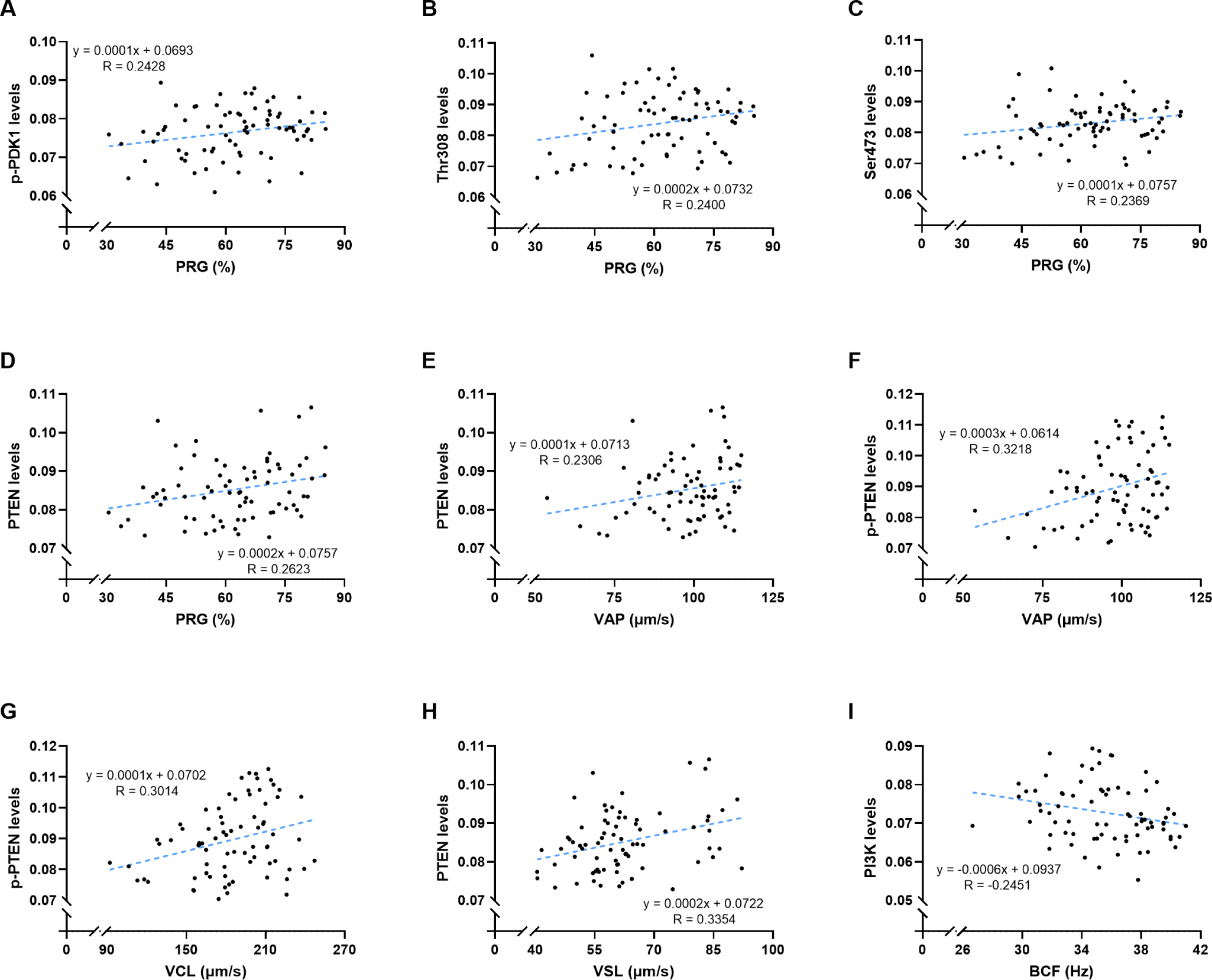
Statistical analyses were conducted using SPSS (version 26.0; IBM). Pearson correlation coefficients were calculated to examine the relationships between sperm motility parameters and the expression levels of PI3K/PDK1/AKT pathway-related proteins. To visualize these correlations, simple linear regression analyses were performed, and corresponding regression equations were presented (Fig. 1). All data are presented as means ± SEM, and statistical significance was defined as p < 0.05.
RESULTS
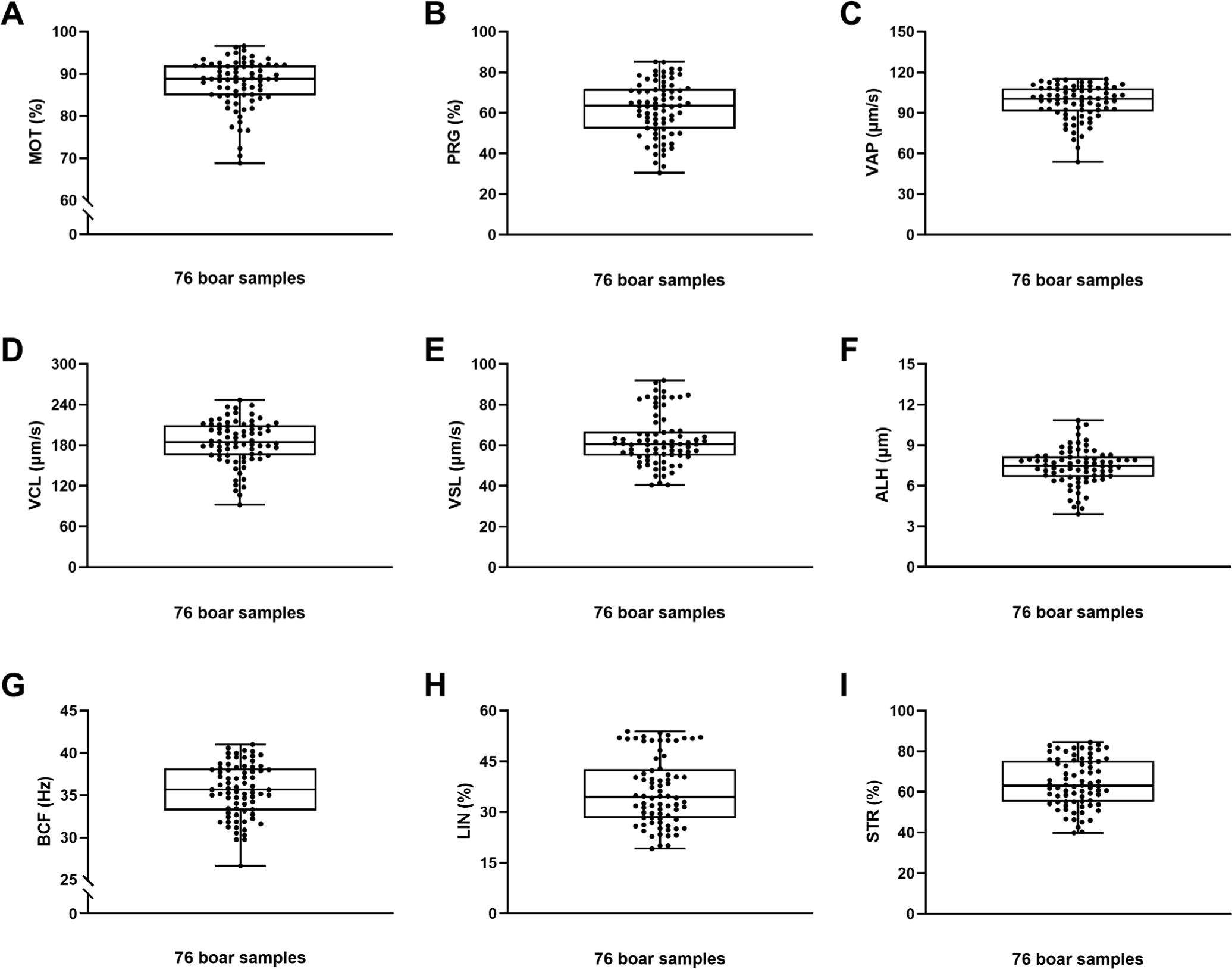
The average values for sperm motility and motion kinematic parameters were as follows: MOT, 87.64 ± 0.66%; PRG, 62.16 ± 1.51%; VAP, 97.74 ± 1.44 μm/s; VCL, 184.26 ± 3.72 μm/s; VSL, 62.59 ± 1.43 μm/s; ALH, 7.43 ± 0.15 μm; BCF, 35.59 ± 0.36 Hz; LIN, 36.21 ± 1.15%; and STR, 64.33 ± 1.35% (Table 1 and Fig. 2).
| MOT | PRG | VAP | VCL | VSL | ALH | BCF | LIN | STR |
|---|---|---|---|---|---|---|---|---|
| 87.6368 ± 0.6599 | 62.1579 ± 1.5143 | 97.7416 ± 1.4330 | 184.2259 ± 3.7191 | 62.5987 ± 1.4321 | 7.4292 ± 0.1542 | 35.5909 ± 0.3601 | 36.2091 ± 1.1491 | 64.3266 ± 1.3530 |
MOT, total sperm motility (%); PRG, progressive sperm motility (%); VAP, average path velocity (μm/s); VCL, curvilinear velocity (μm/s); VSL, straight-line velocity (μm/s); ALH, mean amplitude of head lateral displacement (μm); BCF, beat-cross frequency (HZ); LIN, linearity (%, [VSL/VCL] × 100); STR Straightness (%, [VSL/VAP] × 100).
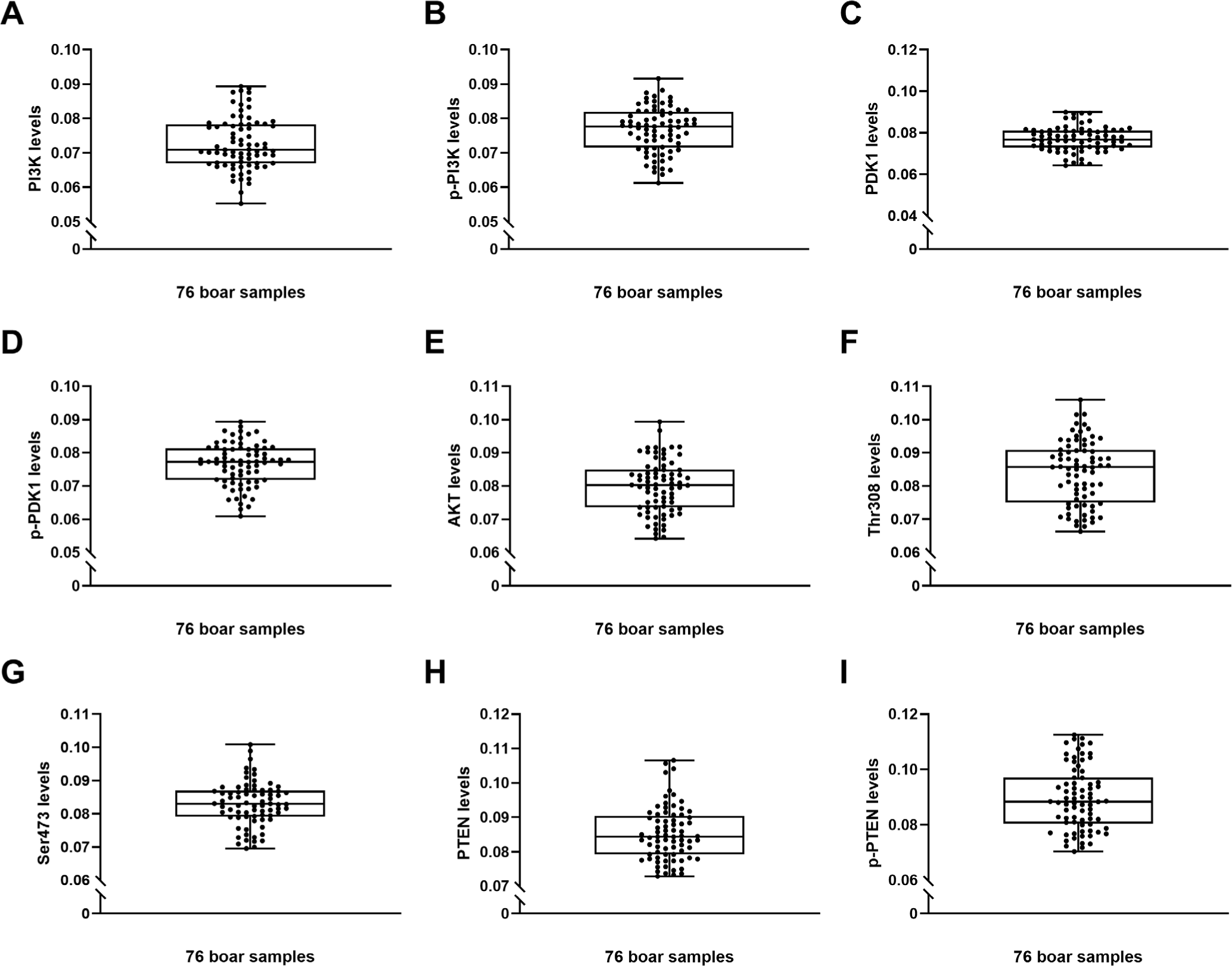
The average expression levels of proteins related to the PI3K/PDK1/AKT signaling pathway were as follows: PI3K, 0.0727 ± 0.0008; phosphorylated PI3K (p-PI3K), 0.0767 ± 0.0007; PDK1, 0.0769 ± 0.0006; phosphorylated PDK1 (p-PDK1), 0.0765 ± 0.0007; AKT, 0.0797 ± 0.0009; AKT phosphorylated at Thr308, 0.0840 ± 0.0001; AKT phosphorylated at Ser473, 0.0830 ± 0.0007; PTEN, 0.0852 ± 0.0008; and phosphorylated PTEN (p-PTEN), 0.0896 ± 0.0012 (Table 2 and Fig. 3).
| PI3K | p-PI3K | PDK1 | p-PDK1 | AKT | Thr308 | Ser473 | PTEN | p-PTEN |
|---|---|---|---|---|---|---|---|---|
| 0.0727 ± 0.0008 | 0.0767 ± 0.0007 | 0.0769 ± 0.0006 | 0.0765 ± 0.0007 | 0.0797 ± 0.0009 | 0.0840 ± 0.0001 | 0.0830 ± 0.0007 | 0.0852 ± 0.0008 | 0.0896 ± 0.0012 |
Significant correlations were observed between PI3K/PDK1/AKT-related protein expression levels and sperm motility parameters. p-PDK1 was positively correlated with PRG (r = 0.243, p < 0.05). Similarly, AKT phosphorylated at Thr308 and Ser473 also showed positive correlations with PRG (r = 0.240, p < 0.05 and r = 0.237, p < 0.05, respectively). PTEN expression was positively correlated with PRG (r = 0.262, p < 0.05), VAP (r = 0.231, p < 0.05), and VSL (r = 0.335, p < 0.01). p-PTEN was positively correlated with both VAP (r = 0.322, p < 0.01) and VCL (r = 0.301, p < 0.01). In contrast, a negative correlation was found between PI3K expression and BCF (r = –0.245, p < 0.05; Table 3 and Fig. 1).
MOT, Total sperm motility (%); PRG, Progressive sperm motility (%); VAP, Average path velocity (μm/s); VCL, Curvilinear velocity (μm/s); VSL, Straight-line velocity (μm/s); ALH, Mean amplitude of head lateral displacement (μm); BCF, Beat-cross frequency (Hz); LIN, linearity (%, [VSL/VCL] × 100); STR, Straightness (%, [VSL/VAP] × 100); PI3K, phosphoinositide 3-kinase; p-PI3K, phospho-PI3K; PDK1, phosphoinositide dependent protein kinase-1; p-PDK1, phospho-PDK1; AKT, protein kinase B; Thr308, phospho-AKT (Thr308); Ser473, phospho-AKT (Ser473); PTEN, phosphatase and tensin homolog; p-PTEN, phospho-PTEN.
DISCUSSION
This study demonstrated significant correlations between PI3K/PDK1/AKT pathway components and specific sperm motility parameters in ejaculated Duroc boar spermatozoa. Our findings revealed both expected and unexcepted relationships, providing new insights into regulation of sperm motility at molecular levels. Notably, PI3K expression exhibited a negative correlation with BCF (r = –0.245, p < 0.05). This finding aligns with previous reports indicating that excessive PI3K activation can impair sperm motility and disrupt energy homeostasis [20,21,27]. The observed negative correlation suggests that elevated PI3K activity may be associated with reduced flagellar beating frequency, possibly reflecting a threshold beyond which PI3K activation becomes detrimental to sperm movement. While PI3K is known to facilitate capacitation by regulating ATP production and flagellar motion via downstream activation of AKT, our findings imply that dysregulation of its activity could influence motility patterns in a dose-dependent manner.
Additionally, PRG was positively correlated with several PI3K/PDK1/AKT pathway components. Specifically, p-PDK1 showed a significant positive correlation with PRG (r = 0.243, p < 0.05). Similarly, positive correlations were observed for phosphorylated AKT at Thr308 (r = 0.240, p < 0.05) and Ser473 (r = 0.237, p < 0.05). The PI3K/PDK1/AKT pathway is a central signaling cascade that regulates cellular processes such as survival, proliferation, and metabolism [10]. Activation of this pathway begins with PI3K, which is located in the sperm plasma membrane and catalyzes the conversion of phosphatidylinositol (4,5)-bisphosphate (PIP2) into phosphatidylinositol (3,4,5)-trisphosphate (PIP3). AKT, a major downstream effector of PI3K, requires dual phosphorylation for full activation: at Thr308 by PDK1, and at Ser473 by other kinases [10,28,29]. Previous studies have shown that inhibition of AKT activity reduces progressive motility in mouse [22] and stallion spermatozoa [23], while its activation enhances motility in human spermatozoa [24]. AKT is known to regulate mitochondrial function, energy metabolism, and cell survival, all of which are intimately linked to flagellar function and sperm motility [10,30]. Therefore, it is reasonable to infer that AKT activation contributes positively to progressive motility in boar spermatozoa.
As PTEN reduces PIP3 levels by dephosphorylating PIP3 back to PIP2, thereby suppressing AKT activation, it is traditionally expected to exert a negative regulatory effect on the PI3K/PDK1/AKT pathway and, consequently, on motility [25]. Interestingly, in our study, PTEN, a known negative regulator of the PI3K/PDK1/AKT pathway, showed a positive correlation with PRG (r = 0.262, p < 0.05). This finding suggests that appropriate levels of PTEN expression may help maintain signaling homeostasis by preventing excessive AKT activation, ultimately promoting PRG. Thus, our results imply that optimal, rather than maximal, AKT signaling may be more beneficial for supporting sperm motility.
Furthermore, PTEN was positively correlated with VSL (r = 0.335, p < 0.01), while p-PTEN showed a positive correlation with VCL (r = 0.301, p < 0.01). Phosphorylation of PTEN at C-terminal residues such as Ser380 is known to stabilize the protein while reducing its enzymatic activity [31]. Thus, increased levels of p-PTEN may reflect reduced PTEN activity, potentially leading to sustained activation of the PI3K/PDK1/AKT signaling pathway. The AKT pathway plays a complex role in regulating sperm motility during capacitation. Activated AKT is associated with hyperactivated motility, which is characterized by vigorous and asymmetrical flagellar beating and results in increased curvilinear velocity [24]. This type of motility is essential for zona pellucida penetration during fertilization [32]. Therefore, the observed positive correlation between p-PTEN and VCL supports the idea that reduced PTEN activity facilitates AKT activation, promoting hyperactivated motility during capacitation.
In contrast, the positive correlation between total PTEN and VSL suggests that higher PTEN levels may help sustain more linear sperm movement by attenuating AKT-mediated hyperactivation and preventing excessive flagellar bending. This more linear motility is advantageous for efficient navigation through the female reproductive tract en route to the oocyte [33]. Taken together, these findings suggest that PTEN may act as a key modulator of sperm motility, helping to balance hyperactivated and linear motility modes depending on the fertilization context. Notably, VAP was positively correlated with both PTEN and p-PTEN. This dual correlation suggests that both the total and phosphorylated forms of PTEN contribute to the regulation of sperm path velocity. It is possible that a finely tuned balance between the active and inactive forms of PTEN, along with other signaling components, orchestrates an optimal motility state suitable for fertilization.
In summary, our findings suggest that the PI3K/PDK1/AKT signaling pathway plays a critical role in male fertility by influencing sperm motility and associated kinematic parameters in Duroc boar spermatozoa. However, several limitations must be considered. First, the analysis was based on correlation, which does not establish direct causality. Second, the correlation coefficients were relatively low, which is generally considered weak correlation. Although the correlations were low, large sample size may have contributed to achieving statistical significance. Third, this study quantified only total and phosphorylated protein levels without directly assessing the enzymatic activity of PI3K/PDK1/AKT pathway components. Therefore, future studies are needed to clarify the functional relevance of these proteins through activity-based assays. Finally, as our findings are specific to boar spermatozoa, species-specific differences should be considered before extrapolating these results to other mammals, including humans. Nonetheless, this study offers valuable insights into the complex relationship between sperm motility and PI3K/PDK1/AKT signaling and provides foundational data to support future research in male reproductive biology.