INTRODUCTION
Weaning is a critical transition in pig production and is characterized by abrupt dietary and environmental changes that may lead to reduced feed intake, impaired growth performance, and intestinal dysfunction [1,2]. During this period, pigs experience substantial stress, immune challenges, postweaning diarrhea, and intestinal inflammation [3,4]. Traditional-feed antibiotics mitigate these challenges; however, increasing concerns over antimicrobial resistance have driven the search for alternative strategies to enhance gut health and growth performance in weanling pigs [5,6]. Among these alternatives, probiotics, synbiotics, bacteriophages, and organic acids have emerged as promising dietary interventions for improving intestinal function and overall health [7].
Probiotics, including Bacillus species, are beneficial microorganisms that modulate the gut microbiota composition, enhance intestinal barrier integrity, and improve nutrient utilization [8–10]. Among these, Bacillus licheniformis has been widely used in swine nutrition owing to its ability to produce various digestive enzymes, inhibit pathogenic bacteria, and maintain a balanced gut microflora [11,12]. Supplementation with B. licheniformis improves nutrient digestibility, enhances intestinal morphology, and upregulates tight junction proteins, thereby promoting gut integrity and reducing intestinal inflammation [8,10–12]. Correspondingly, probiotic supplementation stimulates local immune responses and contributes to the overall health and growth of weanling pigs [13].
Synbiotics are a combination of probiotics and prebiotics that exert synergistic effects by supporting the survival and metabolic activity of beneficial bacteria in the gastrointestinal tract [14,15]. In particular, synbiotic supplementation of B. licheniformis with dietary fiber provides dual benefits: B. licheniformis improves gut microbial balance and nutrient absorption, whereas dietary fiber serves as a fermentable substrate that generates short-chain fatty acids (SCFA). These SCFAs act as energy sources for enterocytes and play key roles in maintaining intestinal barrier integrity [16–18]. In addition to probiotics and synbiotics, organic acids, such as benzoic acid, have been explored as feed additives for improving gut health in pigs. Benzoic acid reduces intestinal pH, thereby creating an unfavorable environment for pathogenic bacteria, while supporting beneficial microbial populations [19–21]. Moreover, organic acids enhance intestinal barrier function by modulating tight junction proteins and reducing inflammatory responses [5,22,23]. Although the individual effects of probiotics, synbiotics, and organic acids have been extensively investigated, research exploring their synergistic interactions—particularly the specific combination of Bacillus licheniformis-based synbiotics and benzoic acid—remains limited. The novelty of this study lies in its comprehensive evaluation of whether acidifying the gastrointestinal environment with benzoic acid can amplify the prebiotic fermentation and probiotic efficacy of a targeted synbiotic in weaned pigs. Furthermore, by simultaneously assessing multifaceted parameters, including physiological stress markers (e.g., hair cortisol), mucosal immunity, tight junction proteins, and microbiome shifts, this study elucidates the complex interactive mechanisms of these combined additives. Therefore, this study aimed to evaluate the effects of dietary supplementation with probiotics, synbiotics, and synbiotics plus benzoic acid on the growth performance, stress response, nutrient digestibility, blood parameters, immune status, gut integrity, and gut morphology of weanling pigs. By assessing these parameters, the study sought to determine whether these specific additive combinations could serve as effective alternatives to in-feed antibiotics and provide a deeper insight into their synergistic mechanisms of action.
MATERIALS AND METHODS
A total of 400 weaned pigs (Landrace × Yorkshire × Duroc) with an average initial body weight (BW) of 6.38 ± 0.13 kg (age: 21 days) were distributed among the following four treatment groups based on BW: a control group fed a basal diet; probiotics (PRO), basal diet supplemented with probiotics (Bacillus licheniformis, 1.0 × 10⁸ colony-forming units [CFU]/g); synbiotics [SYN], basal diet supplemented with synbiotics consisting of B. licheniformis (1.0 × 10⁸ CFU/g) and dietary fiber; and SYN + benzoic acid [SYP], basal diet supplemented with synbiotics consisting of B. licheniformis (1.0 × 10⁸ CFU/g), dietary fiber, and benzoic acid. The synbiotic supplements were experimental products manufactured by WOOGENE B&G and were not commercially available. In the SYN and SYP treatments, dietary fiber and benzoic acid were incorporated at levels equivalent to 2% of the supplement, and all supplements (PRO, SYN, and SYP) were included in the basal diet at a rate of 0.1% (as-fed basis). Consequently, the final inclusion levels of dietary fiber and benzoic acid in the complete diet were 0.002%. Each treatment was replicated across the four pens, with 25 piglets per pen. The pigs were fed pelleted diets (Table 1) in two phases, phase 1 (days 0–14) and phase 2 (days 15–28), formulated according to the National Research Council [24] guidelines. The pigs were housed in metal pens (3 × 4.8 m) with plastic flooring. Environmental conditions were maintained at an initial temperature of 30°C, which was gradually reduced to 25°C from day 8 onward, with humidity controlled between 61%–66%. Each pen was equipped with low-pressure nipple drinkers and self-feeders to ensure ad libitum access to water and food.
1) Supplied per kg of diet: 16,000 IU vitamin A (palmitate), 2.00 mg vitamin B1 (thiamin), 5.00 mg vitamin B2 (riboflavin), 2.00 mg vitamin B6 (pyridoxine), 0.03 mg vitamin B12 (cyanocobalamin), 25.00 mg niacin, 0.40 mg folic acid, 0.05 mg biotin, 5.00 mg ethoxyquin, 2,000 IU vitamin D3 (cholecalciferol), 75.00 mg vitamin E (dl-α-tocopheryl acetate), 2.00 mg vitamin K3 (menadione).
BW was measured at the end of the second and fourth weeks. Growth performance parameters, including average daily gain (ADG), average daily feed intake (ADFI), and gain-to-feed ratio (G:F), were evaluated at the end of each dietary phase, with adjustments made for mortality. Blood samples were collected from four randomly selected pigs per pen at the end of each phase (days 14 and 28), via jugular vein puncture using anticoagulant-free vacutainer tubes (Becton Dickinson). Samples were centrifuged at 3,000×g for 15 min at 4°C and stored at –20°C for subsequent hematological analysis.
Rectal temperature and respiratory rate were measured twice daily (11:30 and 13:00). Rectal temperature was recorded using a digital thermometer (SK-1260, SATO), whereas respiratory rate was determined by counting abdominal movements over 1 min with a stopwatch. The average of these readings was used for the statistical analysis. Hair cortisol concentrations were assessed using the method described by Nejad et al. [25]. Hair samples were collected from the forehead region on days 0 and 28, stored in aluminum foil, and stored in polypropylene tubes (HM Hyundai Micro) for drying at room temperature. To remove contaminants, samples were washed three times with 5 mL of isopropyl alcohol and dried at 23 ± 1°C for seven days. Cortisol extraction was performed using a methanol dilution, and cortisol levels were quantified using an enzyme-linked immunosorbent assay (ELISA) kit (Cayman Chemical) following the manufacturer’s instructions.
To determine the apparent total tract digestibility (ATTD), chromic oxide (0.25%) was included in the diet as an indigestible marker on days 10–14 and 24–28. Fecal samples were collected on the last two days of each phase, pooled within each pen, and dried in a forced-air oven at 60°C for 72 h. The dried samples were ground using a 1-mm screen in a Wiley mill (Thomas Model 4, Thomas Scientific) for further analysis. Analyses were conducted in triplicate following the Association of Official Analytical Collaboration [26] methods: dry matter (DM, method 930.15), crude protein (CP, method 990.03), and ether extract (EE, method 2003.05). The gross energy in the feed and feces was determined using a bomb calorimeter (Model 1261, Parr Instrument). Chromium concentrations were analyzed via spectrophotometry (Jasco V-650, JASCO), following the method described by Fenton and Fenton [27].
Blood samples (10 mL) were collected via jugular vein puncture from four randomly selected pigs per pen on days 14 and 28. Samples were drawn into sodium heparin-containing vacutainer tubes (Becton Dickinson), centrifuged at 3,000×g for 15 min at 4°C, and stored at –20°C for further analysis. Biochemical markers, including aspartate transaminase (AST; Kit #08104719190) and alanine transaminase (ALT; Kit #08104697190), creatinine (Kit # 08057532190), and uric acid (Kit # 08058750190) were measured using spectrophotometric kits (TBA-120FR, Toshiba Medical Systems) and a Cobas 6000 c501 automatic biochemistry analyzer (Roche Diagnostics). Hematological parameters, including white blood cell (WBC) count, red blood cell count, neutrophil count, and lymphocyte count, were assessed using the Hemavet® Hematology System (CDC Technologies, Drew Scientific). The cytokines IL-1β (Cat #MBS700738, MyBioSource), TNF-α (Cat #MBS2019932, MyBioSource), and IL-10 were quantified using ELISA kits (Invitrogen, Thermo Fisher Scientific). For intestinal barrier integrity analysis, jejunal tissue samples (approximately 2 cm) were collected from the mid-jejunum immediately after euthanasia, rinsed gently with cold phosphate-buffered saline (PBS), and stored at –80°C until analysis. The frozen tissues were homogenized in ice-cold PBS (1:9, w/v) and centrifuged at 10,000×g for 10 min at 4°C. The supernatant was collected, and the concentrations of tight junction proteins, ZO-1 (Cat #MBS9310967, MyBioSource) and occludin (Cat #MBS740246, MyBioSource), were determined using ELISA kits (Invitrogen, Thermo Fisher Scientific). The results were normalized to the total protein concentration, which was measured using the Bradford assay, and expressed as ng/mg total protein.
Intestinal tissue samples were collected from the duodenum, jejunum, and ileum of the pigs. A portion of each sample was frozen in liquid nitrogen and stored at –80°C for future analysis, while the remaining sections (5 cm) were fixed in 10% neutral-buffered formalin for 24 h and transferred to a 70% ethanol solution for preservation. The samples were embedded in paraffin, sectioned, and stained with hematoxylin and eosin. Microscopic analysis was performed using a Vanox-S Microscope (Olympus), and image analysis was conducted using SPOT simple imaging software (SPOT Imaging). Villus height (VH) and crypt depth (CD) were measured in five representative villi and crypts per section.
Fecal samples were collected from representative pigs in each treatment group on days 0 and 28 for microbiota analysis. Metagenomic DNA was extracted from the samples, and the V3–V4 region of the 16S rRNA gene was amplified and sequenced using a next-generation sequencing platform. Sequence data quality control was performed using the DADA2 (v1.18.0) package in R (v4.0.3), with trimming of forward (250 bp) and reverse (200 bp) reads, removal of chimeric sequences, and exclusion of sequences with expected errors ≥2 or a quality score <25. Multiple sequence alignments were performed using MAFFT (v7.475). Alpha diversity (Shannon, Chao1, and Simpson indices) and beta diversity (Bray–Curtis dissimilarity) were calculated using R (v4.4.1). Group differences in diversity and relative abundance at the phylum and genus levels were assessed using t-tests and analysis of variance, whereas differentially abundant taxa were identified using linear discriminant analysis effect size (LEfSe), with a linear discriminant analysis (LDA) score threshold of 3.0. Core and unique microbiome analyses were conducted to identify the shared and group-specific taxa.
Statistical analysis of the experimental data was performed using the General Linear Model procedure in SAS (SAS Institute) using a completely randomized design. The pen was the experimental unit for growth performance and nutrient digestibility, whereas the individual pig was considered an experimental unit for stress indicators, blood parameters, and intestinal morphology. The treatment means were separated using Tukey’s multiple range test with p < 0.05 indicating statistical significance.
RESULTS
The effects of dietary probiotic and synbiotic supplementation on the growth performance of weanling pigs are presented in Table 2. No substantial differences were observed in the initial or final BW among the treatment groups. During phase 1, ADG was markedly higher in the SYN and SYP groups than in the control group. No considerable differences were observed in ADFI and G:F among the treatments. In phase 2, ADG and G:F were markedly higher in the SYN and SYP groups than in the control group, whereas ADFI remained unaffected. Overall, ADG and G:F were substantially higher in the SYN and SYP groups than in the control group, whereas ADFI showed no notable differences among the treatments.
Physiological stress indicators, including respiratory rate, rectal temperature, and hair cortisol concentration, are summarized in Table 3. No notable differences were observed among the treatment groups in phase 1. Similarly, in phase 2, no considerable differences in the respiratory rate, rectal temperature, or hair cortisol levels were observed among the treatments.
The effects of the dietary treatments on ATTD are shown in Table 4. In phase 1, no notable differences were observed in the ATTD of GE, CP, or EE among the treatments. However, the ATTD of the DM tended to be higher in the SYP group than in the control group. In phase 2, the ATTD of the GE, CP, and EE remained unaffected across treatments. However, the ATTD of the DM in the SYP group was markedly higher than that in the control group.
The blood parameters are summarized in Table 5. In phase 1, no pronounced differences were observed in terms of WBC and red blood cell count, neutrophil and lymphocyte percentage, and AST, ALT, creatinine, or uric acid levels among the treatments. In phase 2, no notable differences were observed in these blood parameters between the treatment groups.
The effects of dietary treatments on immune status and gut integrity are presented in Table 6. In phase 1, no pronounced differences were observed in the blood IL-10 and TNF-α concentrations among the treatment groups. Moreover, IL-1β concentrations tended to decrease in the SYP group compared with that in the control group. In phase 2, IL-10 and TNF-α concentrations showed no substantial changes among treatments, whereas IL-1β concentrations tended to decrease in the SYP group. Intestinal ZO-1 concentration was markedly higher in the SYP group than in the control group (Table 7), whereas no noteworthy differences were observed in the occludin levels across treatments.
| Item | Control | PRO1) | SYN2) | SYP3) | SEM | p-value4) |
|---|---|---|---|---|---|---|
| Jejunum | ||||||
| ZO-1 (ng/mg protein) | 10.320b | 13.890ab | 15.410a | 15.900a | 1.830 | 0.022 |
| Occludin (ng/mg protein) | 3.510 | 3.900 | 3.630 | 3.720 | 0.440 | 0.743 |
The gut morphology measurements are summarized in Table 8. In the duodenum, VH was substantially higher in the SYN and SYP groups than in the control group, whereas CD remained unchanged. Jejunal VH was markedly greater in the SYN and SYP groups than in the control group, with no pronounced difference in CD. In the ileum, VH and CD showed no considerable differences among the treatments. However, the VH-to-CD ratio (VH/CD) tended to be higher in the PRO, SYN, and SYP groups than in the control group.
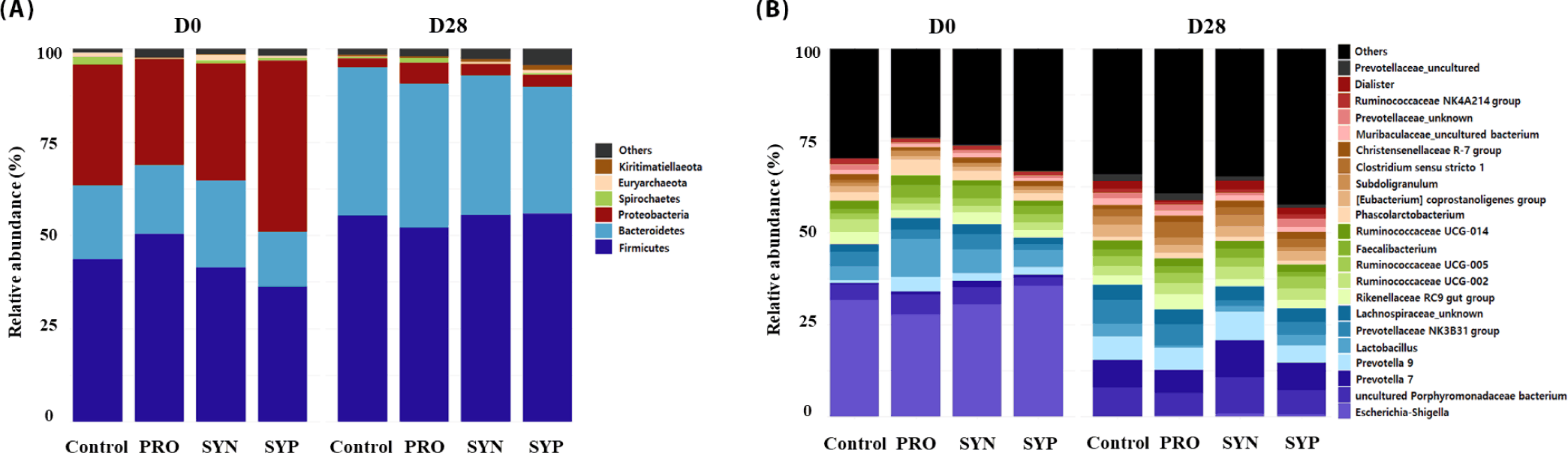
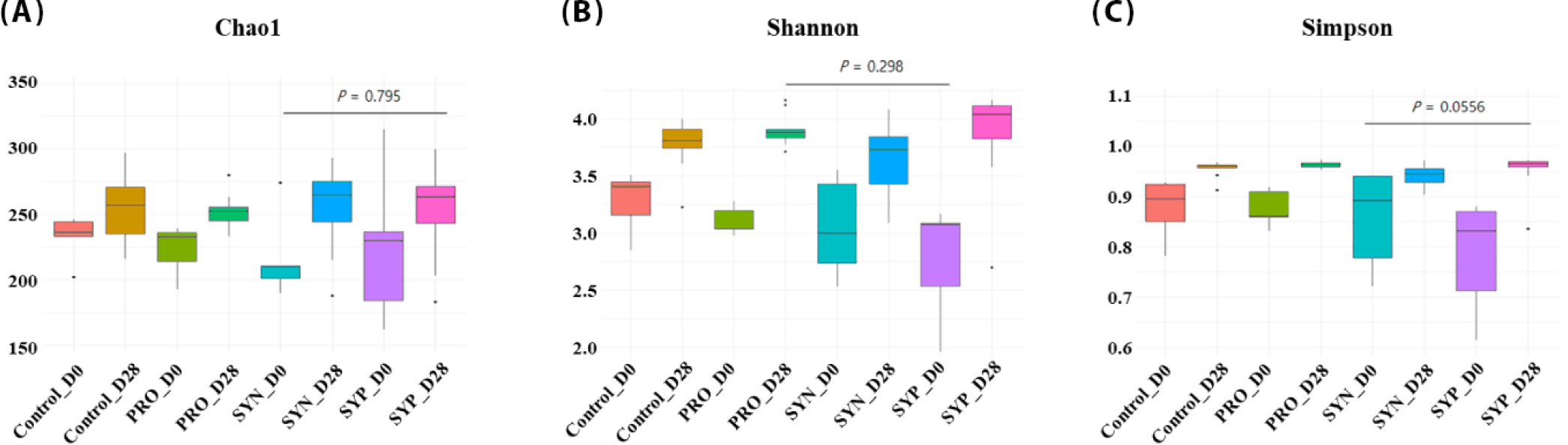
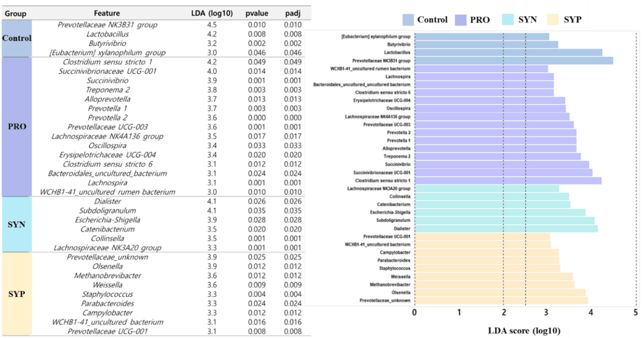
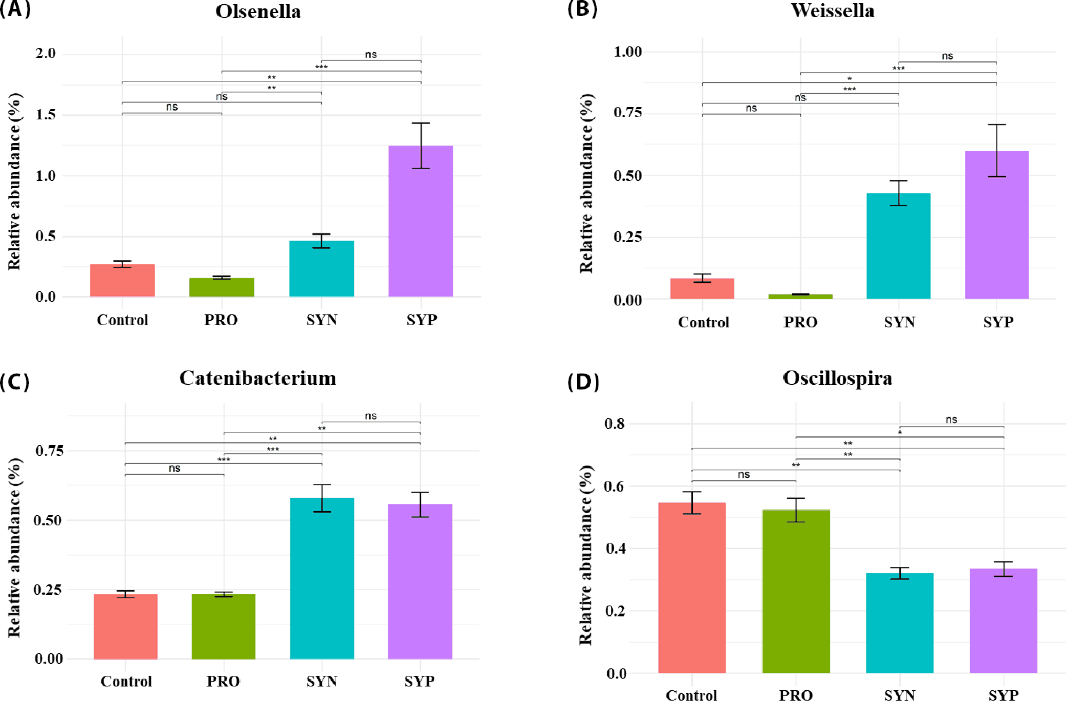
When the microbiome composition of the treatment groups (PRO, SYN, and SYP) was compared with that of the control at the phylum level (Fig. 1A), feeding trials led to a pronounced reduction in Bacteroidetes (–1.3% to –5.7%) across all experimental groups. In contrast, SYP significantly increased the abundance of Firmicutes (55.7%) and Proteobacteria (3.2%; p < 0.01), whereas the abundance of Firmicutes in the PRO group (52.2%) was lower than that in the control group (55.4%). Spirochaete abundance also decreased significantly in the SYN group (0.16%) compared with that in the control group (0.42%; p < 0.01). Subsequently, at the genus level (Fig. 1B), Ruminococcaceae UCG-002 and Christensenellaceae R-7 were consistently enriched in all treatment groups, whereas Prevotellaceae NK3B31, Ruminococcaceae UCG-014, [Eubacterium] coprostanoligenes, and uncultured Muribaculaceae were significantly reduced (p < 0.05). In particular, SYN increased the relative abundance of uncultured Porphyromonadaceae bacterium (9.9%), Prevotella 9 (7.8%), Ruminococcaceae UCG-002 (3.4%), Faecalibacterium (2.6%), Christensenellaceae R-7 (1.8%), and Subdoligranulum (3.2%), while decreasing Prevotellaceae NK3B31 group (1.5%), Ruminococcaceae UCG-014 (2.0%), [Eubacterium] coprostanoligenes group (2.7%), Muribaculaceae uncultured bacterium (1.4%), and uncultured Prevotellaceae (1.1%). Similarly, SYP significantly increased Ruminococcaceae UCG-005 (3.4%) and Christensenellaceae R-7 (1.9%) and decreased Prevotellaceae NK3B31 (3.5%), Ruminococcaceae UCG-014 (1.9%), Coprostanoligenes (2.6%), and uncultured Muribaculaceae (1.4%; p < 0.05). Alpha diversity analysis showed that microbial diversity tended to increase from day 0 to day 28 across all treatment groups, thereby indicating gradual maturation of the gut microbiota after weaning. However, no pronounced differences were observed between treatments at either time point (Fig. 2). In addition, the LEfSe analysis (LDA score > 3.0) identified 34 distinct discriminant taxa in each group (Fig. 3). Subsequently, genera showing significant differences from the control group were selected among the identified 34 taxa. Consequently, Olsenella and Weissella had a significantly higher relative abundance in the SYP group (Fig. 4A and 4B) and Catenibacterium had a significantly higher relative abundance in both the SYN and SYP groups than in the control group (Fig. 4C; p < 0.05). Oscillospira had a significantly lower relative abundance in both SYN and SYP groups than in the control group (Fig. 4D; p < 0.05). However, the relative abundances between the SYN and SYP groups showed no considerable differences in any of the selected taxa, including Olsenella, Weissella, Catenibacterium, and Oscillospira (Fig. 4).
DISCUSSION
The improvements in ADG and G:F observed in the SYN and SYP groups suggested that the inclusion of synbiotics had a beneficial effect on the growth performance of weanling pigs. The lack of notable differences in ADFI among treatments indicated that the improved growth efficiency in the SYN and SYP groups was likely attributable to enhanced nutrient utilization rather than increased feed intake.
It should be noted that the inclusion level of benzoic acid used in the present study (0.002%) was substantially lower than the dosages commonly reported to exert direct antimicrobial or acidifying effects in previous studies (typically 0.5–1.0%) [19,20]. At such a micro-dose level, the standalone direct bactericidal or strong acidifying properties of benzoic acid are likely limited. Therefore, the pronounced improvements observed in the SYP group cannot be attributed solely to the classical mechanisms of benzoic acid. Instead, we hypothesize that these improvements reflect synergistic interactions among Bacillus licheniformis, dietary fiber, and benzoic acid. Even at low concentrations, benzoic acid may act as a mild modulator that complements the probiotic and prebiotic components, collectively optimizing gut microbial ecology and reinforcing intestinal barrier function [18,22]. These findings align with those of previous studies, indicating that synbiotic supplementation positively influences growth performance in weanling pigs [13,14].
The beneficial effects of this combined approach can be attributed to several integrated mechanisms. Specifically, the probiotic Bacillus licheniformis produces extracellular enzymes, such as proteases and amylases, which enhance nutrient digestion and absorption [8,11]. Simultaneously, the fermentation of prebiotic dietary fiber by the gut microbiota generates short-chain fatty acids (SCFAs, such as butyrate, propionate, and acetate), which serve as crucial energy sources for enterocytes, improve intestinal barrier integrity, and reduce inflammation [16]. Furthermore, the micro-dose addition of benzoic acid likely complements this environment by subtly modulating intestinal pH to inhibit pathogenic bacteria (e.g., Escherichia coli) while favoring commensals like Lactobacillus and Bifidobacterium, thereby reducing metabolic energy losses associated with immune activation [5,9,19,28]. Interestingly, although the PRO group did not show notable improvements in growth performance compared with that in the control group, the SYN and SYP groups demonstrated clear advancements. This suggests that the synergistic integration of these three components is necessary to achieve substantial improvements in growth efficiency.
Microbiota analysis revealed that both synbiotic groups (SYN and SYP) exhibited similar shifts, characterized by increased abundances of Weissella, Catenibacterium, and Olsenella, and a reduction in Oscillospira compared with that in the control group. Weissella produces lactic acid and bacteriocins that contribute to microbial homeostasis and pathogen inhibition [29], whereas Olsenella and Catenibacterium participate in SCFA synthesis, thereby enhancing intestinal metabolic stability [22]. Conversely, Oscillospira has been associated with reduced feed efficiency and slower growth [30]. Thus, synbiotic supplementation contributes to a more stable and metabolically balanced post-weaning gut ecosystem. In particular, the increased abundance of Firmicutes and SCFA-producing families, including Ruminococcaceae and Christensenellaceae, in the SYN and SYP groups was consistent with improved fiber fermentation and energy harvesting, contributing directly to the enhanced feed efficiency observed.
The observed improvement in DM digestibility in the SYP group further highlights the enhanced nutrient utilization efficiency driven by these combined additives. As established, the extracellular enzymes produced by B. licheniformis facilitate the breakdown of complex dietary components [10,12]. This enzymatic action, coupled with the improved microbial balance and favorable hindgut environment, leads to superior absorption and increased feed efficiency without the need for additional feed intake [13,16].
Regarding immune status, the tendency for decreased pro-inflammatory IL-1β concentrations in the SYP group suggests anti-inflammatory benefits during post-weaning stress [31,32]. Probiotics like B. licheniformis are known to modulate immune function by promoting regulatory T cell activity and enhancing anti-inflammatory cytokines like IL-10 [8,10]. Furthermore, the enrichment of SCFA-producing taxa in these groups actively downregulates inflammatory responses via increased butyrate availability [16,18]. This enhanced microbial ecosystem also positively influenced gut barrier function, as evidenced by the increased blood ZO-1 concentrations in the SYP group [6,33]. The structural integrity of tight junction proteins is likely supported by the combined stabilization of gut permeability mediated by both SCFA production and organic acid modulation [21,23]. The absence of differences in occludin concentration suggests that the structural core of tight junction proteins remains stable.
The improved intestinal absorptive capacity, indicated by the increased VH in the duodenum and jejunum of the SYN and SYP groups, provides morphological evidence of enhanced gut health [7]. The accelerated enterocyte proliferation and villus development are direct functional outputs of the synergistic mechanisms mentioned earlier, particularly the increased SCFA production in the hindgut which fuels enterocyte differentiation [2,11,34]. The absence of pronounced changes in CD across treatments suggests that cell turnover rates remained stable. The trend toward an increased VH/CD ratio in the ileum in the PRO, SYN, and SYP groups further supports the notion that dietary supplementation contributes to improved gut morphology.
In the SYP group, the pronounced shift toward a community enriched in fiber-fermenting, SCFA-producing bacteria (e.g., UCG-002, UCG-005, and Christensenellaceae R-7) directly supports the observed morphological and immunological improvements, as these taxa degrade complex polysaccharides and strengthen tight junction integrity [1,4,17,18]. The concurrent decline in taxa associated with proinflammatory responses and inefficient energy utilization (e.g., Prevotellaceae NK3B31, Ruminococcaceae UCG-014, and [Eubacterium] coprostanoligenes) further validates the metabolically efficient nature of this microbial shift [3,35,36]. In addition, the detection of unique taxa, such as Methanobrevibacter, suggests improved fermentation efficiency via hydrogen removal, while the enrichment of lactic acid bacteria (Weissella, Olsenella, and Parabacteroides) enhances colonization resistance against pathogens [15,22]. Collectively, these compositional shifts confirm that the specific combination of Bacillus licheniformis, prebiotic fiber, and benzoic acid successfully creates a stable, metabolically efficient, and anti-inflammatory gut ecosystem [8,11,12,14,20,21,23].
CONCLUSION
This study demonstrates that dietary supplementation with synbiotics and benzoic acid improves the growth, nutrient digestibility, intestinal morphology, and immune status of weanling pigs. Although probiotics alone did not confer considerable benefits, the synergistic effects of Bacillus licheniformis with dietary fiber (SYN) enhanced growth efficiency and villus development, whereas the addition of benzoic acid (SYP) further improved DM digestibility and modulated the gut microbiota toward a more beneficial SCFA-producing profile. These findings suggest that combining synbiotics with organic acids provides a multifaceted strategy for enhancing gut health and productivity in piglets, thereby offering a viable alternative to in-feed antibiotics. Accordingly, future studies should investigate the long-term effects of these additives on lifetime productivity, microbiota stability, and resilience to pathogenic challenges.