INTRODUCTION
Weaning is an important part of the pig lifecycle. During the weaning period, piglets encounter various environmental challenges such as separation from the sow, changes in diet, culminating in changes in the intestinal environment [1]. This may induce diarrhea, which results in reduced growth rate and mortality in piglets [2]. Reports from large-scale pig production systems indicate that post-weaning diarrhea can affect up to 50% of piglets, with mortality rates increasing to 15%–20%, underscoring the considerable impact of weaning-related gastrointestinal challenges on herd health and farm profitability [3]. To address these challenges, several studies have been conducted to alleviate weaning stress, reduce diarrhea, and enhance pig growth. Previous studies have shown that probiotic feed additives can improve nutrient utilization, feed efficiency, and growth-related performance in pigs by supporting intestinal function [4]. In particular, Lactobacillus plantarum-based feed additives may support postweaning productivity by improving intestinal morphology and nutrient absorption [5], as well as resilience against diarrhea-associated growth retardation through pathogen suppression and host defense modulation [6]. These effects are closely associated with intestinal environmental changes, including increased villus height, modulation of gut microbiota, suppression of opportunistic pathogens, and stimulation of local immune responses [5,6].
Antibiotics have traditionally been used to control diseases in livestock. However, their continuous application disrupts the gut microbiota and accelerates the spread of antimicrobial resistance, raising significant public health concerns [7,8]. To address these challenges, probiotics have emerged as promising alternatives to promote swine health and performance. Probiotics, including L. plantarum, help maintain balanced intestinal microbiota, suppress opportunistic pathogen growth, and stimulate local immune responses [4,6]. Recent studies have reported that L. plantarum supplementation is associated with increased villus height in the ileum, jejunum, and duodenum, thereby enhancing nutrient absorption and strengthening the gut barrier against pathogens [5]. Collectively, probiotics contribute to the stabilization of intestinal function and resilience, ultimately supporting animal growth and productivity.
Previous studies have investigated the effects of L. plantarum on weaned piglets; however, most have relied on a single-omics approach [5,9]. Although studies provide valuable insights, the complexity and interconnectedness of biological systems in animals make it difficult to fully capture host responses through single-omics analysis alone. To overcome these limitations, this study employed an integrated multi-omics approach, combining transcriptomics, metabolomics, and microbiomics data. This systematic integration enables a more comprehensive characterization of the interplay between gene expression patterns, metabolic alterations, and microbial community dynamics, thereby offering deeper insights into the mechanisms by which L. plantarum influences piglet development and health.
The ileum, which is the terminal segment of the small intestine, is structurally characterized by abundant villi and microvilli that expand its absorptive surface. In addition to its role in nutrient absorption, the ileum contains Peyer’s patches—specialized lymphoid structures—that function as key sites for initiating mucosal immune responses [10]. Although the ileum does not directly secrete digestive enzymes, it depends on enzymes from the jejunum for complete digestion and nutrient uptake. Given its dual roles in nutrient assimilation and mucosal immunity, the ileum is regarded as a critical region for intestinal health evaluation. Previous studies have shown that probiotics, particularly L. plantarum, can influence ileal physiology by enhancing the villus architecture and modulating immune activity, thereby contributing to intestinal barrier integrity and overall gut function [5].
In this study, we aimed to elucidate the biological mechanisms underlying host–microbiome interactions mediated by intestinal metabolites in weaned piglets and identify key molecular pathways and microbial signatures that contribute to gut health and development during the critical weaning transition. Using an integrated multi-omics framework, specifically one that combines transcriptomics, metabolomics, and microbiological data, this study provides a comprehensive framework for understanding how L. plantarum shapes the intestinal ecosystems of weaned piglets. These insights may inform the development of targeted nutritional or microbial interventions to promote intestinal integrity, enhance immune function, and improve growth performance in swine production.
METHODS
Six piglets ([Landrace × Yorkshire] × Duroc) were weaned at 28 days of age and used in this study. The animals were randomly assigned to one of two treatment groups (n = 3 per group): a control group fed a standard corn–soybean meal-based diet, and a treatment group receiving the same basal diet supplemented with 0.02% L. plantarum IDCC 3501 (CON + L. plantarum), providing 2 × 10⁹ CFU/g of probiotic. Each pig was housed individually in a pen (232 × 175 cm) equipped with a slatted plastic floor, feeder, and ad libitum access to water. The ambient temperature was maintained at 28°C–30°C, and relative humidity was controlled at 55 ± 5%, under a 12-h light/dark cycle. Diets were formulated to meet or exceed the nutritional requirements of the weaning pigs [11], and the ingredient and calculated nutrient compositions of the basal diet are provided in Table 1. After a 2-week feeding period, ileal tissue and fecal samples were collected from three pigs per group. Ileal tissues were used for transcriptomic analysis via RNA sequencing, and fecal samples were subjected to 16S rRNA gene sequencing for microbiota profiling and untargeted metabolomic analysis using gas chromatography-mass spectrometry (GC-MS).
1) The vitamin–mineral premix supplied the following amounts per kilogram of diet: vitamin A, 12,000 IU; vitamin D3, 2,500 IU; vitamin E, 30 IU; vitamin K3, 3 mg; D-pantothenic acid, 15 mg; nicotinic acid, 40 mg; choline, 400 mg; vitamin B12, 12 μg; iron, 90 mg as iron sulfate; copper, 8.8 mg as copper sulfate; zinc, 100 mg as zinc oxide; manganese, 54 mg as manganese oxide; iodine, 0.35 mg as potassium iodide; and selenium, 0.30 mg as sodium selenite.
After the 2-week feeding period (day 14 post weaning), fecal samples were collected from all pigs (n = 3 per group) and immediately stored at –80°C for downstream microbiome and metabolomic analyses. Following sample collection, the pigs were euthanized and ileal tissue samples were obtained for transcriptomic analysis. Approximately 3 cm segments of the ileum were excised, gently rinsed with sterile saline, and immersed in RNAlater® solution (QIAGEN GmbH) at room temperature for 24 h. The stabilized samples were then stored at –80°C until RNA extraction.
To characterize the fecal microbiota, the V3–V4 regions of the 16S rRNA gene were amplified using a standard Illumina 16S Metagenomic Sequencing Library Preparation protocol. Genomic DNA (2 ng per sample) was subjected to PCR in a reaction mixture containing 5 × reaction buffer, 1 mM dNTPs, 500 nM of each universal primer, and Herculase II Fusion DNA Polymerase (Agilent Technologies). The thermal cycling conditions were as follows: initial denaturation at 95°C for 3 min; 25 cycles of 95°C for 30 s, 55°C for 30 s, and 72°C for 30 s; and a final extension at 72°C for 5 min. The primer pair used for the amplification included Illumina adapter overhang sequences: V3-F (5’-GTCGGCAGCGTCAGATGTGTATAAGAGACAGCCTACGGGNGGCWGCAG-3’) and V4-R (5’-GTCTCGTGGGCTCGGAGATGTGTATAAGAGACAGGACTACHVGGG TATCTAATCC-3’). The initial amplicons were purified with AMPure beads (Agencourt Bioscience). Two microliters of the purified product was then re-amplified with Nextera XT index primers to incorporate dual indices and Illumina adapters. This indexing PCR was performed under the same thermal profile as the first reaction, but for only 10 cycles. The indexed products were purified again using AMPure beads, after which the library concentration was determined by quantitative PCR using the KAPA Library Quantification Kit for Illumina. Library size distribution and integrity were verified using the TapeStation system with a D1000 ScreenTape (Agilent Technologies). Equimolar pooled libraries were sequenced using an Illumina MiSeq instrument (2 × 300 bp paired-end reads) at Macrogen.
Cutadapt v3.7 [12] was used to trim adapter sequences from raw 16S rRNA gene reads. All sequences were processed using QIIME2 version 2022.8. To correct sequencing errors and remove noise, amplicon sequence variants (ASVs) were inferred using the DADA2 algorithm [13]. The ASVs were taxonomically assigned by aligning them to the SILVA v138_99 reference database with 99% similarity. The V3–V4 hypervariable regions of the 16S rRNA gene were annotated using a Naïve Bayes classifier trained with the q2-feature-classifier plugin.
To assess within-sample microbial diversity, alpha diversity was estimated using multiple indices, including the ACE, Chao1, Shannon, and Simpson indices, which provide complementary measures of species richness and evenness. These metrics were calculated using the QIIME2 core metrics phylogenetic pipeline, and statistical comparisons between groups were performed using the alpha group significance plugin.
Taxonomic classification was further refined by assigning ASVs to known taxonomic groups based on sequence similarity in the SILVA database, enabling detailed characterization of the microbial composition across treatment conditions.
Total RNA was isolated from ileal tissue using TRIzol reagent (Invitrogen, Life Technologies) following the manufacturer’s protocol [14]. The concentration and purity of the RNA were assessed using a NanoDrop ND-1000 spectrophotometer (NanoDrop Technologies, Thermo Fisher Scientific). Library preparation was performed using 1 μg of total RNA with the Illumina TruSeq™ RNA Sample Preparation Kit. Paired-end sequencing (2 × 100 bp) was performed using an Illumina HiSeq 2000 platform (Illumina). Detailed sequencing quality metrics for each sample are provided in Table 2.
Sample ID: Sample name, Total bases (bp), total number of bases sequenced; Total reads, total number of reads (for illumina paired-end sequencing, this value refers to the sum of read1 and read2); GC (%), ratio of GC content; AT (%), ratio of AT content; Q20 (%), ratio of bases that have phred quality score of over 20; Q30 (%), ratio of bases that have phred quality score of over 30.
During library construction, messenger RNA was fragmented into short segments, which were then reverse-transcribed into first-strand cDNA using random primers and reverse transcriptase. Second-strand synthesis was performed using DNA Polymerase I and RNase H, followed by end repair and adapter ligation.
Quality control of raw reads was performed using FastQC v0.12.1 [15], and low-quality bases and adapter sequences were removed with Trimmomatic v0.39 [16], using the parameters SLIDINGWINDOW:4:15 and MINLEN:36. The cleaned reads were re-evaluated for quality using FastQC and aligned to the pig reference genome (Sus_scrofa.Sscrofa11.1.112) obtained from the Ensembl genome browser using HISAT2 v2.2.1, with the default settings [17].
Aligned reads were sorted and converted into BAM format using Samtools v1.19.2 [18]. Gene-level read counts were obtained using the featureCounts function in the Subread package [19], based on exon annotations from the Ensembl GTF file version 112.
Differential gene expression analysis was conducted using the edgeR package v3.40.2 in the bioconductor environment [20]. Genes with fewer than 10 raw counts across all samples were excluded to ensure statistical robustness. Count data were normalized using the trimmed mean of M-values method [20]. Multidimensional scaling analysis was performed using the limma package [21], and visualization was performed using the ggplot2 package [22] to assess sample clustering.
Differentially expressed genes (DEGs) were identified based on a false discovery rate (FDR) threshold of 0.05, adjusted using the Benjamini–Hochberg method, and a minimum absolute Log₂ fold change of 1.
To investigate the biological significance of DEGs, functional enrichment analysis was performed using Gene Ontology (GO) biological processes [23] and Kyoto Encyclopedia of Genes and Genomes (KEGG) pathway annotations [24]. Functional annotation and enrichment were performed using the Database for Annotation, Visualization, and Integrated Discovery (DAVID) v2022 [25].
GO terms were filtered using the “DIRECT” annotation option to ensure specificity, and enrichment analysis was conducted with the following thresholds: p-value < 1 and gene count ≥ 2 for initial filtering, followed by p-value < 0.1 and count ≥ 2 for significance. KEGG pathway enrichment analyses were performed using the same criteria. Enrichment results were reported as –Log₁₀ (p-value) and fold enrichment values for each pathway, with significance assessed directly from the enrichment test p-values rather than corrected q-values.
To improve interpretability, the enriched GO terms were grouped based on their semantic similarity using the REVIGO tool [26]. Tree map visualizations were generated to highlight the representative GO terms from each functional group, emphasizing the most statistically significant terms in each cluster.
For untargeted metabolomic profiling, fecal samples were extracted using ice-cold 100% methanol at a ratio of 0.5 mL per 100 µL of sample volume. The extraction procedure consisted of three cycles of 1-min vigorous vortexing, interspersed at 1-min intervals on ice to enhance metabolite solubilization. Following extraction, samples were centrifuged at 16,000×g for 5 min at 4°C, and the resulting supernatants were filtered using 0.2 µm polyvinylidene fluoride syringe filters. A 400 µL aliquot of the filtered extract was dried using a speed vacuum concentrator and stored at –80°C until analysis.
Prior to GC-MS analysis, the dried metabolites were chemically derivatized under a nitrogen atmosphere using N,O-bis(trimethylsilyl)trifluoroacetamide to improve their volatility and thermal stability. Chromatographic separation was performed on a fused silica capillary column coated with 5% diphenyl/95% dimethylpolysiloxane (20 m × 0.18 mm ID, 0.18 µm film thickness), with helium used as the carrier gas. The oven temperature was programmed to increase from 60°C to 340°C, over a 17.5-min period.
Each sample was spiked with nine internal standards (250 ng each) for normalization and quality control: amylbenzene, 1-phenylhexane, 1-phenyloctane, 1-phenyldecane, 1-phenyldodecane, hexadecylbenzene, octadecylbenzene, tetradecylbenzene, and 2,6-di-tert-butyl-4-methylphenol. Mass spectrometric detection was conducted using a Thermo-Finnigan Trace DSQ fast-scanning single-quadrupole system equipped with electron impact (EI) ionization and operated in full-scan mode over a mass range of 50–750 m/z. Metabolites were annotated by comparing EI mass spectra and retention information against the NIST mass spectral library.
To evaluate the biological significance of the metabolomic alterations, pathway enrichment analysis was performed using MetaboAnalyst 6.0 [27]. A list of annotated metabolites obtained from the GC-MS profiling was uploaded to the Enrichment Analysis module, and the compounds were mapped to KEGG identifiers using the default metabolite mapping function. Ambiguous or unmatched entries were excluded from analysis.
Overrepresentation analysis was performed using the KEGG metabolic pathway library as a reference. Statistical significance was assessed using a hypergeometric test, and pathways were ranked according to their enrichment ratios and corresponding nominal p-values. For visualization, the top 25 enriched metabolic pathways were selected and summarized in a bubble plot, in which the x-axis indicates –Log₁₀ (p-value), bubble size represents the enrichment ratio, and bubble color reflects the degree of significance.
Microbiome and metabolomics data were integrated using Model-based Integration of Metabolite Observations and Species Abundances 2 (MIMOSA2; http://elbo–spice.cs.tau.ac.il/shiny/MIMOSA2shiny/). This framework enables the quantification of community-level metabolic potential (CMP) scores by modeling metabolite turnover within microbial communities based on species-level metabolic capabilities annotated in the KEGG database. MIMOSA2 supports paired microbiome–metabolome datasets and facilitates mechanistic interpretations by estimating the contribution of each taxon to metabolite production or consumption [28]. The resulting scatterplots include trend lines indicating that the directionality of metabolite concentration changes with positive slopes for increases and negative slopes for decreases, along with corresponding R² values that represent the degree of association between CMP and metabolite levels. Contribution bar charts illustrate the proportional impact of individual taxa on metabolite variance, with positive and negative values indicating their potential roles in metabolite accumulation and depletion, respectively (p < 0.1).
To further explore host–microbiome metabolic interactions, a joint pathway analysis was performed using MetaboAnalyst 6.0 [27]. This analysis integrated the DEG-abundant metabolites identified by MIMOSA2 with the host DEGs to uncover convergently modulated metabolic pathways. Pathway impact scores were computed to evaluate the relative significance of each pathway within the network, considering the pathway topology and metabolite–gene associations.
RESULTS
RNA sequencing of the ileal tissue generated 233,587,372 raw reads with an average GC content of 44.75%. After quality trimming, 230,054,530 clean reads were retained, exhibiting a slightly reduced GC content (44.58%). Alignment to the reference Sus scrofa genome resulted in an overall alignment rate of 84.8%, with 74.19% of the reads uniquely mapped (Table 3).
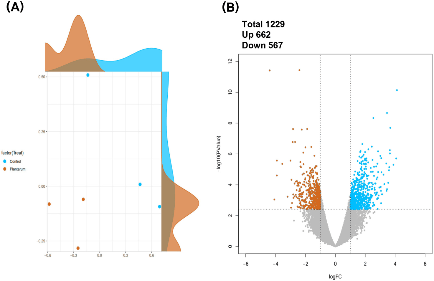
Differential expression analysis between the control and L. plantarum treatment groups identified 1,229 DEGs, including 662 upregulated and 567 downregulated transcripts, based on a threshold of |Log₂FC| ≥ 1 and FDR < 0.05 (Fig. 1).
Functional enrichment analysis of the DEGs in the ileum was performed using the DAVID database. Gene Ontology analysis revealed significant enrichment in immune-related biological processes, including the positive regulation of interleukin-6 production and actin filament organization, indicating the potential activation of immune responses and epithelial remodeling (Fig. 2).
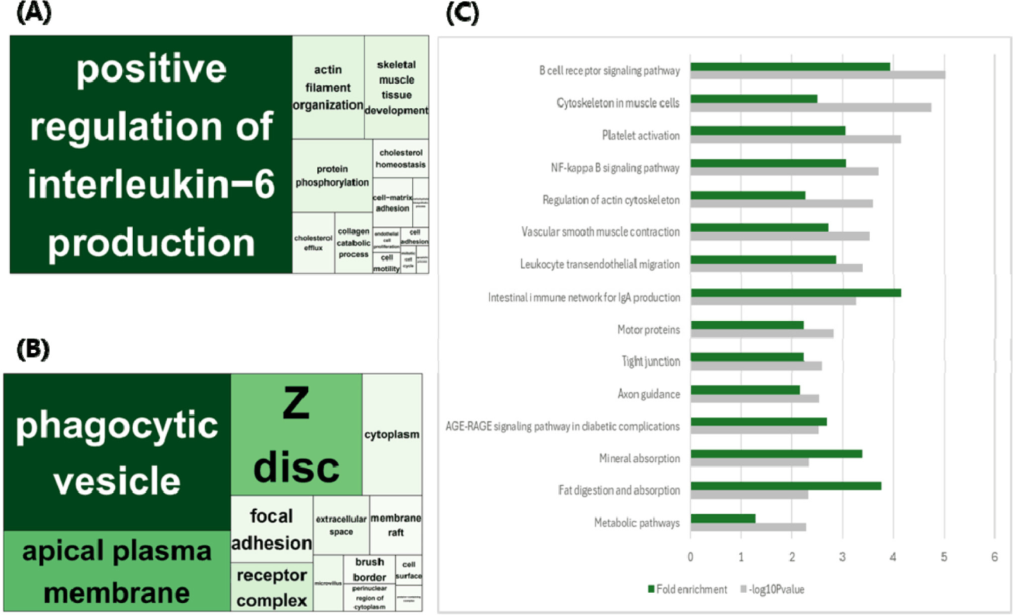
In the cellular component category, DEGs were associated with structures such as phagocytic vesicles, Z discs, apical plasma membranes, and focal adhesions, implying alterations in membrane dynamics and epithelial barrier organization (Fig. 2).
KEGG pathway analysis identified multiple enriched pathways related to immune regulation and barrier function, such as the B-cell receptor signaling pathway, nuclear factor kappa-light-chain-enhancer of activated B cells (NF-κB) signaling pathway, the intestinal immune network for IgA production, and leukocyte transendothelial migration. In addition, enrichment of tight junctions, mineral absorption, fat digestion and absorption, and general metabolic pathways suggests coordinated regulation of mucosal defense and nutrient absorption (Fig. 2).
To explore metabolic alterations induced by L. plantarum supplementation, untargeted metabolomic profiling of fecal samples was performed. Enrichment analysis using MetaboAnalyst6.0 revealed that DEG-abundant metabolites were predominantly associated with amino acid and lipid metabolism pathways (Fig. 3).
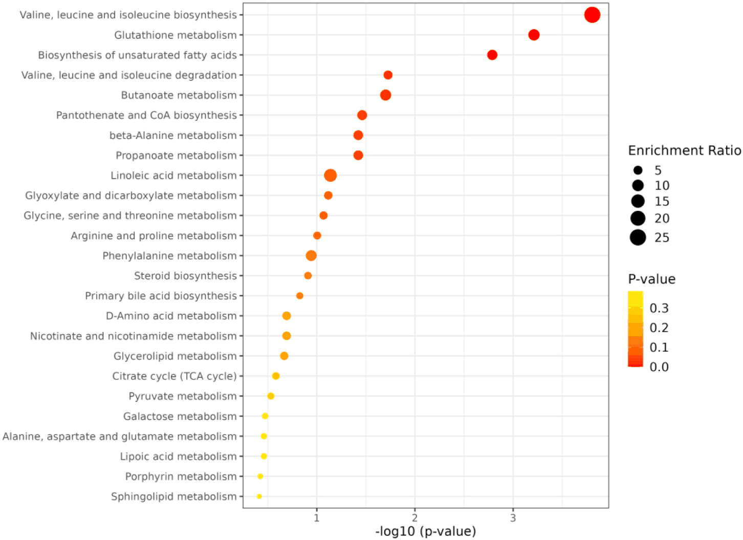
Specifically, valine, leucine, and isoleucine biosynthesis; glutathione metabolism; arginine and proline metabolism; and glycine, serine, and threonine metabolism were among the most enriched pathways. These pathways are closely linked to cellular repair, the oxidative stress response, and mucosal homeostasis.
Furthermore, the enrichment of linoleic acid metabolism, pantothenate and CoA biosynthesis, and butanoate metabolism suggests the potential modulation of fatty acid processing and short-chain fatty acid production. Notably, sphingolipid metabolism, although less prominently enriched, was also observed, implying its potential role in epithelial barrier dynamics.
Overall, metabolite-based pathway analysis indicates that L. plantarum supplementation alters key metabolic pathways in the gut environment, particularly those involved in nutrient absorption, redox balance, and epithelial support.
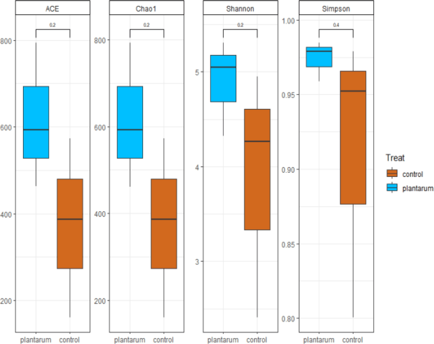
Alpha diversity analysis revealed that dietary supplementation with L. plantarum led to increased microbial richness and diversity in fecal samples, as evidenced by higher ACE, Chao1, Shannon, and Simpson indices than those in the control group (Fig. 4). These results suggest that L. plantarum intake enhances the ecological complexity of the gut microbiota in weaned piglets. This observation is consistent with previous studies reporting that dietary L. plantarum administration increases alpha diversity in the gastrointestinal microbiota, thereby contributing to improved gut microbial stability and resilience [5].
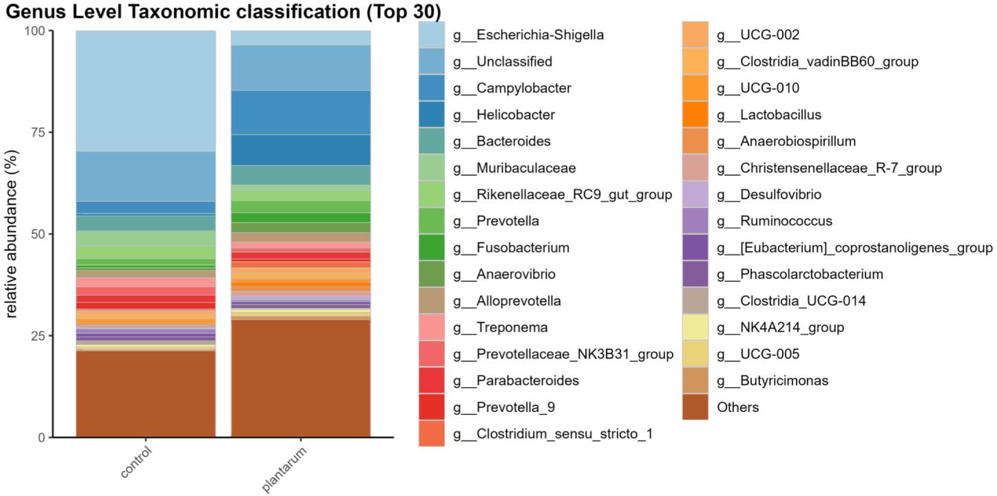
Taxonomic analysis at the genus level revealed compositional differences between the control and treatment groups (Fig. 5). Notably, the relative abundance of Lactobacillus increased in the plantarum-supplemented group, indicating the successful colonization or promotion of commensal lactic acid bacteria. In contrast, potentially pathogenic genera, such as Shigella, were markedly reduced, which may be attributed to competitive exclusion or pH-lowering effects associated with lactic acid production. Interestingly, an increase in the genus Helicobacter was observed in the treatment group. Although Helicobacter includes pathogenic species, its proliferation may reflect transient ecological shifts rather than pathogenic overgrowth.
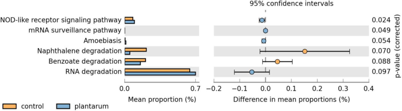
To investigate the functional implications of microbial compositional changes, predictive metagenomic analysis was performed based on 16S rRNA gene data (Fig. 6). Comparison of functional pathway abundance revealed significant enrichment of the NOD-like receptor signaling pathway in the L. plantarum-supplemented group compared to that in the control group (p = 0.024). The NOD-like receptor family is integral to innate immune surveillance, recognizing intracellular microbial components and initiating downstream signaling cascades, including activation of NF-κB and inflammasome formation, ultimately leading to the production of proinflammatory cytokines and antimicrobial peptides.
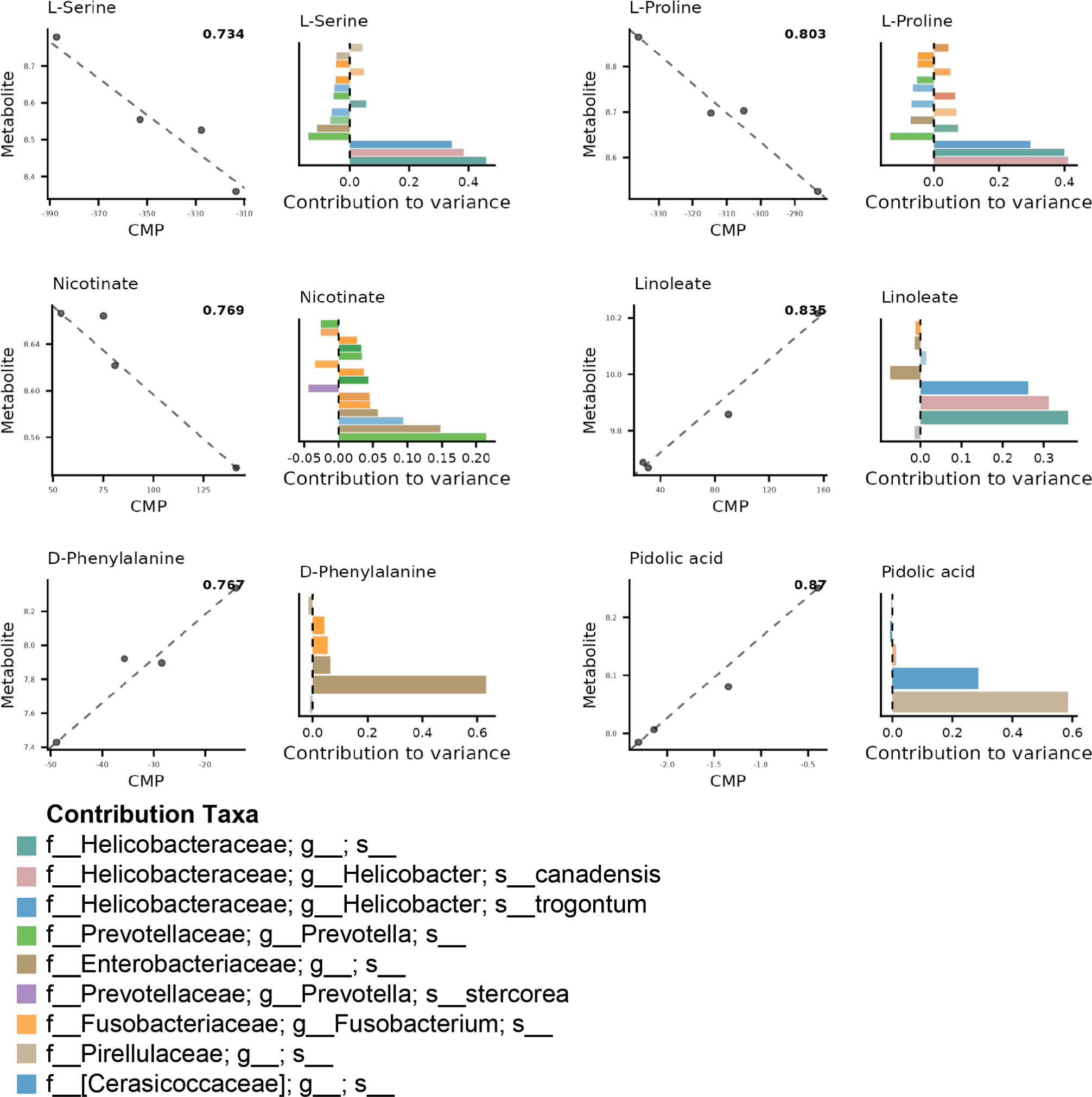
To elucidate the functional contributions of the gut microbiota to metabolite variation, an integrative analysis was performed using MIMOSA2, which infers CMP based on species abundance and genome-scale metabolic reconstructions. The analysis identified six metabolites that were significantly influenced by the microbiome composition: L-serine, nicotinate, L-proline, linoleate, pidolic acid, and D-phenylalanine (Fig. 7). For each metabolite, the CMP scores were calculated and matched to the metabolite abundance profiles, revealing taxa-specific associations (p < 0.1).
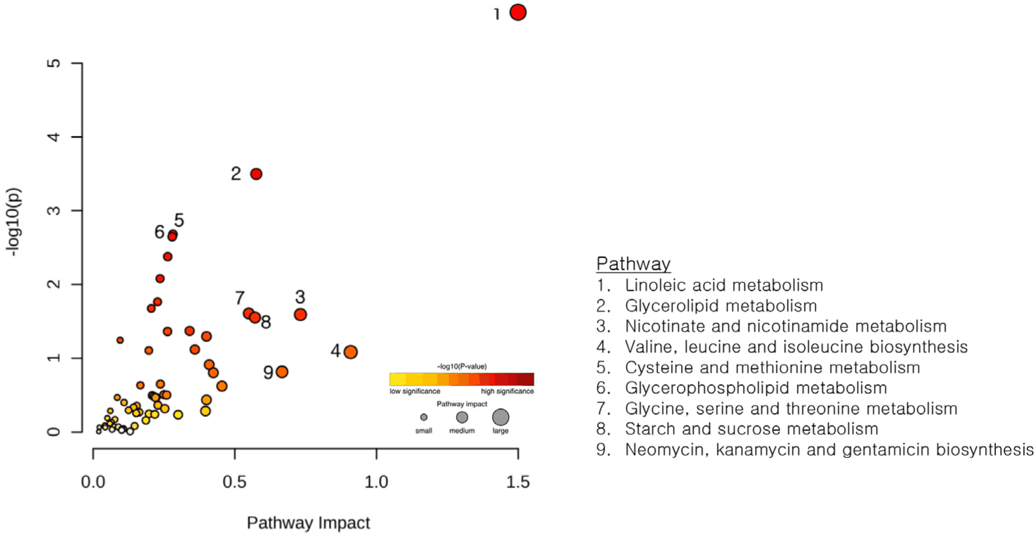
To identify the functional convergence of host gene expression and microbial-derived metabolites, joint pathway analysis was conducted using MetaboAnalyst 6.0. Additionally, KEGG pathway integration was performed by combining the ileal DEGs with metabolites predicted using MIMOSA2. Nine metabolic pathways were identified as functional nodes between the host transcriptome and the microbial metabolome based on both pathway impact and statistical enrichment (Fig. 8). Linoleic acid metabolism exhibited the greatest impact and significance. Glycerolipid metabolism, and cysteine and methionine metabolism also showed high statistical enrichment, whereas glycerophospholipid metabolism displayed a comparatively lower pathway impact but remained statistically significant.
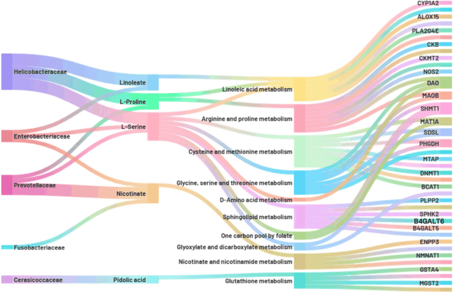
The alluvial diagram constructed from the integrated multi-omics analysis revealed extensive interactions between the ileal microbiota, microbially derived metabolites, host metabolic pathways, and host genes (Fig. 9). At the family level, the distinct microbial taxa were strongly associated with specific metabolites. Helicobacteraceae predominantly contributed to linoleate, L-proline, and L-serine metabolism, whereas Enterobacteriaceae were linked to linoleate, l-serine, and nicotinate metabolism. Additionally, Prevotellaceae was involved in the metabolism of L-proline, L-serine, and nicotinate; Fusobacteriaceae was primarily associated with nicotinate; and Cerasicoccaceae showed a specific relationship with pidolic acid. Integration of these microbial-derived metabolites with the host ileal transcriptome identified several metabolic pathways, notably linoleic acid metabolism; glycine, serine, and threonine metabolism; and sphingolipid metabolism as key nodes influenced by the microbiota during the weaning transition.
DISCUSSION
A comprehensive multi-omics strategy was used to investigate the effects of dietary L. plantarum supplementation on host–microbiome interactions in weaned piglets. First, each omics layer was analyzed independently, including assessments of the ileal transcriptome, fecal microbiome, and fecal metabolome, to characterize the alterations induced by probiotic intake. The relationships between fecal microbiota and microbial-derived metabolites were examined to determine how specific taxa contributed to shifts in metabolite profiles. Metabolites that were significantly modulated by microbial activity were subsequently integrated with the ileal transcriptomic data to explore their associations with host gene expression. This integrative analysis provides a comprehensive understanding of how probiotic-induced changes in the gut microbial ecosystem and its metabolic outputs converge with host intestinal transcriptional responses during the postweaning transition.
Multi-omics integration revealed that L. plantarum supplementation strongly influenced host–microbiome metabolic interactions, particularly within the lipid and amino acid pathways. Linoleic acid metabolism exhibited the greatest impact (Fig. 8). Transcriptomic evidence showed increased CYP1A2 expression, suggesting enhanced conversion of linoleic acid into epoxy-linoleates. Unlike other linoleic acid derivatives that can promote inflammatory signaling, epoxy-linoleates exert anti-inflammatory and cytoprotective effects, including the suppression of reactive oxygen species and preservation of tight junction protein expression [29–31]. These effects are particularly relevant during the postweaning transition, when piglets face high oxidative stress and a heightened risk of barrier disruption.
Alterations in glycine, serine, and threonine metabolism were also evident. Although Helicobacteraceae members were associated with serine consumption, host transcriptional responses appeared to compensate through enhanced serine biosynthesis. Specifically, the upregulation of PHGDH and PGAM1 diverted glycolytic intermediates toward serine production, whereas the downregulation of SDSL limited the conversion of serine to glycine. This coordinated adjustment promotes serine accumulation, which is critical for mucin production and membrane phospholipid synthesis, thereby reinforcing epithelial protection [32–34].
Sphingolipid metabolism further suggested probiotic-driven remodeling. Transcriptomic profiles showed reduced SPHK2 expression, along with increased B4GALT6 expression, suggesting a shift toward ceramide retention and complex glycosphingolipid synthesis (Fig. 9). These molecules are known to cluster within lipid rafts, disperse pathogen receptors, and reduce microbial adherence [35,36]. Such modulation suggests that L. plantarum supplementation may support both metabolic stability and structural defense against pathogenic invasion.
Collectively, these findings highlight a coordinated metabolic adaptation in which L. plantarum supplementation directs lipid and amino acid metabolism toward pathways that reduce oxidative stress, stabilize tight junctions, and enhance mucosal defense. These adjustments are likely central to maintaining intestinal integrity during physiologically stressful weaning periods.
In addition to metabolic adaptations, L. plantarum supplementation modulated immune-related processes at both the transcriptomic and microbiome levels. Gene Ontology and KEGG analyses revealed enrichment of pathways associated with interleukin-6 signaling, NF-κB activation, B cell receptor signaling, and IgA production, indicating that probiotic intake promoted immune responsiveness at multiple levels (Fig. 2). Collectively, these pathways contribute to mucosal defense by coordinating cytokine release, immune cell recruitment, and antibody production. Importantly, such effects are highly relevant in weaned piglets, whose adaptive immune systems remain immature; antigen-specific IgA responses typically become functional only after approximately six weeks of age [37]. Therefore, the activation of immune pathways in response to L. plantarum may represent a compensatory mechanism that reinforces mucosal defense during this immunologically vulnerable period. However, these immune-related changes should be interpreted with caution because the piglets were not experimentally challenged with specific pathogens. Therefore, the observed immune responses may reflect enhanced mucosal preparedness rather than a pathogen-induced inflammatory response.
Microbiome analysis confirmed these immune-related changes. Alpha diversity indices, including ACE, Chao1, Shannon, and Simpson indices, were consistently higher in the L. plantarum group, reflecting a more balanced microbial ecosystem (Fig. 4). Enhanced microbial diversity is widely associated with greater functional redundancy and ecological resilience, allowing the gut microbiota to resist perturbations and outcompete opportunistic pathogens [38,39]. Taxonomic shifts supported this protective role; the abundance of Lactobacillus increased, while that of potentially pathogenic genera such as Shigella were reduced, consistent with probiotic-mediated pathogen suppression via competitive exclusion and lactic acid production (Fig. 5) [2,6].
Interestingly, a transient increase in Helicobacter was observed. This transient shift may also be associated with changes in gut pH, as lactic acid produced by probiotics can contribute to acidification, potentially influencing the relative abundance of acid-tolerant taxa such as Helicobacter. Although certain species within this genus can be pathogenic, previous studies have shown that acid-tolerant Helicobacter strains may expand temporarily when microbial competition decreases after weaning [39]. This transient shift likely represents an ecological adjustment rather than harmful colonization, further underscoring the dynamic interplay between microbial competition and the host environment during the weaning transition.
Predictive metagenomic analysis provided additional evidence of immune modulation, revealing the enrichment of the NOD-like receptor signaling pathway in the L. plantarum-supplemented group (Fig. 6). NOD-like receptors act as critical intracellular sensors that detect microbial motifs and initiate downstream activation of NF-κB and inflammasome pathways, culminating in the production of cytokines and antimicrobial peptides [1]. This suggests that probiotic supplementation may enhance innate immune preparedness, effectively bridging the gap until the adaptive immune system becomes fully functional.
Taken together, these findings demonstrate that L. plantarum supplementation not only reshapes the gut microbiota composition, but also strengthens mucosal immunity by stimulating innate defense pathways and promoting ecological stability. Such changes likely contribute to the reduced susceptibility to postweaning diarrhea and improved intestinal resilience.
The multi-omics framework used in this study revealed how L. plantarum supplementation shapes host–microbiome interactions during the postweaning transition. MIMOSA2 analysis identified taxon-specific contributions to metabolite turnover, with Helicobacteraceae linked to L-serine and L-proline consumption, Prevotellaceae and Enterobacteriaceae influencing both amino acid and nicotinate metabolism, and Cerasicoccaceae associated with pidolic acid production (Fig. 7). Conversely, metabolites such as linoleate, pidolic acid, and D-phenylalanine were predicted to be produced microbially, indicating that distinct bacterial taxa contributed to either consumption or production depending on the metabolic context. These findings highlight the division of metabolic labor within the gut microbiota and illustrate how probiotic-driven shifts reconfigure the luminal metabolite pool.
When host transcriptional responses were overlaid with microbial metabolite profiles, several convergent pathways emerged, including linoleic acid, glycerolipid, cysteine, methionine, and glycerophospholipid metabolism (Fig. 8). These pathways were closely associated with epithelial lipid remodeling, antioxidant defense, and cell membrane turnover, suggesting that probiotic supplementation fosters a metabolic environment conducive to epithelial resilience. Key host genes were identified as central mediators within these processes. CYP1A2 directs linoleic acid toward epoxy-linoleates, reducing oxidative stress and reinforcing tight junctions. PGAM1 and PHGDH enhanced serine biosynthesis, whereas SDSL downregulation limited serine catabolism, collectively ensuring serine retention for mucin and phospholipid synthesis. B4GALT6 promotes glycosphingolipid formation that disperses pathogen receptors within lipid rafts, thereby decreasing the opportunities for microbial adhesion.
Importantly, these host–microbiome adjustments did not operate in isolation but formed a coordinated network of interactions. While Sankey plot visualization reflects correlation rather than direct causation, the associations highlight functionally meaningful links (Fig. 9). For example, even though Helicobacteraceae transiently expanded and correlated with the consumption of L-serine and L-proline, host transcriptional adjustments compensated for the enhanced serine biosynthesis and restricted catabolism. This ensures sufficient serine availability for barrier-supportive functions, thereby maintaining the epithelial integrity in an environment with increased microbial competition. Probiotic supplementation buffers the potential negative effects of transient microbial shifts, with host metabolic compensation and enhanced microbial diversity, thereby preventing long-term destabilization.
Overall, these integrative findings illustrate that L. plantarum supplementation orchestrates a multilayered adaptation in which microbial taxa, metabolite profiles, and host gene expression converge to stabilize the intestinal barrier and promote immune preparedness. This systems-level interplay highlights the value of multi-omics approaches in uncovering mechanisms that would remain hidden under single-omics frameworks, providing a holistic understanding of how probiotics mitigate weaning-associated stress in piglets.
CONCLUSION
This study characterized the effects of L. plantarum supplementation on host–microbiome interactions in weaned piglets using an integrated multi-omics approach. Probiotic supplementation increased microbial diversity, promoted beneficial bacterial populations, reduced potentially pathogenic taxa, and was associated with host transcriptional changes related to immune function, epithelial barrier maintenance, and metabolic regulation. Integrated analysis identified linoleic acid, glycine/serine/threonine, and sphingolipid metabolism as key pathways linking microbial and host responses. These findings indicate that L. plantarum may help improve intestinal resilience during weaning and provide mechanistic insight into its beneficial role in gut health. Practically, the microbial, metabolic, and host transcriptional signatures identified here may guide the development of targeted probiotic-based feed additives aimed at improving antioxidant capacity, mucosal immune preparedness, and metabolic stability during the postweaning period. Such strategies may help reduce weaning-associated intestinal stress and support healthier growth in commercial swine production.