INTRODUCTION
Intestinal disease comprises a group of chronic conditions primarily affecting the colon [1], including Crohn’s Colitis and Ulcerative Colitis et al [2]. These diseases result in immune-mediated gastrointestinal inflammation, often accompanied by a range of symptoms and complications [3], deemed incurable [4]. Treatment modalities involve medication, lifestyle changes, and occasionally surgery [5,6]. While the exact causes of many intestinal diseases remain unclear, genetic, environmental, and immune factors are believed to play a role in its development [7].
The gut microbiota may play a significant role in the pathogenesis of intestinal diseases [8], and maintaining a balanced gut microbiome is crucial for the prevention and treatment of Inflammatory bowel disease (IBD). The balance of the gut microbiota is vital for overall host health [9,10], and imbalance may be associated with various diseases such as IBD, cancer, cardiovascular diseases, and metabolic syndrome [11]. Therefore, factors regulating the gut microbiome are crucial for disease prevention and treatment. Many natural products (NPs) are considered to have immune-regulatory and nutritional effects on patients with intestinal diseases [12], although their application remains controversial.
In the current approaches to treating IBD, NPs are considered potential sources for developing new drugs due to their diverse biological activities [13]. Evodiamine is a NP known for its anti-inflammatory and antioxidant properties [14–16]. Some studies suggest that evodiamine exhibits in vivo therapeutic activity in experimental IBD models, possibly by improving the gut microbiota to alleviate intestinal inflammation [17,18]. Canines serve as valuable animal models for studying diseases, with experimental results providing valuable insights into clinical disease treatment for humans [19]. Additionally, the pet industry has experienced rapid growth in recent years, making research on the improvement of canine intestinal diseases significant for the global pet economy and animal welfare. Although existing studies indicate the physiological activity of evodiamine, its specific impact on canine intestinal improvement after being added to dog food remains unknown. While evodiamine has demonstrated physiological activity in previous research, the question of whether its addition to canine food can lead to improvements in canine intestinal health remains unanswered.
Therefore, we propose a scientific hypothesis that a functional diet enriched with evodiamine could promote canine intestinal health by modulating the gut microbiota, thus promoting the canine health. To validate this hypothesis, we prepared a canine intestinal prescription food containing evodiamine and conducted a series of animal experiments to assess the positive regulatory effects of this functional dietary product on intestinal function and microbiology.
MATERIALS AND METHODS
The canine intestinal prescription food is jointly developed by the Gannan Institute of Innovation and Translational Medicine and Jiangxi Huizhou Pet Technology Co. The formulation of this prescription food is designed with reference to national standards (GB/T 31216-2014), full-priced pet food specifically formulated for dogs, and guidelines provided by the American Association of Feed Control Officials (AAFCO). It is based on the regulatory mechanism of intestinal microorganisms and tailored to meet specific nutritional requirements. The daily dose of evodiamine was added to the daily food of the experimental dogs, which was continuously administered orally for 8 weeks. The ingredients and additives used in the formulations are in accordance with the Feed Ingredients Catalogue and the Feed Additives Catalogue. The crude protein (GB/T 6432-2018), crude fat (GB/T 6433-2006), crude fiber (GB/T 6434-2006), ash (GB/T 6438-2007), moisture (GB/T 6435-2014), calcium (GB/T 6436-2018) and total phosphorus (GB/T 6437-2018) of the intestinal tract prescription diet were determined referring to national standard and the energy was determined by Oxygen-type calorimeter.
Ten healthy Chinese field dogs, weighing 7–10 kg, aged 1–5 years, with no history of disease, were selected for the test and were routinely dewormed and vaccinated in vitro and in vivo before the test. The animals were fed and watered ad libitum in a 12 h alternating light and dark environment at 22°C–26°C. The experiment was approved by the Biomedical Research Ethics Committee of Gannan Medical College (Approval number 2021335), and all animal manipulations were in accordance with the operational guidelines of the Biomedical Ethics Committee of Gannan Medical College.
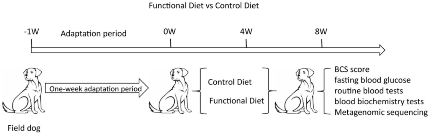
Dogs were randomly divided into a control group feed with control diet (n = 5) and EVO group feed with evodiamine-enriched functional diet (n = 5). All dogs were received a week food adaptation period before formal feeding. The daily feeding amount was calculated according to the formula: maintenance energy requirement (MER) / metabolizable energy (ME), MER = body weight (BW) (kg)0.75 × 70 × life stage factor. The experimental dogs were fed twice a day (at 9:00 am and 4:00 pm) for 8 weeks. Their daily food intake was recorded during the experiment. Physical examinations including fasting blood glucose and body condition score (BCS) were performed at 0 w, 4 w, and 8 w. The blood samples were collected for complete blood count and blood biochemistry examination at 0 w, 4 w, and 8 w. Fecal samples were taken at the 8th week after the start of the experiment. The mid-range feces 5 g were collected with a sterile fecal collector and immediately put into sterilized freezing tubes and labeled, then immediately put into liquid nitrogen for storage, and then put into –80°C for freezing and storage [20].
Measwere measured daily before breakfast, and body condition was scored independently by two researchers. Five milliliters whole blood was collected from the distal cephalic vein. The fasting blood glucose was tested on Blood glucose meter (AccU-Chek Active, Roche) with 20 μL blood. Using a 5-part ethylenediaminetetraacetic acid (EDTA) anticoagulant hematology analyzer (Abaxis VetScan HM5, Zoetis), 1 mL of venous blood was collected in an EDTA anticoagulant tube for routine blood tests. Draw 2 mL of blood and preserve it in heparin lithium anticoagulant tubes. Centrifuge at 1,000×g for 10 minutes to obtain the upper serum layer for blood biochemical testing. The biochemical detection was applied on Automatic biochemical analyzer (Hitachi 3110, Hitachi). Complete blood count and blood biochemical tests were performed by the medical Laboratory of Well Animal Test.
The E.Z.N.A.® Soil DNA Kit (Omega Bio-tek) reagent kit was used for sample DNA extraction. After completion of genomic DNA extraction, DNA degradation degree and potential contamination were monitored on a 1% agarose gel. DNA concentration was measured using the Qubit® dsDNA Assay Kit in Qubit® 2.0 Fluorometer (Life Technologies). Genomic DNA 1 μg was taken and ultrasonically interrupted using a Covaris instrument. The interrupted samples were fragment-selected with magnetic beads so that the sample bands were concentrated around 200–400 bp. Then formulate the reaction system and react for a certain period of time at the right temperature to repair the ends of double-stranded cDNA and add the A base at the 3’ end; formulate the junction reaction system and react for a certain period of time at the right temperature to make the junction connected to the DNA. Then the polymerase chain reaction (PCR) reaction system was prepared and the reaction program was set up to amplify the ligated products. The amplified products were purified and recovered using magnetic beads. After denaturing the PCR product into single-stranded, the cyclization reaction system is prepared, and the single-stranded cyclic product can be obtained by mixing well and reacting at the right temperature for a certain period of time, and the final library can be obtained after digesting the linear DNA molecules that have not been cyclized. The cyclized product was then subjected to a pre-loading concentration test. The tested libraries were arranged for on-board sequencing (DNBSEQ), where single-stranded cyclic DNA molecules were replicated by rolling rings to form a DNA nanoball (DNB) containing multiple copies. The resulting DNBs are added to a mesh of holes on the chip using high-density DNA nano-chip technology and sequenced by co-probe anchored polymerization. To ensure the reliability of the subsequent information analysis results, it is necessary to filter these low-quality data. The high-quality CleanData was obtained using the software SOAPnuke. After quality control, CleanData data were assembled using the software MEGAHIT on the samples, and fragments below 300 bp were filtered out for statistical analysis and subsequent gene prediction.
The data were statistically analysed using SPSS 26.0 statistical software, and the two-tailed t-test or Wilcoxon rank sum test was used for statistical analysis between groups. All values in the text are expressed as mean ± SD, and p < 0.05 was considered a statistically significant difference.
RESULTS
Here, we added evodiamine to a diet and systematically analyzed the chemical composition and of the formulated gut-functional dietary products and the control dietary products. Compared with the control food, the Functional group contained 0.25% of evodiamine, while the control group did not contain this ingredient. To investigate the effect of evodiamine on the intestinal tract after its addition to the diet, we designed an experimental program as shown in Fig. 1.
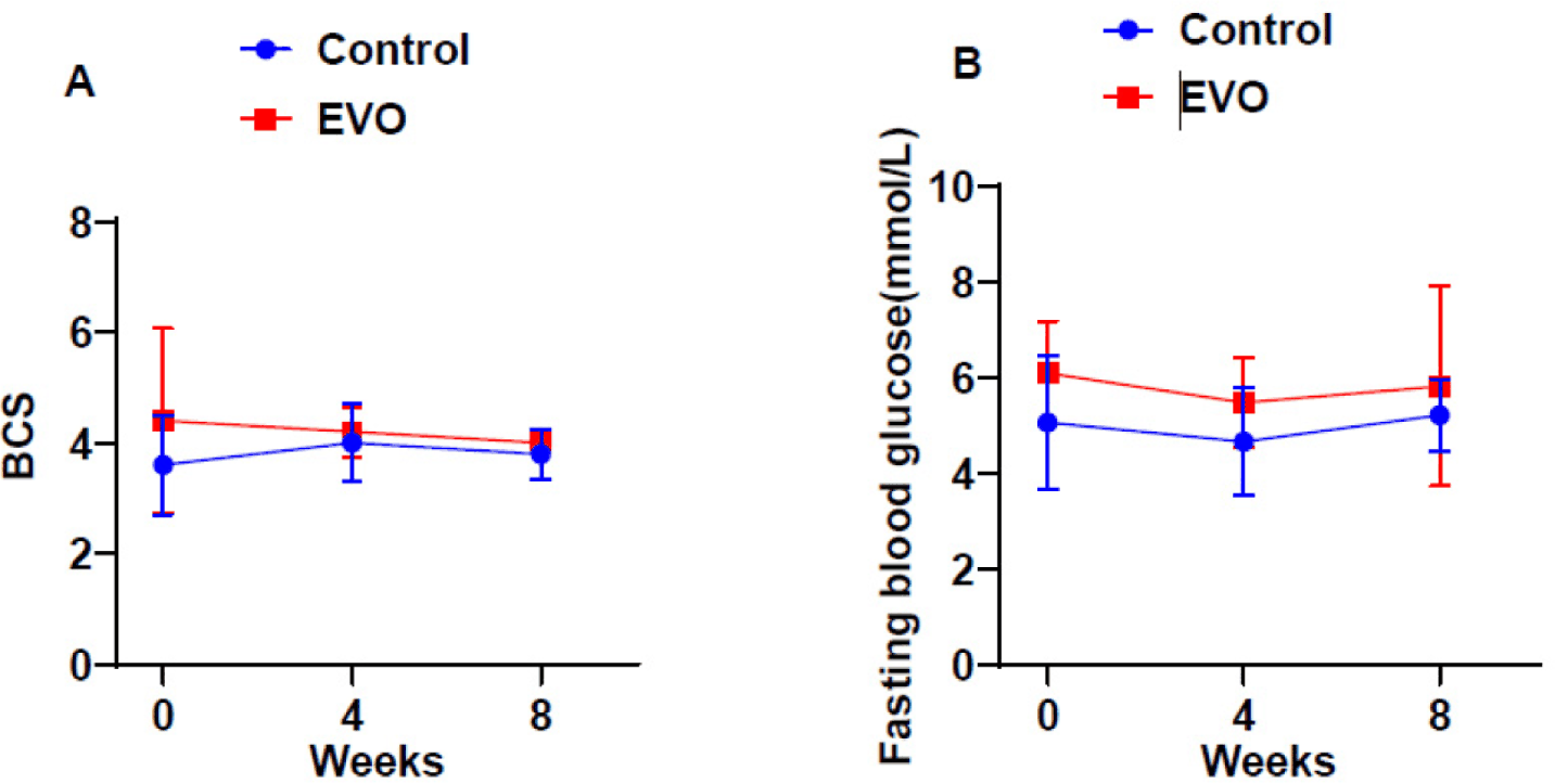
After an 8-week feeding experiment, we analyzed the effects of canine intestinal functional dietary products on canine BCS (Fig. 2A) and fasting blood glucose (Fig. 2B). The results showed that the BCS and fasting blood glucose were relatively stable in the EVO group compared with the control group, and there were no significant differences in BCS score and fasting blood glucose among all groups.
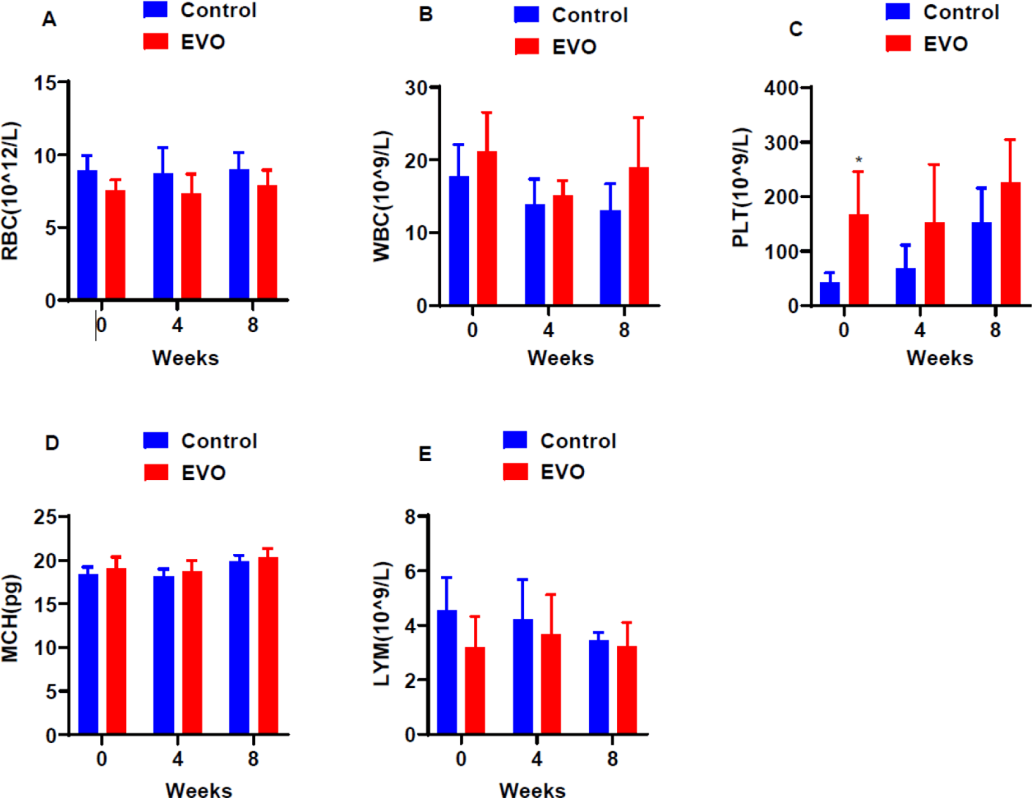
We further analyzed the effect of enteral functional diet on the safety of dogs by hematological tests. As shown in Fig. 3, the platelet count level of EVO group was significantly higher than control group, and no significant differences in the other indices including red blood cells, white blood cells, mean corpuscular hemoglobin and lymphocyte. All of the indices were within the normal reference ranges.
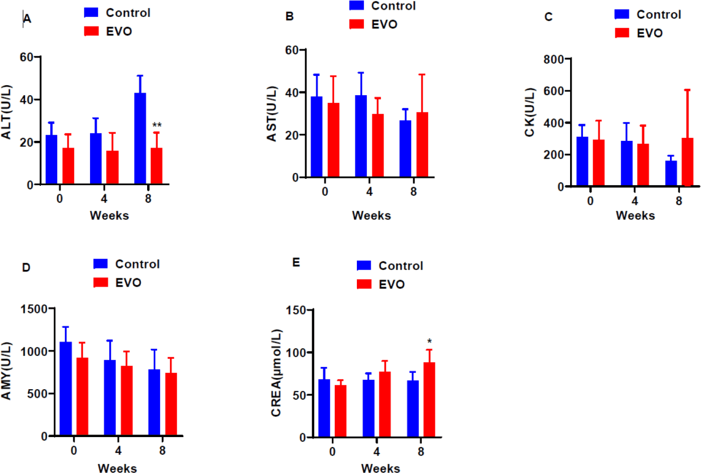
In the context of blood biochemical indices, including alanine aminotransferase (ALT; Fig. 4A), Glutamic oxaloacetic transferase (AST; Fig. 4B), creatine kinase (CK; Fig. 4C), alpha-amylase (AMY; Fig. 4D) and creatinine (CREA; Fig. 4E), a statistical analysis was conducted to compare values in dogs before and after an 8-week testing period with those of the control group. The results revealed that ALT exhibited significantly lower level, while CREA demonstrated a significantly higher level compared to the control group at 8-week. It is noteworthy that these alterations, although statistically significant, remained within the normal range. The reduced levels of ALT are particularly significant as they are associated with a decreased likelihood of developing a spectrum of liver metabolic diseases and their associated complications. No significant differences were observed in the other blood indices, including AST, CK, and AMY. This indicating that the functional diet did not exert a substantial impact on the overall health status of dogs. This further underscored the safety profile of the functional diet.
During the 8-week feeding experiment, fecal samples were collected from the mid-section of fresh feces for metagenomic analysis, as shown in Fig. 5. The analysis focused on examining alterations in the species composition of ccontrol and prediet groups, specifically Firmicutes (F) and Bacteroides (B) at phylum (Fig. 5A) and genus (Fig. 5B) level and through diverse analytical method.
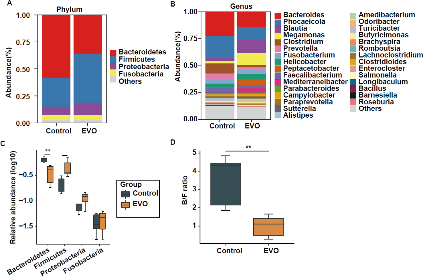
The results revealed a noteworthy increase of the Firmicutes in the EVO group compared to the control group, accompanied by a significant decrease in the Bacteroidetes phylum (Figs. 5A and 5C). This suggests that Firmicutes might play a predominant role in facilitating intestinal mucosal repair, thereby enhancing the intestinal barrier. From a genus level perspective, there is a significant decrease in the relative abundance of Bacteroides, Phocaeicola, and Clostridium in the Prediet group, while Blautia, Megamonas, and Peptacetobacter show an increase in relative abundance (Fig. 5B). Additionally, we computed the ratio of Firmicutes to Bacteroidetes (B/F ratio) and observed a significant reduction in the EVO group compared with control group (Fig. 5D).
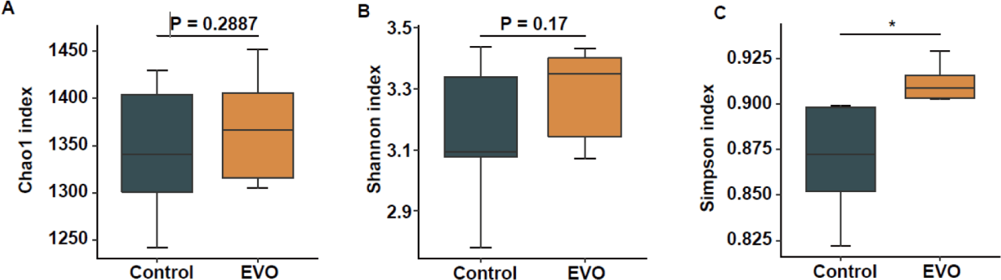
By analyzing the Alpha diversity of intestinal microbiota, Chao1 (Fig. 6A), Shannon (Fig. 6B), and Simpson (Fig. 6C) indices results showed that compared with the control group, Chao1 and Shannon indices of the EVO group did not change significantly, while the Simpson index was significantly elevated in EVO group when compared with control group. It indicated that the functional diet of the intestine can regulate the elevated diversity of intestinal microorganisms, improve intestinal function, and protect intestinal health.
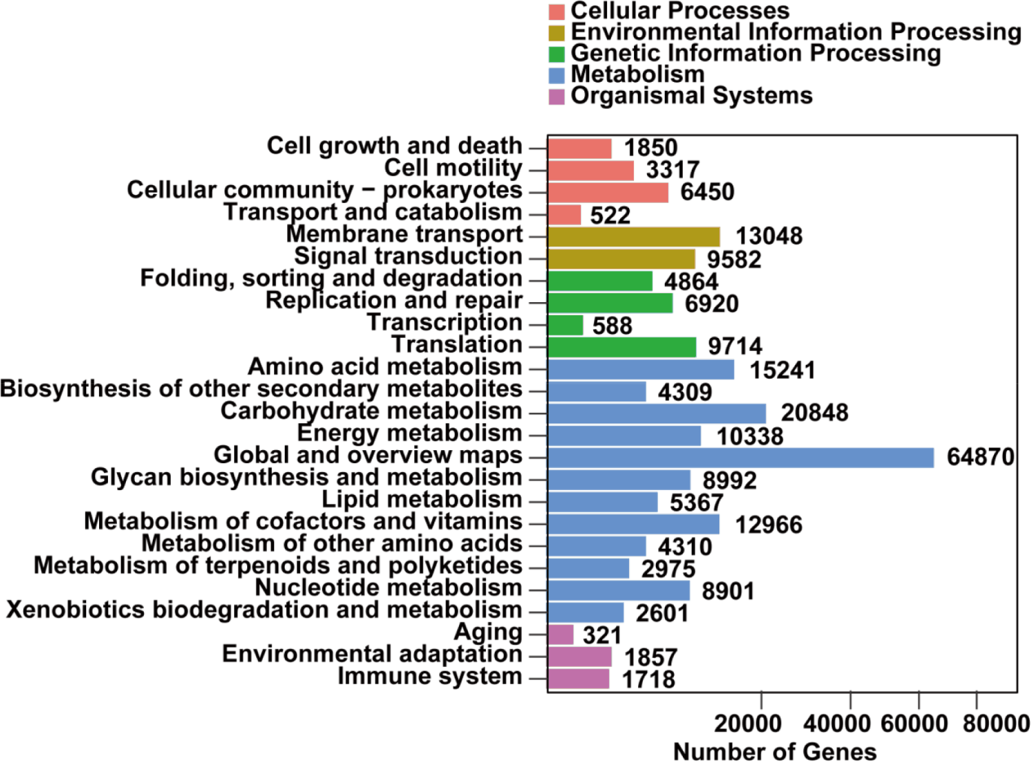
Kyoto Encyclopedia of Genes and Genomes (KEGG) pathway enrichment analysis results (Fig. 7) showed that the classification of biometabolic pathways into Metabolism, Genetic Information Processing, Environmental Information Processing, Cellular Processes, and Organismal Systems. Among them, the metabolic domain has the highest number of genes of 161,718 and the widest distribution of genes. The number of genes in environmental information processing was the second highest, with a total of 22,630. The third largest number of genes is processed for genetic information, with a total of 22,086. The fourth largest number of genes is Cellular Processes, with a total of 12,139. And the Organismal Systems has the smallest number of genes, with a total of 3,896. It is shown that gut functional diets can regulate gut microbial metabolic pathways and promote intestinal health.
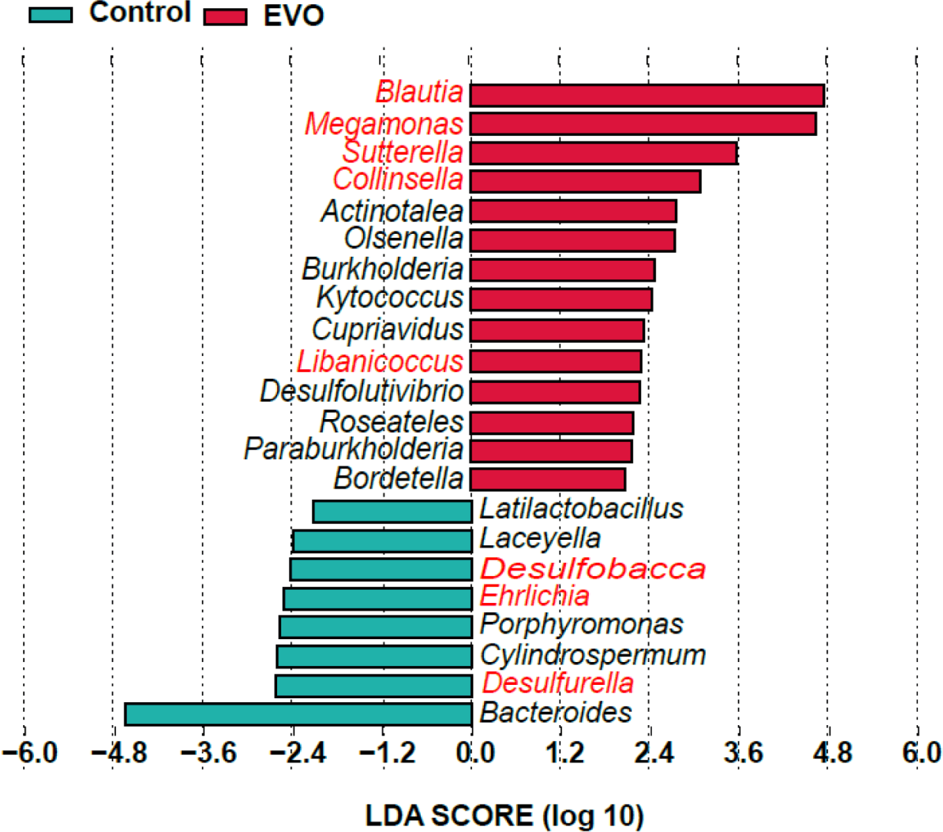
A linear discriminant effect size analysis (LEfSe) was performed on the gut microorganisms (Fig. 8), analyzing the linear discriminant analysis (LDA) bar graphs, where the specific bar corresponded to the specific species, the color of the bar corresponded to the group corresponding to that species, and the length of the bar represented the LDA value, and the larger the LDA value represented the more significant difference. The results revealed a comprehensive screening of 22 distinct strains between the control group and the EVO group. Among these, 14 microorganisms (Blautia,Megamonas, Sutterella, and Collinsella et al.) exhibited higher abundance, while 8 microorganisms (Bacteroides, Desulfurella, Ehrlichia, and Desulfobacca et al.) displayed lower abundance in the EVO group compared to the control group. This indicated that the functional diets were able to modify the species differences in the intestinal flora and protect the intestinal health.
DISCUSSION
Maintaining gut health is a highly complex task influenced by various physiological factors, such as the intricate interplay among the immune system, intestinal mucosal barrier, and gut microbiota [21]. The gut, housing a diverse bacterial community, significantly impacts the host’s physiological responses, potentially disrupting internal equilibrium [22]. Thus, ensuring robust gut health is crucial for animal well-being. The animal’s immune system plays a pivotal role by recognizing gut microbiota and modulating responses. It moderately promotes innate and adaptive immune pathways, enhancing gut barrier function. This enables the immune system to effectively respond to invasive pathogens while maintaining tolerance to beneficial microbiota and food antigens [23–25]. This regulatory role involves mechanisms like mucus production and epithelial layer focus. These immune responses not only alter nutrient substrates in the gut but also contribute to shaping the gut microbiota composition [26]. The key to promoting gut health lies in enhancing immune system functionality, reinforcing the intestinal barrier, and improving gut microbiota composition [27]. Seeking effective feed additives to enhance animal gut health is a beneficial strategy, contributing to overall animal health.
To confirm the functionality of a diet, it is essential to ensure its safety. The experimental diet utilized by our research group includes the addition of evodiamine, a NP. Extensive literature supports the use of evodiamine for its anti-inflammatory and antioxidant properties, indirectly indicating the safety of evodiamine within certain concentration ranges [17,28,29]. For example, studies have shown that evodiamine could significantly reduce the inflammation of AOM/DSS-induced colitis, which may be related to the maintenance of intestinal barrier function [28]. There are also studies that show that evodiamine may act directly on the increase of beneficial bacteria, leading to the reduction of harmful bacteria and the maintenance of intestinal balance of nature, or it may act indirectly on the gut microbiota, leading to a change in the interaction between beneficial bacteria and harmful bacteria, to maintain the intestinal balance of nature [30]. Furthermore, we conducted experiments to directly demonstrate the safety of the Evodiamine concentration added to the food for dogs.
Research indicates that the addition of evodiamine significantly enhances the α-diversity of canine gut microbiota. Specifically, by measuring the Shannon diversity index, we observed a higher microbial diversity in the EVO group, where evodiamine was added to the diet, compared to the control group. This suggests that evodiamine contributes to fostering a more abundant and diverse microbial community in the canine gut. From the perspective of β-diversity, we further analyzed the clustering of gut microbiota after the addition of evodiamine. The results revealed a significant difference in microbial clustering between the EVO group and the control group, indicating that the introduction of evodiamine indeed leads to a noticeable alteration in the overall composition of the canine gut microbiota. This change is evident not only in the types of microorganisms present but also in the relative abundance among them, highlighting the impact of evodiamine on the overall structure of the gut microbiota. Similar studies also suggest that other plant extracts, such as certain plant polyphenols or fiber-related substances [31,32], similarly contribute to increased diversity in gut microbiota. This further emphasizes the potential benefits of plant extracts in modulating the balance of gut microbiota [33–36]. The elevation of this diversity is considered conducive to maintaining the homeostasis of gut microbiota, thereby promoting overall gut health and immune function [37].
To further explore whether the changes in the microbial community induced by evodiamine are beneficial to health, we conducted a detailed analysis of the altered bacterial taxa. We observed a significant increase in the abundance of Firmicutes, a phylum known for its beneficial members such as Lactobacillus, which can promote intestinal mucosal repair and improve gut barrier function. Similarly, it was also found that plant extracts significantly increased the abundance of Firmicutes [38,39]. Additionally, evodiamine markedly reduced the levels of Bacteroidetes. Similar to Firmicutes, Bacteroidetes includes several probiotic species [38]. Therefore, the mechanism through which evodiamine improves gut health involves overall changes in the microbial community rather than alterations in individual species. A similar effect was observed with plant extracts, increasing Firmicutes and decreasing Bacteroidetes levels [12,40–42]. Furthermore, our results indicated that Evodiamine significantly lowered the ratio of Bacteroidetes to Firmicutes (B/F ratio). The B/F ratio is widely recognized as having a crucial impact on maintaining the normal equilibrium of the gut. An elevated or reduced B/F ratio is considered indicative of ecological imbalance, with the former often associated with IBD [43]. Therefore, evodiamine may exert intestinal protective effects by lowering the B/F ratio, contributing to maintaining a healthy gut state. Short-chain fatty acids (SCFAs) are essential in maintaining gut bacterial balance, gut epithelial functional integrity, gut immunology, and inflammation [44]. Evodiamine alleviates intestinal inflammation and maintains the intestinal barrier by promoting bacterial enrichment that produce SCFA. At the same time, evodiamine treatment promotes the functional maturation of the gut microbiota, which is characterized by increased basal metabolism (carbohydrate metabolism, amino acid metabolism, cofactor and vitamin metabolism, energy metabolism, and cell motility). The metabolism associated with SCFAs (e. g. butyrate metabolism and propionate metabolism) is consistent with the increase of bacteria producing SCFAs [28,45]. Evodiamine promotes the enrichment of bacteria that produce SCFAs and reduces the level of pro-inflammatory bacteria, which contributes to changes in microbiota metabolism, especially tryptophan metabolism, which is a new strategy for the treatment of intestinal disease [45].
To further analyze the impact of evodiamine on the microbial community, we conducted an analysis at the genus level. At the specific genus level, evodiamine significantly increased the levels of Blautia. Liu et al. have summarized the probiotic functions of the Blautia genus, playing a crucial role in improving metabolic and inflammatory diseases [46]. The elevation of Blautia may be one of the reasons for the dietary improvement in gut health containing evodiamine. Evodiamine also markedly reduced the levels of the Clostridium genus. Many species within Clostridium are reported to produce toxins, posing a threat to overall health, such as Clostridium difficile, known for producing toxins causing conditions like pseudomembranous colitis. L. acidophilu is a probiotic isolated from human feces and its physiological, biochemical and fermentative properties have been widely investigated, which can improve the epithelial barrier function and transport properties by activating the ERK isoforms [47,48]. Surprisingly, the abundance of L. acidophilus was increased after the addition of evodiamine [30]. In this study, we designed a functional gut diet and investigated the ability of this diet to regulate the gut microbiota in dogs. The functional intestinal diet did not significantly affect the physiological parameters of dogs, indicating that this dietary product has a certain degree of safety for dogs. At the same time, the functional diet for dogs not only has a regulatory effect on canine intestinal microorganisms, which can regulate the abundance and diversity of intestinal microorganisms, increase the proficiency of beneficial bacteria, promote the repair of intestinal mucosa and intestinal barriers, and protect the health of the intestinal tract, but also regulate the metabolic pathway of the canine intestine, and promote the improvement of intestinal function. Studies have shown, evodiamine may act directly on the increase of beneficial bacteria, leading to the reduction of harmful bacteria and the maintenance of intestinal balance of nature, or it may act indirectly on the gut microbiota, leading to a change in the interaction between beneficial bacteria and harmful bacteria, to maintain the intestinal balance of nature [30]. This is consistent with our findings. Although only tested for a short period of time, most of the dogs underwent a dietary intervention that produced significant changes in gut flora, with a significant increase in the species diversity of the gut microbiota Firmicutes and a significant decrease in Bacteroides in the dogs in the gut EVO group compared to the control group. SCFAs are known to enhance regulatory T cells in the intestinal mucosa that support immune tolerance and act as microbial metabolites to reverse the dysbiotic microbiota, while a decrease in the abundance of Firmicutes may reduce the production of SCFAs [49,50]. Some studies have shown that the production of SCFAs was related to the increase of Firmicutes [49].
To further analyze the potential physiological and metabolic impacts of these microbial changes on the host, we conducted KEGG pathway enrichment analysis on differentially expressed genes between the two groups. We found that 1718 differentially expressed genes were enriched in the immune system signaling pathway. Key pathways such as STING, NF-κB, inflammasome, TCR, BCR signaling, JAK/STAT, and TLR signaling play vital roles in immune system signal transduction. These signaling pathways are closely associated with intestinal diseases, especially inflammation, indicating that Evodiamine may regulate the immune system signaling pathways by altering the gut microbiota. This modulation could contribute to the protection and maintenance of intestinal health.
In addition, the analysis of routine blood and blood biochemical indicators of dogs showed that the red blood cell index, white blood cell index, mean hemoglobin index, and lymphocyte index in the dog’s routine blood indicators were not statistically significant before and after the experiment. The platelet index of the Evodiamine group at week 0 was significantly higher than that of the control group, but the results were all within the normal range; the aspartate aminotransferase index, creatine kinase index, and amylase index in the dog’s blood biochemical indicators were not statistically significant before and after the experiment. In the eighth week of the experiment, the aspartate aminotransferase index of the Evodiamine group was significantly lower than that of the control group, and the creatinine index of the Evodiamine group was significantly higher than that of the control group, but the results fluctuated within the normal range. Therefore, the results showed that Evodiamine had no significant effect on the physiological and biochemical indicators of dogs.
Although much research has been done on evodiamine, it is often used to study its effects on weight loss [51], anti-inflammation [52], anti-tumor [53], and treatment of cardiovascular disease [54]. Currently, there are no prescription foods on the market with added evodiamine for modulating canine gut microflora diversity and gut function. In this study, we focused on the functionality and safety of the designed gut functional diets. The formulation, composition and functionality of the canine gut functional diets were also studied in a completely innovative manner. Here, we observed no significant changes in physiological indices in dogs fed with enteric functional diets, but increased species diversity of beneficial flora, decreased abundance of harmful flora, and co-regulated an increase in intestinal microbial metabolites, which served to improve the intestinal barrier and protect intestinal health. A limitation of this study is that we only tested the effects of a gut-functional diet on gut microbes in a single breed of experimental dog, and the effects of this diet have not been further validated in pet dogs. Further clinical studies will be conducted in many clinical animals and the effects of a formulated gut-functional diet will be extensively validated.
CONCLUSION
The addition of evodiamine to the diet significantly increased the diversity of canine gut microbiota and regulated the levels of key microbial groups. This suggests that evodiamine has potential positive effects on intestinal health. These findings provide a foundation for further research on the application of evodiamine in improving canine intestinal health and preventing related diseases.